
The Cardiovascular Plan: The Body's River System
Not one of your roughly 37 trillion cells is more than a hair's breadth from a stream of moving blood. No cell can store enough oxygen to last more than a few minutes, so the body solves the problem the way a great city solves water: it builds a delivery network that reaches every doorstep. Laid end to end, your blood vessels would run for something like 100,000 kilometres — more than twice around the planet. And the pump that drives them never clocks off, beating roughly 100,000 times a day, every day, for a lifetime. This is the master plan of that river system: where the blood goes, why it goes there, and the vessels that carry it.
Follow a single red blood cell for one minute. It leaves the left side of the heart in a violent surge, riding the aorta as it arches over the top of the heart. Within seconds it is swept into a narrowing branch, then a smaller one, then a vessel so fine it must fold itself to squeeze through — a capillary in the wall of your big toe. There, through a wall just one cell thick, it hands over its oxygen to a muscle fibre and takes on the carbon dioxide that fibre wants gone. Now heavy and dark, it drifts into a vein, is helped upward against gravity by the squeeze of your calf muscles, and returns to the right side of the heart. One more short trip — to the lungs and back — to breathe out the waste and pick up a fresh load, and it is ready to start again. The whole journey took about a minute. It will happen tens of thousands of times before you sleep tonight.
One heart, but two pumps
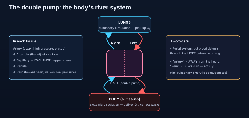
The single most useful idea in the whole subject: the heart is not one pump but two, laid side by side. The right side and the left side of the heart never mix in a healthy adult; a muscular wall, the septum, keeps them apart. And they drive two completely different loops, wired in series so blood must pass through both. The right side receives dark, oxygen-poor blood returning from the body and pushes it the short distance to the lungs — this is the pulmonary circulation, whose whole purpose is to drop off carbon dioxide and pick up oxygen at the air sacs. Freshly oxygenated blood then returns to the left side, which is the powerful pump: it drives that bright red blood out under high pressure to the entire body — the systemic circulation — from your brain to your toes. Body to right heart, right heart to lungs, lungs to left heart, left heart to body, and round again. Everything else in this article is detail hung on that skeleton. (The chambers, valves and coronary supply of the heart itself get their own full treatment in the Thorax wave — here we care only about the plan.)
Picture a figure-eight racetrack with the heart at the crossing point in the middle. The small upper loop runs to the lungs; the big lower loop runs to the whole body. A car (a red cell) that finishes the big loop can't go round again until it has done one lap of the small loop first — the two circuits are locked in series. And because they share a wall in the middle, the two pumps beat in perfect lockstep: one squeeze fills the small loop while the other fills the big one, at the very same instant.
The vessel types, following a drop of blood
The plumbing is not one kind of pipe but a graded series, each shaped for its job. Leaving the left heart, blood enters the arteries — thick-walled, elastic, muscular tubes built to survive the pounding pressure of each heartbeat. The largest, the aorta, has a wall so springy it stretches with each surge and recoils between beats, smoothing the flow like a shock absorber. Arteries branch again and again into arterioles, the small resistance vessels. These are the taps of the system: their muscular walls tighten or relax to decide how much blood reaches each tissue — and, added up across the body, they set your blood pressure. The autonomic nervous system tunes them constantly, which is why the whole circulation answers to the wiring described in the nervous-system plan. Beyond the arterioles lie the capillaries — walls just one cell thick, so fine that red cells pass in single file. This is where the entire system earns its keep: oxygen, glucose and nutrients slip out to the tissues, and carbon dioxide and waste slip in. Everything upstream exists only to deliver blood to these microscopic exchange beds; everything downstream only to carry it away. The capillaries drain into venules, then into veins — thin-walled, low-pressure vessels that carry blood back to the heart. Because the pressure here is so gentle, many veins carry one-way valves, and the squeeze of surrounding muscles acts as a pump to push blood uphill against gravity. The great veins, the superior and inferior venae cavae, finally return everything to the right heart.
Here is the trap that catches every beginner. "Artery" does NOT mean "oxygen-rich blood," and "vein" does not mean "oxygen-poor blood." The definition is purely about direction: an artery carries blood AWAY from the heart, a vein carries it TOWARD the heart — full stop. Usually arteries do carry bright oxygenated blood and veins dark deoxygenated blood, so the shortcut feels safe. But the pulmonary circulation breaks it wide open: the pulmonary artery leaves the right heart carrying dark, oxygen-poor blood to the lungs, and the pulmonary veins bring bright, oxygen-rich blood back. Away versus toward — never the colour — is the rule that never fails.
Special routes: the portal detour and the back-up roads
Most of the body follows the simple plan — artery, capillary bed, vein, home. But there are elegant exceptions. The most important is the hepatic portal system: blood leaving the stomach and intestines, loaded with everything you just absorbed from a meal, does NOT go straight back to the heart. Instead it is gathered into the portal vein and delivered first to the liver, passing through a second capillary bed there before finally draining onward. This is the body's customs checkpoint — the liver inspects, stores, detoxifies and processes the incoming nutrients before they are allowed into the general circulation. Two capillary beds in series, joined by a vein, is the signature of a portal system. A second theme worth knowing is the anastomosis: places where neighbouring arteries join, creating collateral routes so that if one path is blocked, blood can still reach the tissue by a detour — the body's built-in redundancy, richly developed around the brain, the heart, and the gut.
Press two fingers on the thumb-side of your wrist and you feel a pulse — that throb is an artery, the wall snapping outward with each surge of pressure from the heart. You will never feel a pulse over a vein, because venous pressure is too low and steady to throb; this is exactly why the pulse is one of medicine's oldest signs. Now look at the back of your hand: the blue-green cords standing up under the skin are superficial veins, low-pressure and valve-studded, easy to see precisely because they are so gentle. When those valves wear out and fail, blood pools and the veins swell and twist into the ropey, aching cords called varicose veins — a portal into how the venous plan works when it stops working. And the difference in pressure explains injuries too: nick a vein and it wells up darkly, but cut an artery and bright red blood spurts in time with the heartbeat, because it is carrying the full force of the pump.
- The heart is a double pump: right side → lungs (pulmonary), left side → body (systemic), the two loops wired in series.
- The pulmonary loop picks up oxygen and drops off CO₂; the systemic loop delivers oxygen to every tissue.
- Vessel order following the blood: artery → arteriole → capillary → venule → vein.
- Arterioles are the adjustable taps set by the autonomic system; together they control flow and blood pressure.
- Capillaries — one cell thick — are where the actual exchange of gases, nutrients and waste happens; the whole point of the system.
- Veins are thin, low-pressure, valve-bearing, and helped by muscle pumps to return blood to the heart.
A network that answers to the nerves — and to drugs
The circulation is not a passive set of pipes; it is under second-by-second control. The walls of arterioles and of veins contain smooth muscle, and the heart itself is built of cardiac muscle — two of the tissues introduced in muscle and movement — and both answer to the autonomic nervous system. When you are frightened, the sympathetic "fight-or-flight" arm speeds the heart and clamps down arterioles in the skin and gut to shunt blood to the muscles; when you are calm, the "rest-and-digest" arm slows the heart again. This is the same balance drawn out in fight-or-flight vs rest-and-digest, and it is precisely why some of the most-used drugs in medicine target these vessels and this pump. Beta-blockers act on the heart's own receptors to slow it and lower its workload, as covered in beta-blockers and the adrenergic antagonists; calcium-channel blockers relax the arteriolar muscle to widen the taps and drop the pressure, as in calcium-channel blockers. Even a blush is this system on display — sympathetic tone easing off lets the facial arterioles fill; going pale with shock is the opposite, the taps clamping shut. And because blood is meant to stay flowing and never clot inside an intact vessel, the whole machinery of clotting and the drugs that tune it — the subject of the haemostasis drug map — is built around keeping this river both liquid and repairable.
- "Artery" = away from the heart; "vein" = toward the heart — direction, NOT oxygen content.
- The pulmonary artery carries deoxygenated blood; the pulmonary veins carry oxygenated blood — the classic exception.
- The hepatic portal system routes gut blood through the liver first — two capillary beds in series before returning to the heart.
- Anastomoses are joins between arteries that give collateral back-up routes if one path is blocked.
- Arteriolar and cardiac muscle answer to the autonomic system — the target of beta-blockers and calcium-channel blockers.
- Believing all arteries carry oxygen-rich blood and all veins carry oxygen-poor blood — the pulmonary vessels prove the definition is about direction, not colour.
- Thinking of the heart as a single pump. It is two pumps side by side, driving two loops that are wired in series, not parallel.
- Assuming all blood returns straight to the heart. Blood from the gut takes the portal detour through the liver first, passing a second capillary bed on the way.
A student states that "all arteries carry oxygen-rich blood." Which single vessel best proves this statement wrong?
- The heart is a double pump: the right side drives the pulmonary loop (to the lungs for oxygen), the left drives the systemic loop (to the body) — two circuits in series.
- Following the blood: arteries (thick, high-pressure) → arterioles (the taps that set pressure) → capillaries (one cell thick, where exchange happens) → venules → veins (thin, valved, low-pressure) back to the heart.
- "Artery" means away from the heart and "vein" means toward it — direction, not oxygen: the pulmonary artery carries deoxygenated blood.
- Special routes: the hepatic portal system sends gut blood through the liver first (two capillary beds in series), and anastomoses give collateral back-up paths.
- Gray's Anatomy for Students (Drake, Vogl, Mitchell) — Introduction: the cardiovascular system.
- Moore's Clinically Oriented Anatomy — Overview of the cardiovascular system and its vessels.
- Netter's Atlas of Human Anatomy — Plates on the systemic and pulmonary circulation.
- Snell's Clinical Anatomy by Regions — Blood vessels: arteries, veins, and capillaries.
- Last's Anatomy: Regional and Applied — The vascular system and portal circulation.
- TeachMeAnatomy — The cardiovascular system: vessels and circulatory loops.

