
The Carotid and Jugular: The Great Highway of the Neck
Every drop of blood the brain and the face receive travels up one side of a single fascial tube in the neck, and almost all of it comes back down the other. Put two fingers on the front of your neck, beside the voice box, and you are a centimetre from an artery that carries a fifth of the heart's output to the brain — and from the exact point where that artery splits in two. That fork is not just plumbing. It is a pressure sensor wired to the heart, an oxygen sensor wired to your breathing, and, in later life, the single commonest place in the whole body for the plaque that causes a stroke. One tube in the neck holds the artery of the brain, the vein that drains it, and the nerve that governs the heart, all bound together — and knowing what lies where in that tube is the difference between a safe operation and a catastrophe.
A 68-year-old man is on the operating table under local anaesthetic, wide awake, squeezing a child's squeaky toy in his right hand and reciting the days of the week aloud. He had a warning stroke three weeks ago — an hour of slurred speech and a weak arm that recovered — and a scan found a tight, ragged plaque narrowing his left internal carotid artery just above where it branches. The surgeon has opened the neck along the front border of the sternocleidomastoid, parted the fascia, and there is the carotid sheath: a glistening tube with the pulsating common carotid inside it, the great blue internal jugular vein lying alongside, and, tucked in the groove between them, the pale cord of the vagus nerve that must not be touched. The surgeon clamps the artery. The toy keeps squeaking; the days keep coming. As long as the man can talk and squeeze, the other carotid and the ring of vessels at the base of the brain are feeding it enough. The artery is opened, the yellow plaque peeled out whole like a cast of the vessel, the wall closed, the clamps released — and a man who was three weeks from a major stroke walks out two days later. The whole operation is a lesson in one tube in the neck: what is in it, what must be spared, and why the point where the artery divides is the one that kills.
One sheath, three travellers
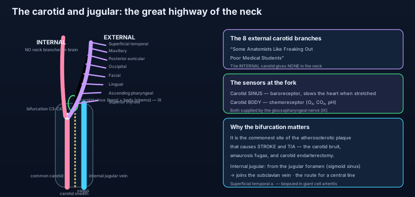
Learn the order of the three structures in the carotid sheath and half of neck surgery is demystified. The CAROTID SHEATH is a tube of condensed deep cervical fascia that runs the length of the neck, deep to the sternocleidomastoid, blending above with the base of the skull around the carotid canal and jugular foramen and below with the root of the neck; its full place among the fascial planes is drawn in the triangles and fasciae of the neck. It carries three great travellers, and their relationship never changes. MEDIALLY and anteriorly lies the artery — the COMMON CAROTID below the bifurcation, the INTERNAL CAROTID above it. LATERALLY, filling most of the sheath, is the INTERNAL JUGULAR VEIN, broad and thin-walled. And BETWEEN and BEHIND the two, in the groove where artery meets vein, runs the VAGUS NERVE (CN X), the tenth cranial nerve, on its way to the thorax and abdomen — one of the twelve surveyed in the cranial nerves overview. Two further structures cling to the sheath rather than lying within it: the ANSA CERVICALIS, the nerve loop to the strap muscles, is embedded in its anterior wall, and the deep cervical lymph nodes run along the internal jugular vein just outside it. Behind the sheath, on the prevertebral fascia, lies the cervical sympathetic chain — near enough that a low neck lesion can catch both the sympathetic trunk and the sheath together.
The common carotid — and the fork that changes everything
Two carotids, two different origins — and one shared point where they divide. The two common carotid arteries begin in different places, and the asymmetry matters. The RIGHT common carotid is a branch of the BRACHIOCEPHALIC TRUNK, arising behind the right sternoclavicular joint. The LEFT common carotid arises directly and higher, as the second branch of the ARCH OF THE AORTA in the superior mediastinum — which is why a left carotid is drawn into the story of the great vessels of the chest, set out in the aorta and great vessels. From there each ascends within the carotid sheath, and here is the identifying feature students forget: the common carotid gives NO BRANCHES in the neck. It runs straight up, and at the UPPER BORDER OF THE THYROID CARTILAGE — roughly the level of the C3–C4 disc — it divides into its two terminal branches: the EXTERNAL and INTERNAL carotid arteries. That bifurcation is the anatomical hub of the whole region. It is where you feel the carotid pulse, where the pressure and oxygen sensors sit, where the surgeon's plaque forms, and where the internal and external carotids part company to supply, respectively, the brain and the face.
The carotid sinus and the carotid body — two sensors at the fork
One measures pressure, one measures chemistry, and both report to the same nerve. Sitting exactly at the bifurcation are two small structures that turn the fork into a control station. The CAROTID SINUS is a slight dilatation in the wall of the proximal INTERNAL carotid artery, just above the bifurcation. Its wall is thin and packed with stretch-sensitive nerve endings: it is a BARORECEPTOR, a pressure sensor, supplied by the GLOSSOPHARYNGEAL nerve (CN IX) through a twig called the nerve of Hering. When blood pressure rises the sinus wall stretches, the receptors fire faster, and a reflex arc through the brainstem SLOWS the heart and DROPS the blood pressure — the negative-feedback loop that keeps pressure stable second to second. This is why firm CAROTID SINUS MASSAGE can slow a racing heart, and why, in some older people, an oversensitive sinus — CAROTID SINUS HYPERSENSITIVITY — can be triggered by a tight collar or a turn of the head and cause a faint. A few millimetres away is the CAROTID BODY, a tiny reddish nodule of chemoreceptor tissue behind the bifurcation, also supplied by the glossopharyngeal nerve. It is a CHEMORECEPTOR: it senses a fall in oxygen, a rise in carbon dioxide, and a fall in pH, and drives the reflex increase in breathing that those changes demand. Pressure at the sinus, chemistry at the body, both wired to CN IX — one fork, two senses.
Think of the carotid bifurcation as the instrumented junction on a motorway where the main road splits toward two cities. Engineers do not scatter their sensors randomly along the road; they cluster them at the junction, because that is where the flow can be read and controlled for everything downstream. The carotid sinus is the pressure gauge built into the junction wall, reporting to a control room that can throttle the pump — the heart — the instant pressure climbs. The carotid body is the air-quality monitor beside it, sampling the blood for oxygen and acidity and calling for faster breathing when the readings drop. And a junction, precisely because it is where flow divides and slows and swirls, is also where debris settles and the road narrows first. That is the bifurcation's double character: the best place in the arterial tree to sense and regulate, and the worst place for turbulence to lay down the plaque that eventually blocks the road to the brain.
The internal carotid — no branches, straight to the brain
The INTERNAL CAROTID ARTERY is the artery of the brain, and it announces itself by a single unmistakable feature: in the neck it gives OFF NO BRANCHES at all. A surgeon who has isolated two arteries at the bifurcation and needs to know which is which uses exactly this — the branchless vessel is the internal carotid; the one throwing off branches is the external. From the bifurcation the internal carotid ascends, usually posterolateral to the external at first, and runs straight up without dividing to the base of the skull, where it enters the CAROTID CANAL in the petrous part of the temporal bone. It then turns forward and medially through the canal, passes above the foramen lacerum, and enters the CAVERNOUS SINUS, where it makes the S-shaped bend called the CAROTID SIPHON with the abducens nerve beside it and the other cavernous nerves in the sinus wall. Emerging beside the optic nerve, it finally pierces the dura and enters the cranial cavity. There it gives the branches that matter: the OPHTHALMIC ARTERY to the eye, the ANTERIOR and MIDDLE CEREBRAL ARTERIES, and the posterior communicating artery — together the ANTERIOR CIRCULATION that feeds the front two-thirds of the cerebral hemispheres and forms the front half of the circle of Willis, traced in full in the vessels of the head and neck. Everything the internal carotid does of consequence, it does inside the skull; in the neck it is a silent conduit.
The external carotid and its eight branches
"Some Anatomists Like Freaking Out Poor Medical Students" — eight branches, in order. The EXTERNAL CAROTID ARTERY is the artery of the face, scalp, jaws and neck, and unlike its silent sibling it is prolific: it gives EIGHT branches before ending, behind the neck of the mandible within the parotid gland, by dividing into its two terminal branches. The classic mnemonic lists them from below upward — Some Anatomists Like Freaking Out Poor Medical Students. The SUPERIOR THYROID artery runs down to the top of the thyroid gland and the larynx. The ASCENDING PHARYNGEAL, the smallest and the only one arising from the deep (medial) surface, climbs to the pharynx and skull base. The LINGUAL artery loops forward to supply the tongue and floor of the mouth. The FACIAL artery hooks over the mandible to snake across the face to the medial angle of the eye, giving the arteries of the lip. The OCCIPITAL artery runs back to the posterior scalp. The POSTERIOR AURICULAR supplies behind the ear. Then come the two TERMINAL branches: the MAXILLARY artery, the larger, which plunges into the infratemporal fossa to supply the deep face and — through two named branches every clinician must know — the MIDDLE MENINGEAL ARTERY to the dura (the vessel torn in an extradural haematoma, discussed with the meninges and dural venous sinuses) and the INFERIOR ALVEOLAR ARTERY to the lower teeth; and the SUPERFICIAL TEMPORAL artery, which continues the line of the external carotid straight up in front of the ear — the pulse you can feel at the temple, and the artery biopsied to diagnose giant cell (temporal) arteritis.
The single fastest way to identify the internal carotid at operation or in an exam is a negative: it has NO branches in the neck. Every other major artery in the region betrays itself by branching — the external carotid throws off eight, the subclavian its own set. So when you must name a vessel at the bifurcation, do not hunt for what the internal carotid gives; hunt for what it does NOT. The one that ascends silently, branchless, usually lying a little posterior and lateral, is the internal carotid heading for the skull base. The one giving the superior thyroid almost immediately, then peeling off branch after branch to the face, is the external. This one fact organises the whole bifurcation: it tells the surgeon which artery to clamp for a stroke operation (the internal, above the plaque), which can be tied off in an emergency without killing the brain (the external, richly collateralised), and which the pathologist calls the culprit when a clot at the fork flicks debris upward into the eye or the brain.
The internal jugular — the great vein coming down
It begins where a dural sinus ends and ends where the arm's vein joins it. The INTERNAL JUGULAR VEIN is the main venous drainage of the brain, face and neck, and it is the direct continuation of the dural venous sinuses. It BEGINS at the JUGULAR FORAMEN in the skull base as the continuation of the SIGMOID SINUS — the last of the dural sinuses that carry blood from inside the skull, whose course from the great sagittal and transverse sinuses is followed in the meninges and dural venous sinuses. It emerges dilated at its origin (the superior bulb), then descends the neck within the carotid sheath, LATERAL to the carotid arteries, all the way to the root of the neck. Along the way it receives the FACIAL, LINGUAL, PHARYNGEAL, and SUPERIOR and MIDDLE THYROID veins. Behind the medial end of the clavicle, at the sternoclavicular joint, it is joined by the SUBCLAVIAN VEIN — the vein of the upper limb — and the two unite to form the BRACHIOCEPHALIC VEIN. Two clinical uses follow directly from its size and position. First, its surface marking — a line from the earlobe to the medial end of the clavicle, deep to the sternocleidomastoid — makes it the preferred route for CENTRAL VENOUS ACCESS, a large catheter passed into it and on toward the heart. Second, because there is no valve between the right atrium and the internal jugular, the column of blood in it transmits the pressure of the right atrium: the JUGULAR VENOUS PULSE, visible in the neck, is a bedside gauge of right heart filling. Superficial to all this, crossing the sternocleidomastoid obliquely in the superficial fascia, runs the smaller EXTERNAL JUGULAR VEIN, draining the scalp and face outside the sheath.
- The CAROTID SHEATH contains three structures: the common/internal carotid artery MEDIALLY, the internal jugular vein LATERALLY, and the vagus nerve (CN X) in the groove BETWEEN and BEHIND them.
- The RIGHT common carotid arises from the brachiocephalic trunk; the LEFT arises directly from the ARCH OF THE AORTA. Each ascends with NO branches and divides at the upper border of the thyroid cartilage (about C3–C4).
- At the bifurcation: the CAROTID SINUS (a baroreceptor in the proximal internal carotid wall, sensing pressure) and the CAROTID BODY (a chemoreceptor sensing O₂, CO₂ and pH) — BOTH supplied by the GLOSSOPHARYNGEAL nerve (CN IX).
- The INTERNAL carotid gives NO branches in the neck (its identifying feature), ascends through the carotid canal and cavernous sinus (the siphon), and supplies the brain and eye (ophthalmic, anterior and middle cerebral — the anterior circulation).
- The EXTERNAL carotid's EIGHT branches — "Some Anatomists Like Freaking Out Poor Medical Students": Superior thyroid, Ascending pharyngeal, Lingual, Facial, Occipital, Posterior auricular, then the terminal Maxillary (→ middle meningeal, inferior alveolar) and Superficial temporal.
- The INTERNAL JUGULAR VEIN begins at the JUGULAR FORAMEN as the continuation of the SIGMOID SINUS, descends in the carotid sheath lateral to the artery, receives the facial, lingual, pharyngeal and superior/middle thyroid veins, and joins the SUBCLAVIAN vein to form the BRACHIOCEPHALIC vein.
- The internal jugular is used for CENTRAL VENOUS ACCESS (surface marking: earlobe to medial clavicle, deep to SCM); with no valve to the right atrium, the JUGULAR VENOUS PULSE gauges right atrial pressure at the bedside.
- The EXTERNAL JUGULAR VEIN crosses the sternocleidomastoid superficially in the superficial fascia, draining the scalp and face OUTSIDE the sheath.
- CAROTID STENOSIS at the bifurcation is a major cause of stroke and TIA; the turbulent narrowing gives a CAROTID BRUIT, and treatment is CAROTID ENDARTERECTOMY — with the hypoglossal (XII), vagus (X) and recurrent laryngeal nerves at risk.
- CAROTID SINUS MASSAGE slows a supraventricular tachycardia by exciting the baroreflex (CN IX → vagal slowing); an oversensitive sinus (carotid sinus hypersensitivity) can cause syncope with a tight collar or head-turn.
- A CAROTID BODY TUMOUR (chemodectoma / paraganglioma) presents as a slowly growing, pulsatile mass at the bifurcation, classically mobile side-to-side but not up-and-down, splaying the carotid fork on imaging.
When the fork narrows — stroke, endarterectomy and the nerves at risk
The carotid bifurcation is the commonest site of atherosclerotic plaque in the whole arterial tree, because the swirl and shear of blood dividing there injures the wall first. A plaque that narrows the internal carotid origin — CAROTID STENOSIS — causes strokes in two ways: by shedding fragments of clot and debris that travel up into the middle cerebral territory (an embolic stroke, or, if the retinal artery is hit, a fleeting monocular blindness called amaurosis fugax), and, less often, by simply throttling flow. The turbulence over the narrowing can often be heard with a stethoscope as a CAROTID BRUIT. When the stenosis is tight and has already caused a TIA or minor stroke, the plaque is removed by CAROTID ENDARTERECTOMY — the operation in the opening scene — and its anatomy is a roll-call of what lies around the sheath. The HYPOGLOSSAL NERVE (CN XII) crosses the internal and external carotids high in the wound and is at risk (injury deviates the tongue toward the damaged side). The VAGUS lies in the sheath itself. Its branch the RECURRENT LARYNGEAL NERVE, and its SUPERIOR LARYNGEAL branch, supply the larynx — so a careless clamp or retractor can leave a patient hoarse, the laryngeal consequences set out with the larynx. Two other everyday clinical threads run through this same anatomy. The SUPERFICIAL TEMPORAL ARTERY, the terminal branch felt in front of the ear, is the vessel inflamed in GIANT CELL (TEMPORAL) ARTERITIS — an older patient with a new headache, scalp tenderness and jaw pain on chewing (jaw claudication), whose feared complication is sudden blindness and whose diagnosis rests on a temporal artery biopsy. And CAROTID SINUS MASSAGE, applied to the sinus at the bifurcation, is used deliberately to slow a supraventricular tachycardia through the baroreflex — the same reflex that, when the sinus is hypersensitive, makes an old man faint when he buttons a tight collar.
A 55-year-old woman notices a painless lump high in the side of her neck, at the level of the hyoid, that has grown slowly over two years. On examination it is firm, it pulsates, and — the classic sign — it can be moved a little from side to side but not up and down, because it is tethered to the artery beneath it. A bruit can be heard over it. Imaging shows a mass sitting in the crotch of the bifurcation, splaying the internal and external carotids apart like a ball pushed into the fork of a slingshot: the "lyre sign". This is a CAROTID BODY TUMOUR, a chemodectoma or paraganglioma arising from the chemoreceptor tissue of the carotid body. It is usually benign and slow, but it is highly vascular and intimately wrapped around the carotids, so it is never simply excised on the ward — it needs imaging, sometimes preoperative embolisation, and careful surgery with the same nerves (vagus, hypoglossal) at risk as in an endarterectomy. It is the anatomy of the bifurcation made into a lump: a sensor that was meant to measure the blood has grown into a mass that grips the very arteries it once monitored.
- Confusing the internal and external carotid at the bifurcation. The INTERNAL carotid gives NO branches in the neck; the EXTERNAL gives eight, beginning with the superior thyroid almost at once. If a vessel is branching in the neck, it is the external — never the internal.
- Mixing up the two sensors. The carotid SINUS is a BARORECEPTOR (pressure) in the arterial wall; the carotid BODY is a CHEMORECEPTOR (O₂/CO₂/pH). Both are supplied by the glossopharyngeal nerve (CN IX) — not the vagus — even though the reflex EFFECT on the heart is carried out through the vagus.
- Placing the vagus nerve outside the carotid sheath. The vagus runs WITHIN the sheath, in the groove BETWEEN and BEHIND the artery and the internal jugular vein — which is exactly why it is at risk in carotid surgery. The sympathetic chain, by contrast, lies BEHIND the sheath on the prevertebral fascia.
During a carotid endarterectomy the surgeon isolates the two branches of the bifurcation. One vessel gives off a branch to the superior pole of the thyroid gland almost immediately; the other gives no branches at all in the neck. Which is which, and which nerve, lying in the sheath itself, is most directly at risk of hoarseness if injured?
- The CAROTID SHEATH binds three travellers the length of the neck: the common/internal carotid artery MEDIALLY, the internal jugular vein LATERALLY, and the vagus nerve (CN X) in the groove BETWEEN and BEHIND them — the arrangement that makes carotid surgery a neurovascular operation.
- The RIGHT common carotid comes from the brachiocephalic trunk, the LEFT from the arch of the aorta; each ascends branchless and divides at C3–C4 (upper border of thyroid cartilage) into the INTERNAL carotid (no neck branches → brain and eye, anterior circulation) and the EXTERNAL carotid (eight branches → face, scalp, jaws).
- At the bifurcation the CAROTID SINUS (baroreceptor) and CAROTID BODY (chemoreceptor) both report to the GLOSSOPHARYNGEAL nerve (CN IX). The external carotid's eight branches: Superior thyroid, Ascending pharyngeal, Lingual, Facial, Occipital, Posterior auricular, then Maxillary (→ middle meningeal) and Superficial temporal.
- The internal jugular vein descends the sheath from the jugular foramen (continuing the sigmoid sinus) to join the subclavian vein — used for central lines and for reading the JVP. Clinically the bifurcation is the commonest site of stroke-causing plaque: carotid stenosis, bruit, and endarterectomy (sparing CN XII, X and the recurrent laryngeal); plus carotid sinus massage, temporal arteritis and the carotid body tumour.
- Drake RL, Vogl AW, Mitchell AWM. Gray's Anatomy for Students — Head and Neck: the carotid sheath, common carotid and its bifurcation, external and internal carotid arteries, and the internal jugular vein.
- Moore KL, Dalley AF, Agur AMR. Clinically Oriented Anatomy — Vasculature of the neck; the carotid system, carotid sinus and carotid body; the internal jugular vein and central venous access.
- Netter FH. Atlas of Human Anatomy — Arteries and veins of the neck; the external carotid artery and its branches; the carotid sheath in cross-section.
- Last RJ. Last's Anatomy: Regional and Applied — The great vessels of the neck and the carotid sheath.
- Snell RS. Clinical Anatomy by Regions — The carotid arteries, the carotid sinus and body, and carotid endarterectomy.
- TeachMeAnatomy — The Common Carotid Artery; The External Carotid Artery; The Internal Jugular Vein.

