
The Diaphragm: The Muscle That Never Rests
Every other muscle in your body is allowed to stop. The quadriceps rest when you sit; the jaw rests between meals; even the heart rests, briefly, between beats. The diaphragm has been contracting every few seconds since before you were born — in the womb it practised, rhythmically, on amniotic fluid — and it will not stop until the moment you die. Somewhere between five and seven hundred million contractions in an average lifetime, and not one of them required your permission. It is a thin dome of muscle, no thicker in places than a few sheets of paper, and it separates the two great cavities of the trunk while letting the aorta, the oesophagus and the inferior vena cava pass through it without ever leaking. Understanding how it does that — three origins, three openings, one nerve — is understanding how you breathe.
A young woman arrives in the emergency department at two in the morning holding her right shoulder. She has not fallen, has not lifted anything, has not slept awkwardly. The shoulder is not tender when pressed and moves through a full range without complaint — and yet the pain is real enough that she cannot lie flat. The registrar spends less than a minute on the shoulder. He asks instead about her last menstrual period, presses gently on her lower abdomen, and orders a pregnancy test and an ultrasound. Blood from a ruptured ectopic pregnancy has tracked upwards and is pooling against the undersurface of her diaphragm. The nerve that carries that irritation entered the spinal cord at the fourth cervical segment — the same segment that supplies the skin over the shoulder — and the brain, receiving a message on a familiar wire, read the return address wrong. She is in theatre within the hour. The shoulder was never the problem; it was the diaphragm calling from four levels away.
A dome, not a plate
The floor of the chest is also the ceiling of the abdomen — and it is not flat. The diaphragm is a musculotendinous sheet shaped like a pair of domes, rising far higher into the thorax than students expect. Its muscle fibres begin all around the lower margin of the thoracic outlet described in the thoracic cage, sweep upwards and inwards, and converge on a flat trefoil-shaped sheet of tendon in the middle — the central tendon. Every fibre therefore pulls towards that tendon rather than towards a bone. The right dome climbs to about the level of the fifth rib in quiet expiration and the left dome to the fifth intercostal space: the right sits roughly half a vertebral level higher because the liver, the heaviest organ in the abdomen, is pushed up under it. The central tendon itself is not free to wander, because its upper surface is fused with the fibrous pericardium above it — the heart quite literally sits on the diaphragm and rides up and down with every breath you take. In quiet breathing the domes descend only about one and a half centimetres, yet that modest travel moves roughly three quarters of the air of a normal tidal breath; in deep inspiration the excursion reaches seven to ten centimetres, and the whole abdominal contents are displaced downwards and forwards to make room. This is why the levels of everything inside the chest are always quoted "in expiration": in full inspiration the diaphragm, the heart resting on it, the lung bases and the openings themselves have all migrated south — and a needle placed safely below the ribs at the end of a breath out can be inside the pleural cavity at the end of a breath in.
Three origins, one muscle
Walk the rim of the diaphragm from front to back and you cross three different territories. The STERNAL part is the smallest: two slips arising from the back of the xiphoid process. The COSTAL part is the largest: fleshy fibres from the inner surfaces of the lower six costal cartilages and their adjacent ribs on each side. These costal fibres interdigitate — interlock like the fingers of two clasped hands — with the fibres of transversus abdominis, which is the anatomical reason the thoracic and abdominal walls behave as one continuous muscular tube rather than two separate boxes. The LUMBAR part is the deepest and most interesting: it arises not only from the vertebral bodies through the two crura, but from two thickened arches of fascia thrown across the muscles of the posterior abdominal wall. The crura are two tendinous pillars gripping the front of the upper lumbar vertebrae: the right crus is the longer and stronger, arising from the bodies and intervertebral discs of L1 to L3, while the left crus arises from L1 and L2 only. Lateral to them, the medial arcuate ligament arches over psoas major from the body of L1 to the transverse process of L1, and the lateral arcuate ligament arches over quadratus lumborum from the transverse process of L1 to the twelfth rib; between the two crura the median arcuate ligament arches across the midline in front of the vertebral column, and it is beneath this arch that the aorta escapes. Each arcuate ligament exists for the same simple reason: a muscle already lies in the way, so the diaphragm vaults over it rather than through it.
Picture a bell jar with a rubber sheet stretched across its open bottom and a small balloon hanging inside on a straw. Pull the rubber sheet downwards and the space inside the jar enlarges, the pressure in it falls, and the balloon inflates without anyone blowing into it. Let the sheet spring back and the balloon empties. That is the whole of quiet breathing, and it is why the lungs are passive passengers — they do not suck air in, they are inflated by a pressure the diaphragm creates around them, a mechanism laid out in breathing mechanics. The one detail the jar model misses is the most elegant part of the real thing: the real sheet has three holes in it, and each hole is engineered differently so that pulling the sheet down does not squeeze shut the tubes running through it.
The three great openings
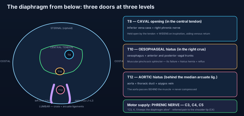
Count the letters and you have the levels: vena cava (8) at T8, oesophagus (10) at T10, aortic hiatus (12) at T12. The CAVAL opening lies at the level of T8, slightly to the right of the midline, and — this is the whole point — it is punched through the CENTRAL TENDON, not through muscle. It transmits the inferior vena cava, whose wall is adherent to the margins of the opening, together with the right phrenic nerve. Because the tendon cannot contract, the opening is held permanently open; and because the tendon is stretched taut as the domes descend, the caval opening actually WIDENS during inspiration. The falling intrathoracic pressure and the widening aperture pull blood upwards together, so every breath in is also a small assist to venous return. A structure whose only requirement is that it must never be squeezed has been placed in the one part of the diaphragm that cannot squeeze.
The OESOPHAGEAL hiatus lies at T10, and its design is the exact opposite. It is a slit in the MUSCLE FIBRES of the right crus, which split and loop around the oesophagus like a sling before rejoining. It transmits the oesophagus itself together with the anterior and posterior vagal trunks — the left vagus rotates to the front and the right to the back as the stomach turns during development — plus the oesophageal branches of the left gastric vessels. Because the aperture is muscular, it TIGHTENS every time the diaphragm contracts, acting as an external pinchcock that helps the lower oesophageal sphincter keep stomach contents where they belong, as discussed in the oesophagus. When that sling weakens and widens, the stomach begins to climb through it: hiatus hernia, and with it heartburn that is worse lying flat, worse after a large meal, worse when bending forward.
The AORTIC hiatus at T12 is a trick of language, because it is not truly an opening in the diaphragm at all: the aorta passes BEHIND the muscle, under the arch of the median arcuate ligament, between the two crura and in front of the vertebral body. Nothing muscular surrounds it, so it is never compressed and aortic flow is unaffected by breathing — a necessity, since the vessel described in the aorta and great vessels carries the entire cardiac output to the lower body. Two other structures share this osteofibrous tunnel: the thoracic duct, which lies just behind and to the right of the aorta, and the azygos vein (often with the hemiazygos), both covered in the thoracic duct and thymus. So the lowest opening is the one the diaphragm does not really own — it simply arches over it and leaves it alone.
The smaller doorways
Beyond the famous three, several smaller structures cross the diaphragm and each has a reason to be remembered. The superior epigastric vessels — the continuation of the internal thoracic artery and vein — pass between the sternal and costal origins, through the small triangular gap known as the sternocostal triangle or foramen of Morgagni, to enter the rectus sheath and supply the anterior abdominal wall. The greater, lesser and least splanchnic nerves pierce the crura on their way from the thoracic sympathetic chain to the coeliac and renal ganglia, which is why abdominal pain is carried on nerves that began in the chest. The sympathetic trunk itself slips behind the medial arcuate ligament, and the subcostal nerve and vessels behind the lateral arcuate ligament. The left phrenic nerve, having no caval opening to use, pierces the muscle of the left dome on its own, just lateral to the pericardium — the two phrenic nerves reach their target by different routes, a small asymmetry with a large consequence in surgery.
"C3, 4, 5 keeps the diaphragm alive"
A muscle in the abdomen taking orders from the neck sounds absurd — until you know where it was built. The entire motor supply of the diaphragm comes from the phrenic nerve, and from nothing else. Each phrenic nerve arises from the anterior rami of C3, C4 and C5 — mainly C4 — descends through the neck on the front of scalenus anterior, enters the thorax, and runs the whole length of the mediastinum in front of the root of the lung to reach the muscle, where it divides into branches that fan out from below. No intercostal nerve, no vagus, no sympathetic fibre can move the diaphragm. The reason for this improbable arrangement is embryological: the muscle begins life as the septum transversum, a plate of mesoderm in the CERVICAL region, and it drags its nerve supply caudally with it as the embryo folds and the muscle descends to its final position. The nerve was recruited in the neck and never let go, a course traced in more detail in the nerves of the thorax. Sensation, by contrast, is divided, and the division is clinically decisive: the central part of the diaphragm — the tendon and the pleura and peritoneum covering it — is supplied sensorily by the phrenic nerve, so its pain is referred to the C3–C5 dermatomes over the shoulder tip and the side of the neck, whereas the peripheral rim receives sensory fibres from the lower intercostal nerves (roughly T5 to T11) and the subcostal nerve (T12), so irritation there is felt locally in the lower chest wall and upper abdominal wall. One muscle, two completely different pain maps, depending on which part of it is touched.
Shoulder-tip pain in a patient with an abdominal problem is one of the most valuable signs in clinical medicine, and it is pure embryology. Blood under the diaphragm after a ruptured spleen (Kehr's sign) or a ruptured ectopic pregnancy, gas left behind after laparoscopic surgery, pus in a subphrenic abscess, or the inflamed gallbladder touching the diaphragm from below — all of them irritate a membrane whose nerve was recruited at C3–C5 in the fourth week of life. The spinal cord has no way of distinguishing a message from the diaphragm from a message from the skin over the shoulder, because they arrive on the same segments. So the patient rubs a shoulder that is entirely healthy, and the experienced clinician looks immediately at the abdomen.
How a dome makes a breath — and a push
The dome has two careers: one of them has nothing to do with air. When the muscle fibres contract they pull the domes down towards the flat central tendon, and the domes flatten. That single movement increases the VERTICAL diameter of the thorax, the intrathoracic pressure falls below atmospheric, and air rushes down the airways to equalize it. Once the central tendon has descended as far as the abdominal contents allow, it becomes a fixed point, and continued contraction of the costal fibres now pulls upwards and outwards on the lower ribs instead, adding to the transverse diameter — the diaphragm quietly changes jobs halfway through the breath. In quiet expiration nothing contracts at all: the muscle simply relaxes and the elastic recoil of the lungs and the pressure of the abdominal viscera push the domes back up. Now contract the same muscle against a CLOSED glottis while the abdominal wall tightens, and instead of drawing air in you raise the pressure inside the abdomen sharply: this is the Valsalva manoeuvre, the engine of coughing, sneezing, vomiting, defecation, micturition, the second stage of labour and the braced trunk of a weightlifter. It is why a person with a painful abdominal wound cannot cough properly, why chest physiotherapy matters so much after surgery, and why a patient told to "take a deep breath and bear down" is really being asked to use their diaphragm as a press rather than a pump.
When the floor gives way
Because it is a partition under pressure from below, the diaphragm fails in characteristic ways. A SLIDING hiatus hernia — about ninety-five per cent of cases — lets the gastro-oesophageal junction itself slide up through a lax T10 hiatus into the chest, destroying the pinchcock and producing reflux. A ROLLING (para-oesophageal) hernia is rarer and more dangerous: the junction stays put while the fundus of the stomach rolls up alongside the oesophagus, where it can twist or strangulate. Congenital defects are different again: a posterolateral gap (foramen of Bochdalek), far commoner on the left, allows abdominal viscera into the chest before birth and prevents the lung from developing — the newborn with a scaphoid abdomen, bowel sounds in the chest, and life-threatening pulmonary hypoplasia. A retrosternal Morgagni hernia through the sternocostal triangle is rarer, usually right-sided, and often silent for years. Blunt trauma can rupture the muscle outright, far more often on the left because the liver shields the right, and abdominal contents herniate into the chest.
Nerve failures complete the picture. Injury to a phrenic nerve anywhere along its long course — a bronchial carcinoma invading the mediastinum, surgical trauma, cold injury during cardiac surgery, or a neck injury — paralyses one dome, which then sits abnormally high on a chest X-ray as a raised hemidiaphragm and moves PARADOXICALLY upwards on sniffing. And the level of a cervical spinal cord injury is a question of life and death for exactly this reason: a lesion above C3 abolishes the phrenic supply and the patient cannot breathe without a ventilator, whereas a lesion below C5 leaves the diaphragm working, so the patient may lose the intercostals and the limbs and still breathe unaided. The whole prognosis turns on three segments of cord.
The gas that hurts the shoulder: a man wakes after a laparoscopic gallbladder removal complaining bitterly of pain in both shoulders. Nobody touched his shoulders; residual carbon dioxide is sitting under his diaphragm, and the phrenic nerve is reporting it to C4. The hiccup that will not stop: a woman develops rhythmic involuntary spasms of the diaphragm with abrupt glottic closure — the sound is the vocal cords slamming shut on an inrushing breath. Most bouts are trivial and self-limiting, but hiccups lasting days demand a search for irritation anywhere along the phrenic or vagal pathway, from the ear canal to the mediastinum to a subphrenic collection. The burn at midnight: a middle-aged man with a sliding hiatus hernia sleeps propped on three pillows because lying flat abolishes the last of his anti-reflux mechanism. The first breath: a newborn with a left Bochdalek hernia is intubated in the delivery room; her abdomen is scaphoid because her bowel is in her chest, and the lung on that side never had room to grow.
- The diaphragm is a musculotendinous double dome separating thorax from abdomen; all its fibres converge on the central tendon, whose upper surface is fused with the fibrous pericardium. The right dome sits higher than the left because of the liver.
- Three origins: STERNAL from the xiphoid process; COSTAL from the lower six costal cartilages (interdigitating with transversus abdominis); LUMBAR from the crura and the arcuate ligaments.
- The right crus arises from L1–L3 and the left from L1–L2; the medial arcuate ligament arches over psoas major, the lateral arcuate over quadratus lumborum, and the median arcuate over the aorta.
- T8 CAVAL opening — in the central tendon; transmits the inferior vena cava and the right phrenic nerve; held open by tendon and WIDENS on inspiration, assisting venous return.
- T10 OESOPHAGEAL hiatus — in the muscle fibres of the RIGHT crus; transmits the oesophagus and the anterior and posterior vagal trunks; acts as a pinchcock sphincter, and its failure is hiatus hernia with reflux.
- T12 AORTIC hiatus — BEHIND the median arcuate ligament, so the aorta is never compressed; transmits the aorta, the thoracic duct and the azygos vein.
- Smaller structures crossing it: superior epigastric vessels through the sternocostal triangle; splanchnic nerves piercing the crura; the sympathetic trunk behind the medial arcuate ligament; the left phrenic nerve piercing the left dome.
- Motor supply is ENTIRELY the phrenic nerve (C3, C4, C5) — "C3, 4, 5 keeps the diaphragm alive" — because the muscle develops from the cervical septum transversum and drags its nerve down with it.
- Sensation: the central part is phrenic (pain referred to the C3–C5 shoulder-tip dermatomes); the peripheral rim is supplied by the lower intercostal (T5–T11) and subcostal (T12) nerves and hurts locally.
- Contraction flattens the domes → increases the vertical thoracic diameter → intrathoracic pressure falls → air rushes in; it produces about three quarters of quiet tidal volume, and quiet expiration is passive elastic recoil.
- Against a closed glottis it raises intra-abdominal pressure instead — the Valsalva engine of coughing, vomiting, defecation, micturition and childbirth.
- Key failures: hiatus hernia (sliding vs rolling), phrenic palsy with a raised hemidiaphragm and paradoxical movement on sniffing, traumatic rupture (usually left), congenital Bochdalek and Morgagni hernias, and apnoea from a cord lesion above C3.
- Saying the aorta passes THROUGH the diaphragm. It passes BEHIND it, under the median arcuate ligament between the two crura — which is precisely why aortic flow is never interrupted by breathing.
- Assuming the oesophageal hiatus lies in the left crus because the stomach is on the left. It lies in the fibres of the RIGHT crus, which loop around the oesophagus as a sling.
- Expecting the intercostal nerves to move the diaphragm because they move the chest wall. They carry sensation from its periphery only; ALL motor supply is phrenic, which is why a high cord lesion stops breathing outright.
A patient's inferior vena cava is not compressed as the diaphragm contracts during inspiration; in fact its opening becomes slightly wider. Which anatomical fact best explains this?
- The diaphragm is a double-domed musculotendinous partition between thorax and abdomen, with all fibres converging on a central tendon fused to the fibrous pericardium; the right dome is higher because of the liver.
- Its three origins are sternal (xiphoid), costal (lower six costal cartilages, interdigitating with transversus abdominis) and lumbar (right crus L1–L3, left crus L1–L2, plus the medial, lateral and median arcuate ligaments).
- The three openings are T8 caval in the central tendon (IVC + right phrenic nerve, widening on inspiration), T10 oesophageal in the right crus (oesophagus + vagal trunks, a pinchcock whose failure is hiatus hernia) and T12 aortic behind the median arcuate ligament (aorta + thoracic duct + azygos vein, never compressed).
- Motor supply is entirely phrenic (C3, C4, C5); contraction increases the vertical thoracic diameter to draw air in and, against a closed glottis, raises intra-abdominal pressure — and its clinical signature is shoulder-tip referred pain, hiccups, hiatus hernia, a raised hemidiaphragm from phrenic palsy, and apnoea after a cord lesion above C3.
- Drake RL, Vogl AW, Mitchell AWM. Gray's Anatomy for Students — Thorax: the diaphragm and its openings.
- Moore KL, Dalley AF, Agur AMR. Clinically Oriented Anatomy — The diaphragm: attachments, apertures, innervation and referred pain.
- Netter FH. Atlas of Human Anatomy — Diaphragm viewed from below; phrenic nerve.
- Last RJ. Last's Anatomy: Regional and Applied — The diaphragm and the posterior abdominal wall.
- Snell RS. Clinical Anatomy by Regions — Diaphragmatic hernia, phrenic nerve palsy and hiccup.
- TeachMeAnatomy — The Diaphragm; The Phrenic Nerve.

