
The Approach to Anaemia: Reading the MCV to Find the Cause
Anaemia is not a diagnosis — it is a clue. A patient is pale and exhausted, the haemoglobin is low, and the temptation is to reach for iron. But which anaemia? The whole differential — a long, frightening list — collapses into three tidy boxes the moment you read one number on the full blood count: the MCV. Learn to read it and the second axis, the reticulocyte count, and the blood film stops being a mystery and becomes a map that points straight at the treatment.
A 34-year-old woman sits on the edge of the couch, out of breath after climbing the single flight of stairs to the clinic. Her lips and the inside of her lower eyelids are pale, almost bloodless. She has been tired for months. The full blood count comes back: haemoglobin 8.2 g/dL — clearly anaemic. A dozen diseases could do this. But your eyes go to a single companion number on the same report — the MCV, the average size of her red cells. It reads 68 (low). In that instant the differential shrinks: the marrow is turning out small, pale cells, and three or four causes rise to the top while the rest fall away. One number, printed for free beside the haemoglobin, has just split the whole problem into three.
What anaemia actually is
Anaemia is simply a low haemoglobin concentration for the patient's age and sex. Haemoglobin is the oxygen-carrier inside red cells, so when it falls the tissues are short of oxygen and the body complains in predictable ways: fatigue, pallor (best seen in the conjunctivae and palmar creases), breathlessness on exertion, and a pounding or racing heart as the circulation works harder to deliver what oxygen remains. These symptoms tell you the patient IS anaemic; they do not tell you WHY. The 'why' is the whole game, because the cause — not the low number itself — decides the treatment.
So the discipline of anaemia is a sorting problem: take one low haemoglobin and route it, by a few cheap tests, to its true cause — and therefore to its correct therapy chapter. The first and most powerful sorter is the MCV.
The first fork: the MCV splits the world in three
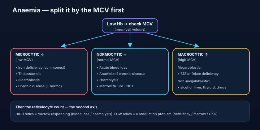
The MCV (mean cell volume) is the average size of the red cells, and it sorts every anaemia into one of three boxes. Microcytic (low MCV, small cells): the marrow is failing to fill each cell with haemoglobin. The classic causes are iron deficiency (by far the commonest anaemia worldwide), the thalassaemias, sideroblastic anaemia, and — sometimes — anaemia of chronic disease. Normocytic (normal MCV, normal-sized cells): acute blood loss (before the marrow adapts), anaemia of chronic disease, haemolysis, primary marrow failure, and the anaemia of chronic kidney disease. Macrocytic (high MCV, large cells): split into megaloblastic causes — vitamin B12 or folate deficiency — and non-megaloblastic causes — alcohol, liver disease, hypothyroidism, myelodysplasia, and certain drugs.
Anaemia of chronic disease is the great shape-shifter: it is usually normocytic but can drift microcytic. That is why it appears in two boxes. When the picture doesn't fit neatly, the chronic-disease pattern is often the reason — check the ferritin and inflammatory markers before you commit.
Two patients, both with haemoglobin 8 g/dL. Patient A has an MCV of 66 — you are now thinking iron deficiency or thalassaemia, and you order a ferritin. Patient B has an MCV of 112 — you are now thinking B12 or folate deficiency, and you order those levels plus a blood film. Same haemoglobin, completely different work-up, decided in seconds by one adjacent number.
- Anaemia = low haemoglobin; symptoms (fatigue, pallor, breathlessness) confirm it but don't explain it.
- The MCV is the first fork: microcytic, normocytic, or macrocytic.
- Microcytic → iron deficiency (commonest), thalassaemia, sideroblastic, sometimes chronic disease.
- Normocytic → acute blood loss, chronic disease, haemolysis, marrow failure, CKD.
- Macrocytic → megaloblastic (B12/folate) vs non-megaloblastic (alcohol, liver, thyroid, MDS, drugs).
The second axis: the reticulocyte count
Once the MCV has sorted by cell size, the reticulocyte count sorts by marrow behaviour. Reticulocytes are the youngest red cells, just released from the marrow — counting them tells you whether the factory is working overtime or has shut down. A high reticulocyte count means the marrow is responding hard, which points to blood loss or haemolysis: the cells are being lost or destroyed and the marrow is replacing them furiously. A low (or inappropriately normal) reticulocyte count means the marrow is NOT keeping up — a production problem: missing raw materials (iron, B12, folate), marrow failure or infiltration, or too little erythropoietin (as in chronic kidney disease).
Put the two axes together and the map is nearly complete. A normocytic anaemia with a HIGH reticulocyte count screams haemolysis or recent bleeding; the same normocytic anaemia with a LOW reticulocyte count points instead to chronic disease, early marrow failure, or renal anaemia. Size tells you the flavour; the reticulocyte count tells you whether the marrow is the victim or the culprit.
The core investigations — and where the map leads
A handful of tests confirm the box and name the cause. Ferritin is the single most useful test in microcytic anaemia: a low ferritin proves iron deficiency, while a normal-or-high ferritin with microcytosis pushes you toward thalassaemia or chronic disease. B12 and folate levels confirm or exclude the megaloblastic macrocytoses. The blood film is the cheap master-key: it shows hypochromic microcytic cells in iron deficiency, target cells in thalassaemia, oval macrocytes and hypersegmented neutrophils in megaloblastic anaemia, and fragments (schistocytes) or spherocytes in haemolysis. And the reticulocyte count, as above, separates a responding marrow from a failing one.
The whole point of the map is that each box opens a door onto a specific therapy chapter. Confirmed iron deficiency leads to the Iron chapter (oral or intravenous iron, and the search for the source of loss). A megaloblastic B12 or folate deficiency leads to the B12 & Folate chapter (replacement, and the crucial rule of never giving folate alone when B12 might be low). The anaemia of chronic kidney disease leads to the ESA chapter (erythropoiesis-stimulating agents, with iron support). A microcytic picture with normal iron leads to the Haemoglobinopathies chapter for thalassaemia. The MCV did not treat the patient — but it decided which treatment page you turn to.
- Reticulocytes high → marrow responding (blood loss or haemolysis); low → production failure.
- Ferritin is the key microcytic test: low = iron deficiency; normal/high = think thalassaemia or chronic disease.
- The blood film names patterns: hypochromic, target cells, oval macrocytes, hypersegmented neutrophils, schistocytes.
- Each box routes to a therapy chapter: iron, B12/folate, ESA, or haemoglobinopathies.
- Order early: FBC with MCV, ferritin, B12/folate, reticulocytes, and a blood film.
- Treating anaemia with iron before confirming iron deficiency. Always prove it with a ferritin first — empirical iron masks and delays the real diagnosis.
- Trusting a 'normal' MCV. A mixed deficiency (iron plus B12/folate) can average out to a normal MCV while hiding two diseases — check the film and the individual levels.
- Giving iron in thalassaemia trait. The microcytosis is not from iron lack; iron loading here is useless and potentially harmful.
- Ignoring the reticulocyte count. Without it you cannot tell a marrow that is bleeding-and-replacing from one that has simply stopped.
A patient has haemoglobin 9 g/dL, an MCV of 105 (high), and a blood film showing oval macrocytes with hypersegmented neutrophils. Which cause fits best?
- Anaemia is a clue, not a diagnosis — the cause decides the treatment, so you must sort it.
- First fork = MCV: microcytic, normocytic, macrocytic — one number splits the differential in three.
- Second axis = reticulocytes: high means marrow responding (loss/haemolysis); low means production failure.
- Confirm with ferritin, B12/folate and a blood film — then route to the iron, B12/folate, ESA or haemoglobinopathy chapter.
- Hoffbrand AV, Moss PAH. Hoffbrand's Essential Haematology — Diagnosis and classification of anaemia; the microcytic, normocytic and macrocytic approach.
- Ralston SH, et al. Davidson's Principles and Practice of Medicine — Anaemia: clinical assessment, the MCV-based classification and reticulocyte response.
- Longo DL, et al. Harrison's Principles of Internal Medicine — Approach to the patient with anaemia; red cell indices and marrow response.
- Bain BJ. Blood Cells: A Practical Guide — Morphological patterns on the blood film in anaemia.
- Kumar P, Clark M. Kumar & Clark's Clinical Medicine — Investigation of anaemia: ferritin, B12/folate and reticulocyte count.

