
Hypercalcaemia: Fluids, Bisphosphonates and the Calcium Axis
Calcium sits under a beautifully tight thermostat — the parathyroids and vitamin D nudging it up, calcitonin trimming it down — because both too much and too little of it stop nerves firing and hearts beating properly. When that thermostat breaks upward, the patient turns to stone: kidney stones, aching bones, a groaning gut, a moaning, confused mind. Two diagnoses cause almost all of it — an overactive parathyroid gland or a cancer — and the treatment is a satisfyingly logical cascade. You rehydrate first, then you turn off the bone. This chapter owns the calcium-homeostasis map, and every drug we use hangs off one of its arms.
A 68-year-old woman with known breast cancer is brought to the ward drowsy and barely making sense. Over a fortnight she has become constipated, thirsty, and is passing large volumes of pale urine; her daughter says she has been "not herself," low and vacant. She is visibly dehydrated. The corrected calcium comes back at 3.6 mmol/L — dangerously high — and the ECG shows a short QT interval. This is not a subtle outpatient finding; this is a hypercalcaemic crisis, and the cancer is driving it. The instinct to reach first for a fancy bone drug is wrong. What this woman needs, before anything else, is litres of intravenous saline — she is dry, her kidneys can barely excrete the calcium they are drowning in, and rehydration alone will pull the number down while the slower drugs take hold.
The thermostat: how the body holds calcium steady
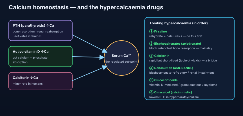
Serum calcium barely moves — because three hormones fight constantly to hold it in place. The figure below is the whole chapter in one picture. When calcium dips, the parathyroid glands release parathyroid hormone (PTH), and PTH raises calcium by three routes: it drives osteoclasts to resorb bone (releasing calcium), it makes the kidney reabsorb more calcium, and it activates vitamin D. Vitamin D — in its active form, calcitriol — is the second arm: its main job is to pull calcium in from the gut, absorbing it from food. PTH and vitamin D are cross-linked, because PTH switches vitamin D on, and together they push calcium up from bone, kidney and gut. The third hormone, calcitonin from the thyroid's C cells, does the opposite — it lowers calcium by quietening osteoclasts — but in humans it is a minor player, a footnote rather than a lever. Read the arms of that map and you can predict every drug: to lower a high calcium you either block the bone, block the gut absorption, or turn down the PTH.
Think of blood calcium as the water level in a bathtub. PTH and vitamin D are two taps pouring in — one filling from the bone-and-kidney supply, one filling from the gut. Calcitonin is a slow, weak drain. Normally the taps and drain balance and the level barely moves. Hypercalcaemia is a tap jammed open — usually an overactive parathyroid gland turning its own tap wide, or a tumour bypassing the plumbing entirely and pouring calcium straight in from dissolving bone. And the very first thing you do for an overflowing bath is not fiddle with the taps — it is open the biggest drain you have. That drain is the kidney, and IV saline is what opens it.
The two causes that matter — and the rest
Ninety per cent of hypercalcaemia is one of two diagnoses, and where you meet the patient usually tells you which. Primary hyperparathyroidism is the classic outpatient cause: an adenoma of a parathyroid gland pumps out PTH regardless of the calcium level. It is usually mild, chronic, and picked up incidentally on a blood test in a well-looking patient. Malignancy is the classic inpatient cause: it tends to be severe, rapid, and the patient is sick, as in our scene. Cancer raises calcium three ways — by secreting PTH-related peptide (PTHrP), a hormone that mimics PTH at its receptor (common in squamous and breast cancers); by direct bone metastases dissolving bone locally; and in myeloma, where malignant plasma cells drive osteoclasts hard. Beyond these two, a longer list: excess vitamin D (supplements or, classically, granulomatous disease such as sarcoidosis, where macrophages activate vitamin D unchecked); thiazide diuretics (they reduce urinary calcium and unmask it); milk-alkali syndrome; prolonged immobilisation; and thyrotoxicosis. The malignant causes connect straight to the Oncology section, where PTHrP, bone metastases and myeloma bone disease are covered in depth.
Recognising it: stones, bones, groans and moans
The symptom rhyme captures it: stones (renal calculi and nephrocalcinosis), bones (bone pain, and the fragility of hyperparathyroid bone), groans (abdominal pain, constipation, nausea, even pancreatitis), and moans (the neuropsychiatric picture — confusion, lethargy, depression, and in a crisis, coma). Two consequences deserve special weight. First, high calcium blocks the kidney's ability to concentrate urine, causing polyuria; the patient pours out dilute urine, becomes dehydrated, and the dehydration cripples the very organ that should be excreting the calcium — a vicious spiral that is the whole rationale for treating with fluids first. Second, calcium is a cardiac ion: hypercalcaemia shortens the QT interval on the ECG, and severe elevations risk arrhythmia. Mild chronic hypercalcaemia may be almost silent; a level above roughly 3.5 mmol/L, or any confusion or arrhythmia, is an emergency.
- Calcium is held steady by PTH and vitamin D (raise it) and calcitonin (lowers it — minor in humans).
- PTH raises calcium via bone resorption, renal reabsorption, and activating vitamin D; vitamin D pulls calcium from the gut.
- The two dominant causes are primary hyperparathyroidism (outpatient, mild) and malignancy (inpatient, severe).
- Malignant hypercalcaemia works via PTHrP, bone metastases, or myeloma.
- Symptoms: stones, bones, groans, moans — plus polyuria/dehydration and a short QT on ECG.
- Always use the corrected (albumin-adjusted) calcium; high calcium causes dehydration that worsens the calcium.
Treatment step 1: rehydrate — fluids come first
Before any bone drug, refill the patient and open the kidney. The single most important first step in acute severe hypercalcaemia is aggressive intravenous rehydration with 0.9% sodium chloride (normal saline). It does two things at once: it corrects the dehydration that the hypercalcaemia caused, and, by restoring renal blood flow and delivering sodium to the tubule, it promotes the excretion of calcium in the urine (calciuresis) — sodium and calcium are handled together, so a saline load drags calcium out. Several litres over the first 24 hours is typical, watching for fluid overload in the elderly or those with heart failure. This alone can drop the calcium by a useful amount within hours, buying time for the slower, definitive drugs to work. Note the historical shift: for decades teaching added a loop diuretic (furosemide) to "force" more calciuresis. That dogma is gone — loops are no longer used routinely for this, and are reserved for the patient who becomes fluid-overloaded from the saline. Fluids first, diuretic only if the tank overflows.
Treatment step 2: turn off the bone — bisphosphonates
Once the patient is rehydrated, the mainstay for lowering calcium — especially in malignancy — is an intravenous bisphosphonate: zoledronate (zoledronic acid) or pamidronate. Bisphosphonates bind avidly to bone mineral and are taken up by osteoclasts, whom they poison, shutting down bone resorption — they clamp the bone arm of the figure. Their catch is timing: they act slowly, taking two to four days to reach full effect, which is precisely why you cannot rely on them alone in a crisis and must rehydrate first. They need dose adjustment and caution in renal impairment (which frequently coexists with hypercalcaemia), and long-term use carries the rare but important risks of osteonecrosis of the jaw and atypical femoral fractures. These are the same agents used for osteoporosis and for cancer bone disease, linking this chapter to the Endocrine section (osteoporosis) and the Oncology section (bone metastases and myeloma).
The rest of the toolkit: calcitonin, denosumab, steroids, cinacalcet
Each remaining drug hangs off a different arm of the same figure. Calcitonin can be given for its speed: it lowers calcium within hours by quietening osteoclasts and increasing renal calcium loss, which makes it a useful bridge while the bisphosphonate is still ramping up. Its weakness is that the effect is short-lived — the body rapidly stops responding (tachyphylaxis) within a couple of days — so it is a bridge, never a solution. Denosumab is a monoclonal antibody against RANKL, the signal osteoclasts need to form and survive; by blocking RANKL it shuts down bone resorption even more completely than bisphosphonates, and crucially it is not cleared by the kidney, making it the agent of choice when hypercalcaemia is bisphosphonate-refractory or the patient has significant renal impairment. Glucocorticoids (prednisolone) are the specific tool for vitamin-D-mediated hypercalcaemia — they cut the active vitamin D that granulomatous disease (sarcoidosis), vitamin D toxicity and some lymphomas/myeloma generate, throttling gut absorption. Cinacalcet is a calcimimetic: it fools the parathyroid's calcium-sensing receptor into thinking calcium is high, turning down PTH — the targeted answer when the driver is hyperparathyroidism that can't be operated on. And for extreme levels or established renal failure, haemodialysis against a low-calcium bath removes calcium directly.
The whole management sequence is just the homeostasis figure read backwards. Calcium is too high, so you attack its sources one arm at a time: first flush it out through the kidney (saline), then silence the bone that is releasing it (bisphosphonate, denosumab, calcitonin), then cut the gut absorption if vitamin D is the culprit (steroids), then quieten the gland if PTH is the driver (cinacalcet). If you can picture the arrows on that diagram, you never have to memorise the drug list — each drug simply blocks one arrow. And underneath all of it: always treat the underlying cause, because a parathyroidectomy or effective cancer therapy is the only durable fix.
IV 0.9% saline — first and most important; rehydrates and drives calciuresis; onset hours. Zoledronate / pamidronate (bisphosphonates) — mainstay, especially in malignancy; block osteoclasts; onset 2–4 days; caution in renal impairment; rare jaw osteonecrosis. Calcitonin — rapid but short-lived (tachyphylaxis); a bridge, not a cure. Denosumab (anti-RANKL antibody) — for bisphosphonate-refractory cases or renal impairment; not renally cleared. Glucocorticoids (prednisolone) — for vitamin-D-mediated causes: sarcoidosis/granulomatous disease, vitamin D toxicity, myeloma. Cinacalcet (calcimimetic) — for hyperparathyroidism. Haemodialysis — for extreme levels or renal failure.
- Step 1 and most important: aggressive IV 0.9% saline to rehydrate and promote calciuresis.
- Step 2 mainstay (especially malignancy): IV bisphosphonate (zoledronate/pamidronate) — onset 2–4 days.
- Calcitonin is a fast but short-lived bridge (tachyphylaxis); denosumab suits bisphosphonate-refractory or renal-impaired patients.
- Glucocorticoids target vitamin-D-mediated/granulomatous/myeloma causes; cinacalcet targets hyperparathyroidism.
- Loop diuretics are NOT routine — only for fluid overload from the saline; thiazides raise calcium and must be stopped.
- Always treat the underlying cause — parathyroidectomy or cancer therapy is the only durable fix.
- Reaching for a bisphosphonate before rehydrating. Fluids are the first and most important step; the bisphosphonate takes 2–4 days and a dehydrated kidney can't excrete calcium anyway.
- Adding a loop diuretic routinely for "calciuresis." That old dogma is abandoned — loops are only for the patient who becomes fluid-overloaded from the saline, not a standard calcium-lowering step.
- Continuing a thiazide diuretic in a hypercalcaemic patient. Thiazides reduce urinary calcium and raise serum calcium — they must be stopped, not overlooked.
A dehydrated, drowsy woman with metastatic breast cancer has a corrected calcium of 3.6 mmol/L and a short QT on ECG. What is the single most important first step in her management?
- Calcium is controlled by PTH and vitamin D (raise it, via bone, kidney and gut) and calcitonin (lowers it — minor); the two dominant causes of hypercalcaemia are primary hyperparathyroidism and malignancy.
- It presents as stones, bones, groans and moans, with polyuria/dehydration and a short QT; the dehydration cripples calcium excretion.
- Treat stepwise: IV saline first (most important), then a bisphosphonate (mainstay, onset 2–4 days), with calcitonin as a bridge, denosumab for refractory/renal cases, steroids for vitamin-D-mediated causes, and cinacalcet for hyperparathyroidism.
- Loop diuretics are no longer routine (only for fluid overload), thiazides must be stopped, and the definitive fix is always treating the underlying cause.
- Rang & Dale's Pharmacology — Bone metabolism and calcium homeostasis; bisphosphonates and denosumab.
- Katzung's Basic & Clinical Pharmacology — Agents that affect bone mineral homeostasis.
- British National Formulary (BNF) — Bisphosphonates, denosumab, calcitonin, cinacalcet; management of hypercalcaemia.
- Guyton & Hall Textbook of Medical Physiology — Parathyroid hormone, calcitonin and calcium regulation.
- Endocrine Society Clinical Practice Guideline — Management of hypercalcaemia of malignancy.
- UK Kidney Association / NICE guidance — Acute kidney injury and IV fluid therapy in adults (CG174).

