
Vitamin D: Activation, Deficiency and Which Form to Prescribe
We call it a vitamin, but vitamin D behaves like a hormone — one your skin manufactures from sunlight and then hands to the liver and the kidney to switch on in two deliberate steps. Get those steps and everything else follows: why we measure one form but the body uses another, why a child's legs bow and an adult's bones ache when it is missing, and — the whole point at the bedside — why one patient needs a cheap capsule of plain vitamin D while another needs an expensive activated analogue. The prescribing decision is not "how much?" so much as "which form?", and the answer is written in the activation pathway.
A 73-year-old woman is brought to clinic because she has stopped getting out of her chair without pushing on the armrests. She lives alone, rarely goes outside, eats little. Her hips and thighs ache; climbing the stairs has become a two-handed job. Blood tests show a low calcium, a low phosphate, a raised alkaline phosphatase, and a 25-hydroxyvitamin D that is barely detectable. Her PTH is high — the parathyroids straining to defend the calcium. This is not simple "weakness of old age." It is osteomalacia: bone that is being laid down but never properly mineralised, and a proximal myopathy that vitamin D deficiency causes directly. The treatment is not a tonic. It is the right form of vitamin D, at the right dose — and knowing which form turns on a single fact about her kidneys.
Why vitamin D is really a hormone
A true vitamin is something the body cannot make. Vitamin D, given sunlight, it can. Ultraviolet light striking the skin converts a cholesterol derivative into cholecalciferol — vitamin D3. We can also swallow it: D3 from animal sources, or ergocalciferol (D2) from plants and fortified foods. But whichever way it arrives, the molecule is inert. It is a prohormone that must be chemically switched on before it can do anything. That activation, and the fact that the final product circulates in the blood to act on distant organs, is exactly what makes vitamin D a hormone rather than a nutrient in the ordinary sense. Understanding it means following two hydroxylation steps, in two different organs.
The activation pathway: skin → liver → kidney
Follow the molecule. First stop, the LIVER: it adds a hydroxyl group to make 25-hydroxyvitamin D — calcidiol. This is the storage form, the one that circulates in bulk with a long half-life, and — crucially — the form we MEASURE when we ask "is this patient vitamin D deficient?" A serum 25-hydroxyvitamin D is our window on total body stores. But calcidiol is still only weakly active. Second stop, the KIDNEY: the enzyme 1-alpha-hydroxylase adds a second hydroxyl to make 1,25-dihydroxyvitamin D — calcitriol — the fully ACTIVE hormone. So the pathway has a clean shape: skin or diet makes cholecalciferol, the liver turns it into the calcidiol we measure, and the kidney turns that into the calcitriol that actually works.
The kidney step is not automatic — it is regulated, and that regulation is the story. The 1-alpha-hydroxylation in the kidney is the rate-limiting, tightly controlled step, and this is where the calcium axis reaches in. Parathyroid hormone (PTH), released when calcium falls, STIMULATES the enzyme — driving production of active calcitriol to pull calcium back up. Pushing the other way, a high phosphate load and the bone-derived hormone FGF23 INHIBIT it, restraining activation when phosphate is already plentiful. This is why the kidney is the master switch: it decides, moment to moment, how much active vitamin D the body makes. And it is why a patient who cannot perform this step — through kidney disease, or through a lack of the PTH that drives it — cannot activate ordinary vitamin D no matter how much they swallow. Hold that thought; it becomes the whole prescribing decision. (The PTH–calcium loop itself is told in full in the Endocrine section's calcium axis chapter.)
Think of vitamin D as a two-key safe. Sunlight (or a supplement) puts the money in the safe, but the cash is locked behind two locks. The liver turns the first key — reliably, almost never the problem — leaving the money in a holding compartment (the calcidiol we can peer in and count). The kidney turns the second key to release the spendable cash (calcitriol), and it only turns that key when PTH tells it the body is short of calcium. Give a person plain vitamin D and you are handing them money for the safe — useless if their second key is broken. For them you must skip the safe entirely and hand over cash already unlocked: an active analogue.
What active vitamin D actually does
Calcitriol's job is to raise calcium and phosphate in the blood, and its single most important action is in the gut: it increases the absorption of both calcium AND phosphate from the diet. That is the main event — without active vitamin D, dietary calcium is poorly taken up no matter how much is eaten. Alongside the gut, calcitriol works hand in hand with PTH on bone (helping mobilise and, in health, mineralise it) and on the kidney (favouring calcium reabsorption). The two hormones are partners: PTH raises calcium and lowers phosphate, while vitamin D raises both, and together they keep serum calcium in its narrow safe band. This is why vitamin D deficiency and disorders of the calcium–PTH axis are inseparable — a theme the Endocrine chapters and the Hypocalcaemia chapter develop further.
- Vitamin D is a prohormone: skin makes cholecalciferol (D3) from sunlight; D2/D3 also come from diet.
- LIVER adds the first hydroxyl → 25-hydroxyvitamin D (calcidiol) — the storage form we MEASURE.
- KIDNEY 1-alpha-hydroxylase adds the second → 1,25-dihydroxyvitamin D (calcitriol) — the ACTIVE hormone.
- The kidney step is rate-limiting: STIMULATED by PTH, INHIBITED by high phosphate and FGF23.
- Main action: increases gut absorption of calcium AND phosphate; partners with PTH on bone and kidney.
- We measure calcidiol (25-OH-D) but the body acts through calcitriol (1,25-(OH)2-D).
Deficiency: rickets, osteomalacia and who is at risk
Without enough active vitamin D, the gut cannot absorb enough calcium, and bone cannot mineralise the matrix it lays down. In a growing child the soft, unmineralised bone deforms under load — this is rickets: bowed legs, swollen wrists and the beaded rib "rosary," with delayed growth. In an adult, whose skeleton is already built, the same failure of mineralisation is osteomalacia — dull bone pain, tenderness, a waddling proximal myopathy (weak hips and shoulders, exactly the woman who cannot rise from her chair), and low-trauma fractures. Because low calcium drives the parathyroids, chronic deficiency also worsens secondary hyperparathyroidism, and the raised PTH slowly leaches the skeleton. Certain people are far more exposed: those with limited sun exposure (housebound, covered, institutionalised), darker skin (melanin filters UV), fat malabsorption (coeliac disease, cholestasis, bariatric surgery — a fat-soluble vitamin needs fat to be absorbed), obesity (vitamin D is sequestered in fat), the elderly (thinner skin makes less), and exclusively breastfed infants, whose milk is a poor source — which is why routine infant vitamin D supplementation exists.
The prescribing decision: which form?
This is the high-yield node of the whole topic — match the form to the patient's ability to activate it. For ORDINARY deficiency — the housebound elderly woman, the malabsorbing patient, the deficient child — give plain, inactive vitamin D: colecalciferol (D3) or ergocalciferol (D2). Their own liver and kidney will activate it perfectly well; you are simply refilling an empty tank, and the body's own regulation then decides how much to switch on (which is also why plain vitamin D is very safe). But where the ACTIVATION machinery itself is broken, plain vitamin D fails — you must give an ACTIVE analogue that bypasses the missing step. When the KIDNEY cannot 1-alpha-hydroxylate — chronic kidney disease being the classic case — give alfacalcidol (1-alpha-hydroxyvitamin D, needing only a liver step) or calcitriol (already fully active). The same logic applies in hypoparathyroidism, where there is no PTH to drive the kidney enzyme, and in inherited 1-alpha-hydroxylase deficiency. For the secondary hyperparathyroidism of CKD specifically, paricalcitol (a vitamin D receptor activator designed to suppress PTH with less tendency to raise calcium) is often chosen. The Phosphate & CKD chapter and the Hypocalcaemia chapter carry this reasoning further — but the rule is portable: intact activation → plain colecalciferol; broken kidney or absent PTH → active alfacalcidol or calcitriol.
Regimens follow the same common sense. A markedly deficient patient is often given a LOADING course first — a large total dose over several weeks to refill stores fast — then switched to a lower MAINTENANCE dose to keep them full. In osteoporosis and in the frail elderly, vitamin D is usually given combined with calcium, because supplying the hormone is pointless if there is no calcium for it to absorb; this combination is a mainstay of fracture prevention and dovetails with the bisphosphonate and osteoporosis material in the Endocrine section. Throughout, remember what you are correcting: plain vitamin D corrects a stores problem, active analogues correct an activation problem.
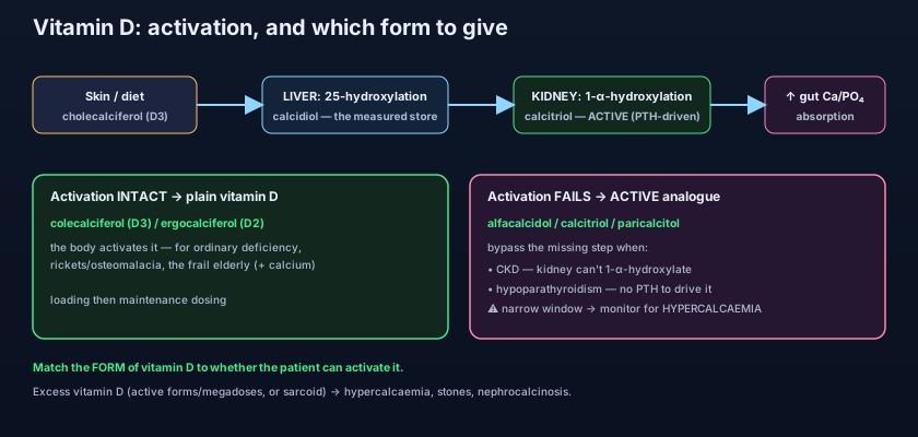
Here is the exam trap that catches people every year. A patient with chronic kidney disease has a low calcium and needs vitamin D — and someone reaches for a big dose of colecalciferol. It won't work: their kidneys cannot perform the 1-alpha-hydroxylation, so the plain vitamin never becomes active. The right answer is an active analogue — alfacalcidol or calcitriol — which is already past the broken step. The single question that unlocks the whole prescription is: can THIS patient activate vitamin D? If the kidney works and there is PTH, give the cheap plain form. If the kidney is failing or PTH is absent, give the active form that bypasses them.
Plain (inactive, needs full activation): colecalciferol (D3), ergocalciferol (D2) — first line for ordinary deficiency and for combined calcium/vitamin D in osteoporosis. Active / analogues (bypass a step): alfacalcidol (1-alpha-hydroxyvitamin D — needs only liver 25-hydroxylation), calcitriol (1,25-dihydroxyvitamin D — fully active) — for CKD, hypoparathyroidism and 1-alpha-hydroxylase deficiency. Paricalcitol — a vitamin D receptor activator used for the secondary hyperparathyroidism of CKD. Rule of thumb: the more of the activation pathway the patient has lost, the further "down" the pathway the drug you give must already be.
Toxicity: too much of a good hormone
Because vitamin D raises calcium, its overdose does exactly that to a dangerous degree: HYPERCALCAEMIA and hypercalciuria, with thirst, polyuria, constipation, confusion, and — over time — kidney stones and nephrocalcinosis (calcium deposited in the kidney itself). Plain colecalciferol has a wide safety margin because the body's own regulation throttles activation, so toxicity from it usually needs sustained megadoses. The active analogues are the real hazard: alfacalcidol, calcitriol and paricalcitol have skipped that regulatory brake, so they have a NARROW therapeutic window and can push calcium up quickly — which is precisely why anyone on an active analogue needs their serum calcium monitored. This is the same hypercalcaemia machinery the Hypercalcaemia chapter examines in full, where vitamin D excess sits alongside primary hyperparathyroidism and malignancy as a leading cause. One elegant natural example makes the mechanism vivid: in granulomatous diseases such as sarcoidosis, the granuloma macrophages express their own 1-alpha-hydroxylase and manufacture active vitamin D unregulated — producing hypercalcaemia from the inside, with no dietary excess at all.
- Deficiency → poor gut calcium absorption + failed bone mineralisation → rickets (children), osteomalacia (adults).
- Osteomalacia = bone pain, proximal myopathy, fractures; deficiency also worsens secondary hyperparathyroidism.
- At risk: limited sun, dark skin, fat malabsorption, obesity, elderly, exclusively breastfed infants.
- Prescribe plain colecalciferol/ergocalciferol when activation is intact; active alfacalcidol/calcitriol when it is not.
- CKD / hypoparathyroidism / 1-alpha-hydroxylase defects → active analogue (alfacalcidol, calcitriol; paricalcitol for CKD secondary HPT).
- Excess → hypercalcaemia/hypercalciuria, stones, nephrocalcinosis; active forms have a narrow window — monitor calcium.
- Prescribing plain colecalciferol in CKD or hypoparathyroidism, where the kidney can't activate it — an active analogue (alfacalcidol/calcitriol) is required.
- Starting an active analogue (calcitriol/alfacalcidol) and not monitoring serum calcium — their narrow window causes hypercalcaemia and nephrocalcinosis.
- Giving vitamin D alone for osteoporosis in a frail patient with poor intake — without calcium there is little for the hormone to absorb.
A 58-year-old man with chronic kidney disease has a low serum calcium and clinical features of vitamin D deficiency. Which is the most appropriate vitamin D preparation to prescribe?
- Vitamin D is a prohormone activated in two steps: liver → 25-hydroxyvitamin D (calcidiol, the form we measure) → kidney 1-alpha-hydroxylase (driven by PTH) → active calcitriol.
- Active vitamin D raises calcium and phosphate — chiefly by increasing gut absorption of both — working with PTH on bone and kidney.
- Deficiency causes rickets (children) and osteomalacia (adults: bone pain, proximal myopathy, fractures) and worsens secondary hyperparathyroidism.
- Match the FORM to activation: plain colecalciferol when the kidney and PTH can activate it; active alfacalcidol/calcitriol in CKD and hypoparathyroidism — and beware hypercalcaemia with the active forms.
- Rang & Dale's Pharmacology — The endocrine pancreas and the control of blood glucose; bone metabolism and vitamin D.
- Katzung, Basic & Clinical Pharmacology — Agents That Affect Bone Mineral Homeostasis (vitamin D, analogues, and calcium).
- British National Formulary (BNF) — Vitamin D substances: colecalciferol, ergocalciferol, alfacalcidol, calcitriol, paricalcitol.
- NICE guidance — Vitamin D: supplement use in specific population groups; chronic kidney disease (bone-mineral disorder).
- Guyton & Hall Textbook of Medical Physiology — Parathyroid hormone, calcitonin, calcium and phosphate metabolism, vitamin D.
- Holick MF. Vitamin D Deficiency. New England Journal of Medicine.

