
Body-Fluid Compartments and IV Fluids: Where Does the Bag Go?
Two patients get a litre of fluid, fast, into a vein. One gets 5% dextrose, the other 0.9% saline. An hour later almost none of the dextrose is left in the circulation — it has scattered through every cell in the body — while roughly a third of the saline is still holding up the blood pressure. Same volume, same vein, completely different destinations. Nobody chose that; the chemistry did. Prescribing fluid without knowing where it lands is like posting a letter with no address — and the physiology that writes the address is the most quietly important thing in acute medicine.
It is 3 a.m. on the ward and a 70-year-old man is hypotensive after a bout of vomiting and diarrhoea — dry mucous membranes, a thready pulse, urine barely trickling. The night doctor reaches for a fluid bag. On the shelf sit three that look almost identical: 5% dextrose, 0.9% saline, and a bag of Hartmann's solution. They cost pennies and they are handed out like water, yet the choice between them will decide whether the litre she hangs actually refills his empty circulation or simply soaks uselessly into his tissues and cells. The same decision, made carelessly on a hundred wards every night, quietly causes swollen legs, breathless lungs and deranged blood chemistry. To prescribe fluid well you have to answer one question first: when it leaves the bag, where does it go?
The body is mostly water — divided into rooms
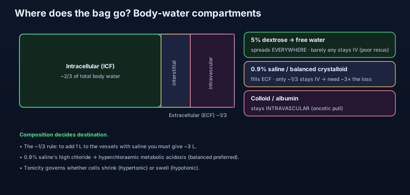
Roughly 60% of an adult's weight is water, and that water is not one pool but several, walled off from each other. Think of total body water as a house with rooms of fixed relative size. About two-thirds of it sits inside the cells — the intracellular fluid (ICF), the largest room by far. The remaining third is outside the cells — the extracellular fluid (ECF). And the ECF itself is split again: about a quarter of it is the intravascular fluid — the plasma inside blood vessels that carries the blood pressure — and the other three-quarters is the interstitial fluid that bathes the cells between the vessels. So of every three litres of body water, two are locked inside cells, and of the one litre outside, only a small fraction is actually in the bloodstream. That last fact is the whole reason a bag of fluid behaves the way it does. Hold on to one iconic number: only about a third of what stays in the ECF remains intravascular.
The walls between the rooms decide everything
What separates these compartments is not brick but two very different kinds of membrane, each with its own rules about what can cross. The cell membrane, dividing intracellular from extracellular, is freely permeable to water but not to solute — water crosses it instantly, but ions like Na+ are largely kept out by pumps. So the cell membrane responds to osmosis: water moves across it toward wherever the dissolved particles are more concentrated. The capillary wall, dividing plasma from interstitium, follows a different rule: it is permeable to water and small electrolytes (Na+, Cl-, K+ slip through freely) but not to large plasma proteins like albumin. So an electrolyte given intravenously spreads across the whole ECF without difficulty, but a big protein molecule stays trapped in the vessel. Two walls, two permeabilities — and between them they explain why every fluid finds a different home.
Picture the body's water as a building with two kinds of internal doors. The cell-membrane door lets only water through, never the furniture (the solutes). The capillary-wall door is a wider gate that lets water and small parcels (electrolytes) pass but stops the grand pianos (proteins). Now pour in a bucket. Plain water seeps through every door and ends up everywhere. Salty water is stopped at the cell doors and spreads only through the outer rooms. A load of grand pianos can't get past the capillary gate at all and piles up in the front hall — the bloodstream. You never had to push the water anywhere; the doors sorted it for you. An IV fluid is just a bucket, and its ingredients decide which doors it can pass.
The key principle: composition decides destination
Fluids are divided into two great families — crystalloids and colloids — by what is dissolved in them. A crystalloid is a solution of small molecules — salts and/or sugar — that pass freely across the capillary wall: saline, dextrose, and the balanced solutions. A colloid contains large molecules — most importantly albumin — too big to cross that wall, so it exerts an oncotic (colloid osmotic) pull that holds water in the vessel. This single distinction, small-particle versus large-particle, is the master switch. It, together with whether the particles can also enter cells, dictates whether a given litre ends up spread through all the water in the body, confined to the extracellular space, or held almost entirely in the bloodstream. The same idea — where a substance distributes according to its physicochemistry — is exactly the volume of distribution taught in the Principles of Pharmacology chapter for drugs; a fluid is just a drug whose only action is where it goes.
5% dextrose: free water that goes everywhere
A bag of 5% dextrose is glucose dissolved in water, and it is isotonic in the bag only because of that glucose. But the glucose is a decoy. Once infused, cells take it up and metabolise it, and what is left behind is pure water — "free water" with no effective solute to hold it anywhere. That water then distributes across every compartment in proportion to their sizes: two-thirds vanishes straight into the cells, and of the third left outside, only a third of that stays intravascular. Do the arithmetic on a litre and barely a tenth of it is still in the circulation. That makes 5% dextrose almost useless as a resuscitation fluid — you cannot refill an empty tank with it. But it is the right tool when the problem is a pure water deficit or simple maintenance, and it connects directly to the Sodium & Water chapters, where giving free water is exactly how you treat hypernatraemia. Its job is to deliver water to the cells, not to prop up the blood pressure.
Saline and balanced crystalloids: filling the extracellular room
Put salt in the bag and the water can no longer enter cells — it is trapped in the extracellular space. 0.9% saline and the balanced solutions — Hartmann's, Ringer's lactate, Plasma-Lyte — are sodium-based crystalloids. Sodium is confined to the ECF, so the water follows it there and stays out of the cells. But the ECF, remember, is only about a third intravascular; the rest is interstitium. So of a litre of saline, only around 300 mL actually remains in the bloodstream — which is the origin of the classic teaching that you need roughly three times the volume of crystalloid to replace a given loss of blood or intravascular volume. It works for resuscitation, but inefficiently, and the two-thirds that spills into the interstitium is exactly the tissue oedema that troubles the over-filled patient. This is the physiology behind the resuscitation fluid choices discussed in the Cardiovascular section, and behind the Fluid-prescribing chapter's insistence on matching volume to genuine deficit.
"Balanced" versus 0.9% saline: the chloride problem
Here is the twist that catches people out: 0.9% saline is not physiological. Plasma holds about 100 mmol/L of chloride, but 0.9% saline holds 154 — a supraphysiological chloride load. Pour litres of it in and the plasma chloride climbs, and by simple acid–base bookkeeping a rising chloride drives down bicarbonate, producing a hyperchloraemic metabolic acidosis. High chloride also constricts the renal afferent arteriole and may worsen kidney perfusion, which is why large-volume saline has been linked to acute kidney injury signals in critically ill patients. The balanced solutions were designed to fix this: they trade some of that chloride for buffer anions (lactate in Hartmann's, acetate/gluconate in Plasma-Lyte) that the liver converts to bicarbonate, giving an electrolyte profile much closer to plasma. The modern preference, reflected in current guidance, is to favour balanced crystalloids over 0.9% saline for most resuscitation and replacement — reserving saline for specific situations such as hypochloraemic alkalosis or hyponatraemia. This is the same hyperchloraemic acidosis flagged in the Acid–Base chapters, met here at its commonest iatrogenic source.
- Total body water ≈ 60% of weight: 2/3 intracellular, 1/3 extracellular (itself ~1/4 intravascular, ~3/4 interstitial).
- Cell membranes pass water but not solute (osmosis); capillary walls pass water + electrolytes but not large proteins.
- Where a fluid distributes depends on its composition — the master principle.
- 5% dextrose = free water → spreads across all compartments; little stays intravascular (poor resuscitation, good maintenance).
- Saline/balanced crystalloid stays extracellular; only ~1/3 stays intravascular → need ~3× the lost volume.
- Colloid (albumin) is held intravascular by oncotic pull → the most volume-efficient, litre for litre.
Colloids: keeping the fluid in the vessel
A colloid carries large molecules that cannot cross the capillary wall, so they linger in the plasma and, by oncotic pressure, hold water alongside them in the vessel. Litre for litre, a colloid expands the intravascular volume more than a crystalloid and does it for longer — the theoretical dream of a resuscitation fluid. The natural colloid is human albumin. The synthetic colloids — the starches (hydroxyethyl starch) and the gelatins — were once popular but have been largely abandoned: the starches were found to increase acute kidney injury and mortality in critically ill patients, and the gelatins carry anaphylaxis risk with little proven benefit, so guidelines no longer recommend them for routine resuscitation. Albumin retains niche roles — for instance in decompensated liver disease and spontaneous bacterial peritonitis — but as a general first-line resuscitation fluid it has never convincingly beaten balanced crystalloid, at a fraction of the cost. The Cardiovascular chapter revisits this crystalloid-versus-colloid debate at the bedside.
Tonicity: why cells shrink or swell
Tonicity is the effective osmotic pull a fluid exerts across the cell membrane — and it governs cell size. Because the cell membrane lets water cross but not solute, water moves toward the side with more effective (impermeant) particles. An isotonic fluid — 0.9% saline, Hartmann's — matches the cell's interior, so no net water shifts and cells keep their size; that is why isotonic crystalloids are the workhorses for expanding the ECF without disturbing cells. A hypotonic fluid — effectively what 5% dextrose becomes once its glucose is burnt — is more dilute than the cell, so water flows inward and cells swell; pushed too far, and too fast, this can swell brain cells dangerously (the hazard behind over-rapid correction of hypernatraemia, cerebral oedema). A hypertonic fluid — such as hypertonic saline — is more concentrated than the cell, so water is drawn out and cells shrink; that osmotic pull is harnessed deliberately to draw water out of a swollen brain, the logic shared with the osmotic agents (like mannitol) covered elsewhere. Tonicity, in short, is the lever that decides whether a fluid leaves cells alone, inflates them, or deflates them.
The single most useful shortcut in all of fluid therapy: to resuscitate — to refill an empty circulation — reach for something that stays in the extracellular space, a balanced isotonic crystalloid, and remember you'll need about three times the volume you think, because two-thirds leaks into the interstitium. To give free water — to correct a pure water deficit or provide maintenance — reach for 5% dextrose, and never expect it to hold up a blood pressure. Match the bag to the compartment you are trying to fill, and half of fluid prescribing solves itself.
5% dextrose: glucose 50 g/L in water; becomes free water; distributes across all compartments; use for free-water/maintenance, not resuscitation. 0.9% saline: Na+ 154, Cl- 154 mmol/L; stays extracellular; ~1/3 intravascular; high chloride → hyperchloraemic acidosis with large volumes. Hartmann's / Ringer's lactate: Na+ ~131, Cl- ~111, plus K+, Ca2+ and lactate buffer; balanced, plasma-like, first choice for most resuscitation. Plasma-Lyte: Na+ ~140, Cl- ~98, with acetate and gluconate buffers; the most physiological balanced crystalloid. Human albumin (4.5% / 20%): a colloid held intravascular by oncotic pressure; niche use (e.g. liver disease), not routine resuscitation.
- Isotonic (0.9% saline, Hartmann's) leaves cell size unchanged — the workhorse for ECF expansion.
- Hypotonic fluid swells cells; hypertonic fluid shrinks them — tonicity governs cell volume.
- 0.9% saline's chloride (154 vs plasma's ~100) drives hyperchloraemic metabolic acidosis in volume.
- Balanced crystalloids (Hartmann's, Plasma-Lyte) are now preferred over saline for most resuscitation.
- Synthetic colloids (starches, gelatins) are largely abandoned; albumin keeps only niche roles.
- Volume of distribution is one idea shared by fluids and drugs: composition/physicochemistry decides destination.
- Resuscitating a shocked, hypovolaemic patient with 5% dextrose — it becomes free water, scatters into cells, and barely a tenth stays in the circulation.
- Pouring litres of 0.9% saline without a thought — the supraphysiological chloride causes a hyperchloraemic metabolic acidosis and may worsen kidney perfusion.
- Forgetting the ~1/3 rule — expecting a litre of crystalloid to restore a litre of intravascular volume, when two-thirds leaks into the interstitium as oedema.
A 65-year-old man is in hypovolaemic shock after gastrointestinal bleeding — cold, tachycardic, hypotensive. Which fluid, and why, is the most appropriate initial resuscitation choice while blood is prepared?
- Total body water is 2/3 intracellular and 1/3 extracellular (intravascular plasma + interstitium); cell membranes pass water not solute, capillaries pass water + electrolytes not proteins.
- Composition decides destination: 5% dextrose = free water spread everywhere; saline/balanced crystalloid stays extracellular (~1/3 intravascular → need ~3× the volume); colloid/albumin stays intravascular.
- 0.9% saline's supraphysiological chloride (154) causes hyperchloraemic metabolic acidosis and possible renal harm — balanced crystalloids are now preferred for most resuscitation.
- Tonicity governs cell volume: isotonic leaves cells alone, hypotonic swells them (cerebral oedema risk), hypertonic shrinks them (the osmotic-agent principle).
- Rang & Dale's Pharmacology — Fluid and electrolyte balance; drugs and body compartments.
- Ganong's Review of Medical Physiology / Guyton and Hall Textbook of Medical Physiology — body-fluid compartments and osmosis.
- NICE Clinical Guideline CG174 — Intravenous fluid therapy in adults in hospital.
- Semler MW, et al. Balanced Crystalloids versus Saline in Critically Ill Adults (SMART trial). New England Journal of Medicine.
- Myburgh JA, Mythen MG. Resuscitation fluids. New England Journal of Medicine.
- British National Formulary (BNF) — Fluids and electrolytes; intravenous fluid preparations.

