
How Cancer Drugs Work: The Cell Cycle, Log-Kill and Why We Combine Them
Cancer is one idea gone wrong: a cell that divides when it should not, and refuses to die. Almost every classical cancer drug is a counter-move to that single idea — it attacks cells while they divide. But that strategy carries three hard truths with it: each dose kills a fraction, not a number; one drug is rarely enough; and the poison cannot tell a tumour cell from your gut lining. Understand those three and the whole rhythm of chemotherapy — the cycles, the combinations, the toxicities — stops looking like a mystery and starts looking like arithmetic.
A young woman with lymphoma finishes her second cycle of chemotherapy. The scan comes back and the radiologist can no longer see the mass — it has, to the eye, disappeared. She asks the obvious question: if it's gone, why do I need four more cycles? Her oncologist draws a single number on the paper: 99.9%. That is the fraction of tumour cells each cycle destroys — not a fixed count, a fraction. Start with a billion cancer cells; after one cycle a million survive; after another, a thousand; then one; then, with luck, none. The scan went blank when the count dropped below what an image can resolve — around a hundred million cells — but a hundred million survivors is not a cure. The extra cycles are the difference between "invisible" and "gone." That one number explains the entire rhythm of chemotherapy.
Cancer is a disease of division
Strip cancer down to one sentence: cells that proliferate without a brake. Normal tissue divides on demand and stops on cue. A cancer cell has broken both signals — it grows when it shouldn't and ignores the orders to die (apoptosis). Because the defining behaviour is uncontrolled division, the oldest and still-central drug strategy is to attack cells while they are dividing. That is why classical chemotherapy hits DNA and the machinery of cell division — and, unavoidably, why it also hits the normal tissues that divide fastest: bone marrow, gut lining, hair follicles. The strategy is powerful precisely because it is not fully selective.
The cell cycle: four phases, and where drugs strike
A dividing cell moves through an ordered loop. The cell cycle has four working phases: G1 (the cell grows and prepares), S (it copies its DNA), G2 (it checks the copy and readies for division), and M (mitosis — it splits into two). Cells not currently cycling rest in a quiet state called G0. This map matters clinically because different drug classes ambush the cell at different points. Some drugs only work if the cell is in a particular phase; others do their damage no matter what the cell is doing. That single distinction — cell-cycle-specific versus cell-cycle-non-specific — organizes the entire cytotoxic pharmacy.
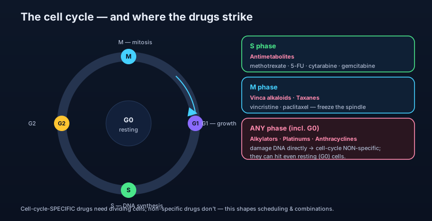
Cell-cycle-specific (CCS) drugs need the cell to be actively cycling, and usually in one phase. Antimetabolites — methotrexate, 5-fluorouracil, cytarabine — mimic the building blocks of DNA and jam replication, so they strike in S phase. The vinca alkaloids (vincristine, vinblastine) and the taxanes (paclitaxel, docetaxel) attack the mitotic spindle, so they strike in M phase. Because they need dividing cells, CCS drugs are most effective against rapidly proliferating tumours and are often given by infusion over time, to catch cells as they enter the vulnerable phase.
Cell-cycle-non-specific (CCNS) drugs do not wait for a phase. Alkylating agents (cyclophosphamide, cisplatin — the platinums behave similarly) crosslink and damage DNA directly; anthracyclines (doxorubicin) intercalate DNA and poison topoisomerase II. Because their damage does not depend on the cell being in S or M, they can injure even slow-dividing and resting (G0) cells — which makes them valuable against bulky, slow-growing tumours where much of the mass is not actively cycling. Their dose-response tends to rise with the size of the dose, which is why they anchor high-dose regimens.
- Cell cycle: G1 (grow) → S (copy DNA) → G2 (check) → M (divide); G0 = resting.
- Cell-cycle-SPECIFIC drugs need dividing cells: antimetabolites → S, vinca/taxanes → M.
- Cell-cycle-NON-SPECIFIC drugs hit any phase, even G0: alkylating agents, anthracyclines.
- CCS agents suit fast tumours and time-based infusions; CCNS agents suit bulky, slow tumours.
The log-kill hypothesis: a fraction, not a number
This is the idea from the scene, made formal. The log-kill hypothesis, from the classic experiments of Skipper and Schabel, states that a given dose of a drug kills a constant FRACTION of the tumour cells present — not a constant number. If a dose achieves "3-log kill," it destroys 99.9% of the cells whether there are a billion or a thousand. The consequence is profound: because you are always killing a percentage of what remains, you can never subtract your way to zero in a single blow. Ten billion cells, knocked down by 99.9%, still leaves ten million. That is why treatment comes in repeated cycles, each one carving the same fraction off a smaller and smaller remnant.
Two practical rules fall straight out of log-kill. First, cure requires driving the burden very low — ideally to zero, but realistically low enough that the body's own immune surveillance can mop up the last few cells; that takes multiple cycles, continued past the point of visible disease. Second, timing matters: cycles are spaced to let normal tissues (marrow especially) recover, but not so far apart that the tumour regrows its losses between doses. The entire schedule of chemotherapy is a negotiation between killing the maximum fraction of tumour and giving the patient's healthy dividing cells time to recover.
This is why oncologists press on after a scan looks clean. "Complete remission" on imaging can still hide up to a hundred million cells — below the resolution of a scan but far above zero. Minimal residual disease is the target of those "extra" cycles. Stopping when the tumour becomes invisible is how a curable cancer relapses.
Growth fraction: why fast leukaemias yield and slow tumours resist
The growth fraction is the proportion of cells in a tumour that are actively dividing at any moment. It is the hidden variable behind why some cancers melt away and others barely flinch. A high growth-fraction tumour — many acute leukaemias, some lymphomas — has most of its cells cycling, so cell-cycle-specific drugs find plenty of targets and responses can be dramatic. A bulky solid tumour, by contrast, often has a low growth fraction: its core is poorly perfused and many cells sit quietly in G0, out of reach of phase-specific drugs. Paradoxically, shrinking a big tumour can raise its growth fraction — surviving cells re-enter the cycle — which is one rationale for giving cell-cycle-specific drugs after debulking.
- Log-kill: each dose kills a constant FRACTION (e.g. 99.9%), never a fixed number of cells.
- Because you kill a fraction, cure needs repeated cycles continued past visible remission.
- Growth fraction = the share of cells actively dividing; it predicts chemo-responsiveness.
- Fast leukaemias (high growth fraction) respond well; bulky slow solid tumours resist.
Why we combine drugs
A single agent almost never cures a cancer. Combinations do. Combination chemotherapy follows a few disciplined rules. Choose drugs each active against the tumour on its own. Choose different mechanisms of action, so they attack the cell in independent ways. Choose non-overlapping toxicities, so each can be given at full dose without stacking the same side effect. And, where possible, hit different phases of the cell cycle, so cells escaping one drug are caught by another. Above all, combinations delay resistance: the chance that a single cell carries resistance to three unrelated drugs at once is vanishingly small compared with resistance to one.
Regimens are named by acronym, one letter per drug. CHOP for aggressive lymphoma = Cyclophosphamide (alkylator), Hydroxydaunorubicin/doxorubicin (anthracycline), Oncovin/vincristine (vinca, M-phase), and Prednisone (steroid). FOLFOX for colorectal cancer = FOLinic acid, Fluorouracil (antimetabolite, S-phase) and OXaliplatin (platinum alkylator). Notice the pattern in each: different mechanisms, different cycle phases, and toxicities that do not all pile onto the same organ. The acronym is the combination principle written down.
Drug resistance: how tumours fight back
Resistance is the reason a tumour that shrank at first can return unmoved by the same drug. It arises because tumours are genetically unstable and mutate constantly, so a resistant clone can be selected and expand. The mechanisms are worth knowing by name. Efflux pumps: the MDR1 gene makes P-glycoprotein, a membrane pump that ejects many structurally unrelated drugs (anthracyclines, vinca alkaloids, taxanes) — a single change conferring multidrug resistance. Altered or amplified targets: methotrexate resistance from amplified dihydrofolate reductase; a mutated target the drug no longer fits. Enhanced DNA repair: cells that repair alkylator and platinum damage faster survive it. Apoptosis evasion: a cell with a broken death programme (e.g. loss of p53) absorbs the damage without dying. Decreased activation or increased inactivation of the drug rounds out the list.
- Combine drugs with different mechanisms, non-overlapping toxicities, and different cycle phases.
- Combinations delay resistance: simultaneous resistance to several unrelated drugs is rare.
- P-glycoprotein (MDR1) pumps out many drugs at once → multidrug resistance.
- Other resistance routes: altered/amplified targets, faster DNA repair, evaded apoptosis.
- Regimens (CHOP, FOLFOX) encode all these principles in their acronyms.
From poison to precision: cytotoxic → targeted → immune
Everything above describes classical cytotoxic chemotherapy. The field did not stop there. Cytotoxic drugs attack division itself, and pay for it in toxicity to every dividing tissue. The next generation, targeted therapy, aims at a molecular lesion the cancer depends on — imatinib blocking the BCR-ABL kinase in chronic myeloid leukaemia, trastuzumab against HER2-amplified breast cancer — sparing much of the collateral damage. The newest wave, immunotherapy, does not attack the tumour directly at all; it releases the brakes on the patient's own immune system — the checkpoint inhibitors against PD-1 and CTLA-4 — so the body's T cells destroy the cancer. These do not replace the principles above so much as build on them; combinations, resistance, and dosing logic all reappear in new forms. The chapters ahead in this Oncology section take each family in turn.
- Thinking chemo kills a fixed NUMBER of cells. It kills a fraction (log-kill) — which is exactly why repeated cycles are needed and why treatment continues after the scan clears.
- Assuming one drug is enough. Single agents select for resistant clones; combinations with different mechanisms and non-overlapping toxicities are the rule for cure.
- Believing chemotherapy is selective for cancer. Classical cytotoxics hit ALL rapidly dividing cells — marrow, gut, hair — which is the source of their toxicities (the next chapter).
- Confusing cell-cycle-specific with non-specific. Only the specific agents need the cell to be in a given phase; alkylators and anthracyclines damage resting cells too.
A tumour of 10^10 cells is treated with a drug producing a 3-log kill per cycle. Roughly how many cells remain after one cycle?
- Cancer is uncontrolled division; classical drugs attack dividing cells — hence toxicity to marrow, gut and hair.
- Cell-cycle-specific drugs need a phase (antimetabolites → S, vinca/taxanes → M); non-specific drugs (alkylators, anthracyclines) hit any phase, even resting cells.
- Log-kill: each dose kills a constant fraction, so cure needs repeated cycles past the point of visible disease.
- We combine drugs (CHOP, FOLFOX) for different mechanisms, non-overlapping toxicities and to delay resistance (efflux pumps, altered targets, DNA repair, apoptosis evasion).
- The field is shifting cytotoxic → targeted → immune therapy, explored in the chapters ahead.
- Katzung BG. Basic & Clinical Pharmacology — Cancer Chemotherapy: cell-cycle kinetics, cycle-specific vs non-specific agents, log-kill & combination principles.
- Brunton LL, et al. Goodman & Gilman's The Pharmacological Basis of Therapeutics — General Principles of Cancer Chemotherapy: growth fraction, log-kill, drug resistance.
- DeVita VT, Lawrence TS, Rosenberg SA. DeVita, Hellman & Rosenberg's Cancer: Principles & Practice of Oncology — Skipper–Schabel model, combination chemotherapy design.
- Skipper HE, Schabel FM, Wilcox WS. Experimental evaluation of potential anticancer agents — the log-kill hypothesis.
- Whalen K. Lippincott Illustrated Reviews: Pharmacology — Anticancer drugs: cell-cycle specificity & mechanisms of resistance.
- Weinberg RA. The Biology of Cancer — proliferation, apoptosis evasion & multidrug resistance (P-glycoprotein/MDR1).

