
The Eye as a Drug Target: Barriers and Routes
The eye is a strange place to deliver a drug. It is tiny, it is sealed off from the bloodstream by barriers as tight as the ones guarding the brain, and it is divided into front and back compartments that almost nothing crosses freely. A drop that floods the surface of the eye may reach the iris beautifully and never touch the retina an inch behind it. Almost every puzzle in ocular pharmacology — why anti-VEGF is injected through the wall of the eye, why glaucoma is treated with drops but macular degeneration is not, why an oral antibiotic barely dents an infection inside the globe — comes back to one question: can the molecule get to the compartment where the disease lives?
Two patients sit in the same waiting room. The first has red, gritty eyes and a diagnosis of bacterial conjunctivitis; she leaves with a bottle of antibiotic drops, and by the week's end the infection is gone. The second, an 78-year-old man, has wet age-related macular degeneration — new, leaky blood vessels growing under the centre of his retina. He is not given drops. He is not given a tablet. Instead a doctor numbs his eye, cleans it with iodine, and passes a fine needle through the white of the sclera to inject a drug directly into the jelly that fills the back of the eye. He will return for that injection again and again, every few weeks. Same organ, same clinic, same afternoon — and two completely different delivery problems. The difference between them is not the drug. It is the address.
An organ built to keep things out
Everything that makes the eye good at seeing makes it hard to medicate. The eye is small, transparent, and jealously guarded. It is an immune-privileged site — it deliberately dampens ordinary inflammation, because a full immune response inside a clear optical instrument would scar it and blind it. It is defended by physical barriers at every surface, and by an unusually efficient washout: blink, tears, and drainage sweep the surface clean within minutes. And it is functionally two eyes in one. The anterior segment — cornea, aqueous humour, iris, lens, ciliary body — sits at the front. The posterior segment — vitreous, retina, choroid, optic nerve — sits at the back. A wall of barriers separates them, and a drug that reaches one does not automatically reach the other. Hold on to that anterior-versus-posterior divide: it is the single most useful idea in the whole of ocular pharmacology, and everything below hangs off it.
The cornea: a lipid–water–lipid sandwich
For a topical drug, the cornea is the gatekeeper of the front of the eye — and it is built like a sandwich that punishes any drug that is only good at one thing. The outer epithelium is a layer of tightly joined, fat-loving (lipophilic) cells: a lipid barrier. Behind it lies the stroma, a thick, waterlogged, water-loving (hydrophilic) layer. Behind that sits a thin lipophilic endothelium. So a molecule must dissolve through fat, then through water, then through fat again to cross. A purely fat-soluble drug races through the epithelium and then stalls in the watery stroma; a purely water-soluble drug can't get past the epithelium in the first place. The molecules that cross best are amphipathic — comfortable in both fat and water, existing in a mix of ionized and un-ionized forms at the tear-film pH. This is the same lipophilicity-versus-water-solubility balancing act taught in the Principles of Pharmacology section on membrane permeability — the cornea simply makes you solve it twice, in opposite directions.
Think of the cornea as an airport with three checkpoints in a row: a fat-only lane, then a water-only lane, then a fat-only lane again. A traveller who can only pass fat lanes clears the first, gets stuck at the second, and never boards. A traveller who can only pass water lanes is turned back at the door. Only the traveller carrying both kinds of pass — the amphipathic drug — walks all the way through to the aqueous humour on the other side. Drop formulators exploit exactly this by shipping drugs as inactive prodrugs that are lipophilic enough to cross the epithelium, then are converted to the active, water-friendly form by enzymes once inside.
Around the cornea, a softer route. The cornea is not the only way in. The conjunctiva — the membrane lining the white of the eye and the inner lids — and the sclera beneath it are far more permeable and far more forgiving of large, water-soluble molecules. Their catch is that they are leaky in both directions: a drug entering through the conjunctiva sits right on top of a rich bed of blood vessels, so much of it is swept straight into the systemic circulation instead of into the eye. That conjunctival absorption is not a minor footnote — it is the main reason a drop meant for the eye can end up acting on the heart and lungs, a theme the Eyedrop pharmacology chapter builds an entire safety lesson around.
The blood–ocular barriers: why the back of the eye is a fortress
If the cornea guards the front from the outside, the blood–ocular barriers guard the inside from the bloodstream — and they are the reason systemic drugs struggle to treat the retina. There are two. The blood–aqueous barrier, formed by tight junctions in the ciliary epithelium and iris vessels, limits what leaks from blood into the aqueous humour at the front. The blood–retinal barrier, formed by tight junctions between retinal capillary endothelial cells and the retinal pigment epithelium at the back, is the eye's equivalent of the blood–brain barrier — anatomically and functionally almost identical. Just as the blood–brain barrier keeps most drugs out of the central nervous system, the blood–retinal barrier keeps most drugs out of the retina. Swallow a tablet, and only a small, lipophilic fraction of it will ever cross into the back of the eye at a useful concentration. This is exactly the logic taught in the Principles of Pharmacology section on the blood–brain barrier, transplanted to the retina.
Here is the payoff. The blood–retinal barrier is not a bug to be worked around — it is the whole reason intravitreal injection exists. Because you cannot reliably reach the retina from the bloodstream or from a drop, the only dependable way to deliver a high concentration to the back of the eye is to put the needle through the wall and inject the drug into the vitreous, on the retina's doorstep. That is why anti-VEGF drugs for macular degeneration and diabetic retinopathy are injected, not swallowed or dropped. The barrier that frustrates you when you want the drug in is also, conveniently, the barrier that keeps it there once injected — a locally injected drug clears slowly, so a single injection can last weeks.
- The eye is small, immune-privileged, barrier-protected, and split into anterior and posterior segments.
- The cornea is a lipid–water–lipid sandwich (epithelium–stroma–endothelium); crossing it favours amphipathic drugs.
- The conjunctiva and sclera are more permeable but drain drug into the systemic circulation.
- Two blood–ocular barriers: blood–aqueous (front) and blood–retinal (back).
- The blood–retinal barrier is the eye's blood–brain barrier — it keeps systemic drugs out of the retina.
- Anterior versus posterior is the organising principle of every delivery decision.
The routes of delivery, and what each one reaches
Choosing a route is really choosing a compartment. Topical (drops and ointments) is the workhorse — cheap, painless, self-administered — and it is essentially an anterior-segment route. Drops treat the cornea, conjunctiva, anterior chamber, iris and ciliary body well, and can lower intraocular pressure in glaucoma; but only a trickle reaches the vitreous and retina, so drops are a poor choice for posterior disease. Subconjunctival and sub-Tenon injections (placing drug just under the conjunctiva or into the space around the eye) bypass the surface barriers and create a depot that gives much higher local concentrations to the anterior and mid segments — useful for stubborn inflammation. Intracameral injection puts drug directly into the anterior chamber, most often at the end of cataract surgery (an intracameral antibiotic to prevent infection, or a miotic to constrict the pupil). Periocular injection sits drug around the globe for a slower, more posterior-leaning effect.
Intravitreal injection is the route that changed modern ophthalmology. A needle passed through the pars plana of the sclera delivers drug straight into the vitreous cavity — the only reliable way to achieve therapeutic levels at the retina. This is how anti-VEGF agents (ranibizumab, aflibercept, bevacizumab), intravitreal steroids, and intravitreal antibiotics are given. It carries real risks — most feared is endophthalmitis, infection seeded inside the eye — which is precisely why the same intravitreal route is used to treat it, injecting antibiotics directly into the vitreous when infection strikes; the Ocular infections chapter covers that emergency in detail. Finally, systemic delivery (oral or intravenous) reaches the whole body and floods the eye's blood supply — but the blood–ocular barriers throttle how much actually crosses, especially into the retina. Systemic therapy earns its place when the disease is in the orbit or the outer coats, when the barrier is already broken by heavy inflammation, or when the eye is only one front of a body-wide disease. For an isolated posterior problem, a tablet is usually the weakest tool in the box.
Topical drop → cornea, conjunctiva, anterior chamber, iris/ciliary body (e.g. glaucoma drops, antibiotic drops for conjunctivitis). Subconjunctival / sub-Tenon → high local levels to the anterior and mid segments (e.g. steroid for severe uveitis). Intracameral → anterior chamber directly (e.g. antibiotic at the end of cataract surgery). Intravitreal → the vitreous and retina (e.g. anti-VEGF for wet AMD, antibiotics for endophthalmitis). Systemic → orbit, outer coats and inflamed eyes, but throttled at the retina by the blood–ocular barrier (e.g. oral acetazolamide, IV antivirals for retinitis).
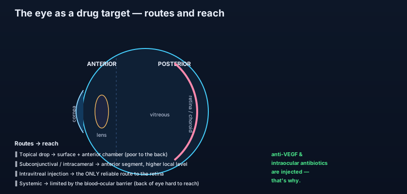
- Topical drops = anterior-segment route; they barely reach the retina.
- Subconjunctival/sub-Tenon and periocular injections give higher local levels, bypassing surface barriers.
- Intracameral injection targets the anterior chamber, classically during cataract surgery.
- Intravitreal injection is the only reliable route to the retina — the reason anti-VEGF is injected.
- Systemic therapy is limited at the back of the eye by the blood–ocular barrier.
- First ask which compartment the disease is in; the route follows from the answer.
- Expecting eye drops to treat retinal disease. Topical drug reaches the anterior segment; posterior conditions like wet AMD need intravitreal delivery.
- Forgetting that topical and subconjunctival drug drains systemically — a "local" eye drop can still cause systemic effects (e.g. a beta-blocker drop causing bronchospasm).
- Assuming an oral antibiotic will sterilise an infection inside the globe. The blood–ocular barrier blunts systemic penetration, which is why endophthalmitis is treated with intravitreal antibiotics.
A 74-year-old man with wet age-related macular degeneration needs an anti-VEGF drug delivered to the retina. Which route reliably achieves a therapeutic concentration at the retina?
- The eye is small, immune-privileged and barrier-protected; the central challenge is getting a drug into the right compartment, front or back.
- The cornea is a lipid–water–lipid sandwich that favours amphipathic drugs; the conjunctiva/sclera are more permeable but leak drug systemically.
- The blood–aqueous and blood–retinal barriers keep systemic drugs out of the eye; the blood–retinal barrier is the eye's blood–brain barrier.
- Match route to compartment: topical/intracameral for the front, intravitreal as the only reliable route to the retina, systemic limited at the back — the anterior-versus-posterior divide runs the whole show.
- Kanski's Clinical Ophthalmology: A Systematic Approach — Introduction and principles of ocular pharmacology.
- Bartlett & Jaanus, Clinical Ocular Pharmacology — Ocular drug delivery and pharmacokinetics.
- American Academy of Ophthalmology, Basic and Clinical Science Course (BCSC) — Fundamentals and Principles of Ophthalmology.
- Rang & Dale's Pharmacology — Routes of administration and drug distribution across barriers.
- Gaudana R, et al. Ocular drug delivery. The AAPS Journal.
- Royal College of Ophthalmologists — Intravitreal injection therapy guidelines.

