
Mydriatics and Cycloplegics: Controlling the Pupil
The pupil is not a passive hole — it is a muscle ring under a tug-of-war between two nervous systems, and a single drop can decide which side wins. Ophthalmologists dilate pupils dozens of times a day to see the retina, to measure a child's true refraction, and to rest an inflamed eye. It looks trivial. But the same drop that opens a pupil for a clean fundus view can slam a narrow angle shut and blind the eye by morning, or send a toddler into an anticholinergic fever. This chapter is really applied autonomic pharmacology — learn the two muscles and their nerves, and every mydriatic, cycloplegic and miotic falls into place.
A busy Tuesday clinic. A 68-year-old woman with long-standing diabetes needs her retina examined, so a technician instils tropicamide and phenylephrine and sends her to wait. An hour later she is back — but with a red, rock-hard, painful eye, a mid-dilated pupil that will not react, and haloes around the lights. She is vomiting. This is not a reaction to the dye; it is acute angle-closure glaucoma, triggered by dilating a shallow-angled eye. Two rooms away, a mother is coaxing her squinting four-year-old to hold still for cyclopentolate drops so the optometrist can measure his true glasses prescription — and the nurse is watching the child's cheeks for the tell-tale flush of anticholinergic absorption. One class of drug, two very different eyes, and in both the danger lives in the pharmacology, not the procedure.
Two muscles, two nerves, one pupil
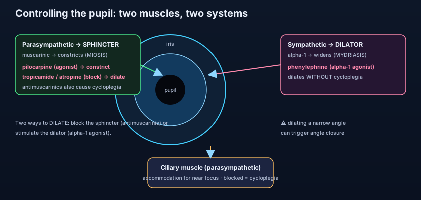
The size of the pupil is set by a pair of antagonist muscles pulling against each other. The iris contains two smooth muscles. The iris SPHINCTER is a circular ring around the pupil margin; when it contracts the pupil gets smaller (miosis). It is driven by the PARASYMPATHETIC system through muscarinic (M3) receptors — the same acetylcholine wiring that runs the near reflex. The iris DILATOR is a set of radial fibres running like spokes; when it contracts it pulls the pupil open (mydriasis). It is driven by the SYMPATHETIC system through alpha-1 adrenergic receptors. So the resting pupil is a balance: parasympathetic tone constricts, sympathetic tone dilates. Tip the balance either way — by adding a drug or blocking a nerve — and the pupil follows. Everything in this chapter is just four levers on those two muscles. The full receptor map lives in the Autonomic Nervous System chapter; here we put it to work in the eye.
There is a third muscle to keep in mind: the CILIARY muscle, which controls the lens for near focus (accommodation). It too is parasympathetic and muscarinic. When it contracts, the lens becomes rounder and the eye focuses up close; when it relaxes, the eye is set for distance. This matters enormously, because any drug that blocks the muscarinic receptors of the iris sphincter will block the ciliary muscle's receptors too. That is why antimuscarinics do two things at once — they dilate the pupil AND paralyse accommodation. The paralysis of accommodation has its own name: CYCLOPLEGIA. Hold onto that link; it explains both the great usefulness and the classic side effect of this drug class.
Think of the pupil as a camera aperture worked by two opposing crews. The parasympathetic crew winches it closed (and simultaneously turns the focus ring to "near"); the sympathetic crew hauls it open. To widen the aperture you can either fire the crew that closes it (antimuscarinics — but they also jam the focus ring, so near vision blurs), or you can give the opening crew a shot of adrenaline (phenylephrine, an alpha-1 agonist — which pulls it open but leaves the focus ring untouched). Same wide pupil, two completely different mechanisms, and the difference between them is exactly whether near focus survives.
Route one: block the sphincter with antimuscarinics
Take the parasympathetic brake off the sphincter and the unopposed dilator swings the pupil open. Antimuscarinic (anticholinergic) eye drops block M3 receptors on the iris sphincter, so it can no longer contract; the sympathetically driven dilator now acts unopposed and the pupil widens. Because the same block hits the ciliary muscle, they also produce cycloplegia. The members of the class differ mainly in how long they last — a spectrum from a quick afternoon dilation to a paralysis lasting weeks. Tropicamide is the short-acting workhorse (onset ~20–30 minutes, wearing off in about 4–6 hours) — ideal for a routine dilated fundus examination. Cyclopentolate is stronger and lasts about a day, the usual choice for cycloplegic refraction in children. Homatropine sits in the middle (a day or two). Atropine is the giant at the end of the spectrum: a single drop can dilate and paralyse accommodation for one to two weeks, which is exactly why it is reserved for therapy — resting an inflamed eye — rather than a quick look.
Tropicamide 0.5–1% — shortest (~4–6 h); the everyday dilator for fundus exams. Cyclopentolate 0.5–1% — about a day; the standard for cycloplegic refraction in children. Homatropine 2–5% — one to two days; an intermediate therapeutic mydriatic. Atropine 0.5–1% — one to two weeks; the strongest cycloplegic, reserved for uveitis (to rest the eye and break adhesions) and, at very low concentration, for slowing myopia progression. Rule of thumb: the longer it lasts, the stronger the cycloplegia — and the bigger the risks if it is misused or absorbed systemically.
Route two: stimulate the dilator with phenylephrine
The second way to open a pupil ignores the sphincter entirely and instead pushes on the dilator. Phenylephrine is a direct alpha-1 adrenergic agonist: it mimics sympathetic tone, the radial dilator fibres contract, and the pupil widens. Crucially, phenylephrine does nothing to the muscarinic ciliary muscle — so it dilates WITHOUT cycloplegia. Near focus is preserved. This is why it is often paired with tropicamide for fundus exams: the antimuscarinic and the alpha-1 agonist push the pupil open by two independent mechanisms, giving a wider, more reliable dilation than either alone. It comes in two strengths — 2.5% for routine dilation and a 10% preparation that dilates powerfully but carries a real risk of systemic blood-pressure spikes. The mirror image of phenylephrine — using muscarinic and alpha mechanisms to CONSTRICT the pupil and lower pressure — is the subject of the Alpha-agonists and miotics chapter, where pilocarpine is the star.
- Iris sphincter = parasympathetic/muscarinic → constriction (miosis); iris dilator = sympathetic/alpha-1 → dilation (mydriasis).
- The ciliary muscle (accommodation) is also muscarinic — so antimuscarinics dilate AND paralyse accommodation (cycloplegia).
- Antimuscarinics by duration: tropicamide (hours) < cyclopentolate (~a day) < homatropine < atropine (1–2 weeks).
- Phenylephrine (alpha-1 agonist) dilates WITHOUT cycloplegia — near vision preserved; often combined with tropicamide.
- Two independent ways to dilate: block the sphincter, or stimulate the dilator.
- Atropine's very long action makes it a therapeutic drug (uveitis), not a diagnostic one.
Why we dilate: three clinical jobs
Dilating a pupil is never idle — it does one of three specific jobs. First, the DILATED FUNDUS EXAMINATION: a small pupil is like peering into a room through a keyhole. Widen it and the whole retina, macula and optic disc come into view — essential in diabetes, in retinal detachment, and in any unexplained visual loss. A short-acting drop (tropicamide, often with phenylephrine) is perfect here. Second, CYCLOPLEGIC REFRACTION in children: a child's powerful ciliary muscle constantly accommodates, which hides their true refractive error and can fake or exaggerate a spectacle prescription. Paralysing accommodation with cyclopentolate (or atropine in strong accommodative squints) freezes the focusing system so the real refraction can be measured — the only way to prescribe glasses accurately in a young child. Third, THERAPY in uveitis: here atropine earns its long duration. In inflammation the sphincter is in painful spasm and the iris tends to stick to the lens, forming adhesions called posterior synechiae. Atropine relaxes the spasm (relieving pain and photophobia) and keeps the pupil moving and wide, breaking existing synechiae and preventing new ones — the reasoning covered in full in the Uveitis chapter.
The dangers every prescriber must know
The most feared complication is one you cause with a single well-meant drop. ACUTE ANGLE-CLOSURE GLAUCOMA is the emergency. In an eye with a narrow, crowded drainage angle, dilating the pupil bunches the peripheral iris into the angle and physically blocks aqueous outflow. Pressure rockets, and the patient gets the picture from the opening scene: a red, painful eye, a fixed mid-dilated pupil, haloes, blurred vision, headache and vomiting. It threatens sight within hours and its management is its own chapter — the Acute angle-closure chapter — but the lesson here is prevention: assess the angle (or at least the anterior chamber depth) before dilating anyone with a shallow-looking eye, hypermetropia, or a family history. If in doubt, use the shortest-acting agent and warn the patient about the red-flag symptoms.
SYSTEMIC ANTICHOLINERGIC TOXICITY is the second great danger, and it belongs to atropine and cyclopentolate above all — especially in infants and small children, whose small body mass and thin, absorptive tissues let a topical drop reach the whole body. The toddler turns into the classic anticholinergic mnemonic: "hot as a hare, red as a beet, dry as a bone, mad as a hatter" — fever, flushing, dry mouth, tachycardia, and agitation or delirium. This is why cycloplegic refraction in a child is done with the lowest effective concentration, why pressing on the inner corner of the eye (punctal occlusion) after instilling reduces systemic absorption, and why atropine drops must be handled with great care in the very young. Third, PHENYLEPHRINE can raise blood pressure — its alpha-1 agonism is not confined to the eye once absorbed. The 10% strength is the culprit; it should be avoided in patients with significant hypertension or cardiovascular disease, in whom the 2.5% preparation is preferred. Finally, the everyday nuisance: after any dilation the patient has BLURRED NEAR VISION and PHOTOPHOBIA, sometimes for hours (tropicamide) to weeks (atropine). Always warn them they must not drive until the vision clears, and offer sunglasses.
The single most exam-worthy distinction: BOTH tropicamide and phenylephrine dilate, but only the antimuscarinic (tropicamide) also blurs near vision, because only it paralyses the ciliary muscle. So if you want to see the retina in an adult but keep their reading vision as intact as possible, lean on phenylephrine; if you need to freeze accommodation to refract a child, you specifically want a strong, longer cycloplegic like cyclopentolate. "Mydriasis" and "cycloplegia" are not synonyms — one drug class gives you both, the other gives you dilation alone. Knowing which is which is the whole point of the chapter.
- Before dilating, screen for a narrow angle — mydriatics can precipitate acute angle-closure glaucoma.
- Atropine/cyclopentolate can cause systemic anticholinergic toxicity — highest risk in infants and children.
- Punctal occlusion and the lowest effective concentration cut systemic absorption of ocular drops.
- Phenylephrine 10% can spike blood pressure — prefer 2.5% in hypertensive or cardiac patients.
- After dilation: blurred near vision and photophobia — warn against driving and offer sunglasses.
- Cyclopentolate is the workhorse for paediatric cycloplegic refraction; atropine is for uveitis.
- Dilating a shallow-chambered eye without checking the angle, then mistaking the resulting red painful eye for conjunctivitis instead of drug-induced acute angle-closure glaucoma.
- Using atropine for a routine fundus exam — a one-to-two-week paralysis of accommodation for a look that needed only a few hours of tropicamide.
- Over-dosing a child with cyclopentolate or atropine drops and missing the early systemic signs — flushing, fever, tachycardia and agitation from anticholinergic absorption.
You need to examine the retina of a healthy 30-year-old but she is driving home afterwards and wants her reading vision affected as little as possible. Which agent best achieves dilation while sparing accommodation?
- The pupil is set by two muscles: the parasympathetic/muscarinic sphincter (constricts) and the sympathetic/alpha-1 dilator (dilates); the muscarinic ciliary muscle drives accommodation.
- Two ways to dilate: antimuscarinics (tropicamide→short, cyclopentolate, homatropine, atropine→weeks) which also cause cycloplegia, or phenylephrine (alpha-1) which dilates WITHOUT cycloplegia.
- Uses: dilated fundus exam (tropicamide ± phenylephrine), cycloplegic refraction in children (cyclopentolate), and uveitis therapy to break synechiae and ease pain (atropine).
- Dangers: acute angle-closure in narrow angles (check first), systemic anticholinergic toxicity in children, phenylephrine 10% hypertension, and post-dilation blurred near vision/photophobia — warn against driving.
- Bartlett JD, Jaanus SD. Clinical Ocular Pharmacology — Mydriatics and cycloplegics.
- Kanski's Clinical Ophthalmology: A Systematic Approach — pharmacology of the pupil; uveitis management.
- American Academy of Ophthalmology, Basic and Clinical Science Course (BCSC), Section 2: Fundamentals and Principles of Ophthalmology.
- Rang & Dale's Pharmacology — muscarinic antagonists and adrenoceptor agonists.
- Katzung. Basic & Clinical Pharmacology — cholinoceptor-blocking drugs; sympathomimetics.
- British National Formulary (BNF) — mydriatics and cycloplegics; eye chapter.

