
Retinal Drug Toxicity: When Systemic Drugs Poison the Retina
The retina is a piece of the brain pushed out to the surface of the eye, and it pays for that exposure. Drugs swallowed for lupus, for schizophrenia, for breast cancer, for hepatitis — drugs that never come near the eye on purpose — can quietly accumulate in the retina and destroy the very cells you read these words with. The damage is often painless, sometimes irreversible, and frequently preventable if you know to look. This chapter is about the drug history that explains an unexplained loss of vision.
A 58-year-old woman comes to the eye clinic saying words on the page have started to "fade in the middle." Her central vision is not black — it is smudged, as though a thumb had wiped across each letter. She has had systemic lupus erythematosus for eleven years and has taken hydroxychloroquine faithfully the whole time. Her general acuity still measures well, which is exactly the trap. When the optometrist runs an automated visual field and an OCT scan, the picture is unmistakable: a ring of thinning around the fovea, the earliest signature of a bull's-eye maculopathy. The drug that has protected her joints and skin for a decade has been slowly poisoning her retina — and stopping it now will not fully reverse what has already been lost.
Why the retina is uniquely vulnerable
High metabolism, no regeneration, and a pigment layer that loves to hoard. The photoreceptors and their support layer, the retinal pigment epithelium (RPE), are among the most metabolically active tissues in the body — and, being neural, they do not regenerate once lost. Two features make them a magnet for drug toxicity. First, the RPE is rich in melanin, and melanin binds many drugs avidly; a molecule can lodge in the pigment and be released slowly over months and years, so tissue levels far outlast the last dose. Second, the retina lives behind a blood–retinal barrier that concentrates certain compounds. Put these together and you get the defining feature of retinal drug toxicity: it is usually dose- and duration-dependent, cumulative, and slow to appear — and, because melanin keeps releasing the drug, it can keep progressing even after the drug is stopped.
Think of the pigmented retina as a sponge left in a bucket of dye. For a long while nothing shows — the sponge simply soaks up colour. Then, long after you have stopped adding dye, the sponge keeps weeping it back out, staining everything it touches. That is why a drug like hydroxychloroquine can damage the retina years into treatment, and why the injury can march on for months after the last tablet: the pigment is still wringing out what it stored.
Hydroxychloroquine: the exam classic
The single most important drug on this page, and the reason screening programmes exist. Hydroxychloroquine (Plaquenil) and its older cousin chloroquine are antimalarials repurposed as disease-modifying anti-rheumatic drugs. They are workhorses of rheumatology — first-line for systemic lupus erythematosus and widely used in rheumatoid arthritis — which is exactly why they cross paths with so many patients. This links directly to the Inflammation & Joints / Rheumatology chapters, where the same drug is prized for controlling disease; the ocular monitoring is the price of that long-term use. The retinal toxicity concentrates the drug in the RPE of the macula and, over years, kills the photoreceptors around the fovea in a ring, sparing the very centre at first. The result is the classic "bull's-eye maculopathy": a doughnut of damage encircling a preserved centre.
The danger is that early toxicity is silent. Central acuity — the number on the letter chart — stays normal until damage is advanced, because the fovea is spared last. By the time the patient notices a blur, irreversible photoreceptor loss has already happened, and it may progress even after stopping. So the whole discipline is built around catching the paracentral ring before the patient can feel it. Modern screening uses tests far more sensitive than acuity: automated visual fields (looking for a paracentral scotoma, classically on a 10-2 field), spectral-domain OCT (showing early thinning and the "flying saucer" loss of the outer retina around the fovea), and fundus autofluorescence (which lights up the stressed RPE as a ring). A baseline eye exam is done within the first year of starting the drug, and regular screening begins after about five years — sooner if extra risk factors are present.
The risk factors are the exam's favourite detail — and they drive prescribing. Toxicity is driven above all by daily dose relative to body size and by total duration of use. Modern guidance doses hydroxychloroquine by ideal (lean) body weight — keeping the daily dose at or below roughly 5 mg/kg of real weight sharply lowers risk — because dosing by actual weight in an overweight patient overshoots the retina's tolerance. Cumulative years matter enormously: risk climbs steeply after five years and after a large cumulative dose. Renal impairment raises risk, because the kidney clears the drug, so a failing kidney lets levels build. Concurrent tamoxifen and pre-existing macular disease add further risk. Notice how this is a genuine drug-monitoring programme, not a one-off warning: right dose by lean weight, watch the kidneys, count the years, and screen with the sensitive tests before the patient can feel anything.
Normal visual acuity does not reassure you here. Hydroxychloroquine attacks the retina in a paracentral ring first, sparing the fovea, so the letter chart stays perfect while a scotoma is already forming just off-centre. The whole point of screening — 10-2 visual fields, OCT, autofluorescence — is to see the damage the patient (and the acuity chart) cannot yet feel. If you wait for symptoms, you have waited too long: the loss is permanent and can even progress after stopping.
- The retina doesn't regenerate and its melanin-rich RPE hoards drugs — toxicity is cumulative, dose/duration-dependent, and can progress after stopping.
- Hydroxychloroquine/chloroquine cause bull's-eye maculopathy: a paracentral ring of damage sparing the fovea at first.
- Early toxicity is silent — central acuity stays normal, so you screen, you don't wait for symptoms.
- Screen with 10-2 visual fields, spectral-domain OCT, and fundus autofluorescence — not acuity.
- Dose by ideal body weight (≈≤5 mg/kg/day), watch renal function and cumulative duration; baseline exam then regular screening from ~5 years.
- Risk factors: high daily dose, long duration, renal impairment, concurrent tamoxifen, pre-existing macular disease.
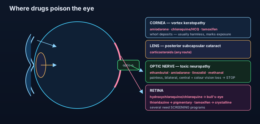
Thioridazine and the phenothiazines: pigmentary retinopathy
Thioridazine, an older antipsychotic of the phenothiazine class, is the second classic retinal toxin. At high doses it causes a pigmentary retinopathy: the RPE clumps and mottles, and patients report blurred vision, dyschromatopsia (colour disturbance), and difficulty seeing at night. Unlike hydroxychloroquine's tidy paracentral ring, thioridazine produces coarse pigment clumping across the mid-periphery and posterior pole, and severe cases can progress to widespread RPE atrophy. Chlorpromazine, another phenothiazine, more often deposits in the lens and cornea than the retina, but the lesson is the same: high-dose, long-term phenothiazines can poison the pigmented layers of the eye, so exceeding the dose ceiling for thioridazine is the thing to avoid. This connects to the Psychopharmacology material, where antipsychotic adverse-effect profiles are compared class by class.
The wider gallery: tamoxifen, interferon, oncology drugs, poppers
Once you know the pattern, you start recognising it under many drug names. Tamoxifen, the anti-oestrogen used in breast cancer, can deposit tiny refractile crystals in the inner retina — a crystalline retinopathy — and, less often, a cystoid maculopathy that blurs central vision; it is classically dose-related and links to the Oncology chapters (and, as above, compounds hydroxychloroquine's risk). Interferon, used for hepatitis and some cancers, causes interferon retinopathy — cotton-wool spots and small haemorrhages from a microvascular insult, usually reversible on stopping. Modern oncology has added its own signatures: the MEK inhibitors (for example trametinib) cause a distinctive serous retinal detachment/retinopathy that is typically reversible, while the checkpoint inhibitors covered in the Oncology immunotherapy chapter can trigger immune-mediated ocular inflammation such as uveitis (a MEK-inhibitor and checkpoint story that belongs beside the irAEs). Two odder causes round out the list: "poppers" (inhaled isobutyl/alkyl nitrites, used recreationally) cause a foveal maculopathy with a yellowish central spot, and canthaxanthin, an oral tanning and food-colour pigment, deposits as glistening crystals in a ring around the macula.
Hydroxychloroquine / chloroquine → bull's-eye maculopathy (screen; may progress after stopping). Thioridazine / high-dose phenothiazines → pigmentary retinopathy. Tamoxifen → crystalline retinopathy / cystoid maculopathy. Interferon → cotton-wool spots and haemorrhages (usually reversible). MEK inhibitors (e.g. trametinib) → serous retinopathy (usually reversible); checkpoint inhibitors → immune uveitis. Poppers (isobutyl nitrite) → foveal maculopathy. Canthaxanthin → glistening crystalline ring around the macula. The unifying thread: take a drug history in any unexplained visual loss.
The unifying lessons
Stand back and the same principles recur — the principles the Toxicology chapter teaches for any drug-induced organ injury. First, most retinal drug toxicity is dose- and duration-dependent: it is the cumulative exposure, not a single tablet, that does the harm, so the prescriber's arithmetic (dose per body weight, years of use, renal clearance) is protective medicine. Second, some of these injuries need formal screening programmes — hydroxychloroquine above all — because the damage is silent until it is permanent. Third, reversibility splits the list in two: interferon and MEK-inhibitor retinopathy often recover when the drug stops, whereas hydroxychloroquine and thioridazine damage is largely irreversible and can even progress. And fourth, tying it all together: a careful drug history is the single most useful test in unexplained visual loss. The bottle in the patient's bathroom cabinet may be the diagnosis.
- Most retinal drug toxicity is cumulative and dose/duration-dependent — prescribing arithmetic is prevention.
- Some drugs (above all hydroxychloroquine) demand formal screening programmes because damage is silent until permanent.
- Reversible if caught early: interferon and MEK-inhibitor retinopathy. Largely irreversible: hydroxychloroquine and thioridazine.
- Thioridazine/phenothiazines → pigmentary retinopathy; tamoxifen → crystalline maculopathy; poppers and canthaxanthin have their own signatures.
- Checkpoint and MEK inhibitors bring oncology-era ocular effects — immune uveitis and serous retinopathy.
- A drug history is the most useful test in unexplained visual loss.
- Relying on visual acuity to screen for hydroxychloroquine toxicity — acuity stays normal until damage is advanced; you need 10-2 fields, OCT and autofluorescence.
- Dosing hydroxychloroquine by actual body weight in an overweight patient (overshoots the retina's tolerance) and ignoring renal function and cumulative years.
- Assuming stopping the drug reverses the injury — hydroxychloroquine and thioridazine damage is often permanent and can progress after cessation.
A 55-year-old woman on hydroxychloroquine for lupus for 7 years has 6/6 acuity but a subtle paracentral scotoma on 10-2 visual field testing and early parafoveal outer-retinal thinning on OCT. What is the single most important next step?
- The retina hoards drugs in its melanin-rich RPE and cannot regenerate, so toxicity is cumulative, dose/duration-dependent, and can progress even after stopping.
- Hydroxychloroquine (and chloroquine) cause bull's-eye maculopathy — the exam classic — needing dosing by ideal body weight, attention to renal function/duration, and a real screening programme (10-2 fields, OCT, autofluorescence).
- Thioridazine/high-dose phenothiazines cause pigmentary retinopathy; tamoxifen a crystalline maculopathy; interferon, MEK inhibitors and checkpoint inhibitors, poppers and canthaxanthin round out the gallery.
- Some injuries reverse on stopping (interferon, MEK), others are largely permanent (hydroxychloroquine, thioridazine) — and a careful drug history is the most useful test in unexplained visual loss.
- Kanski's Clinical Ophthalmology: A Systematic Approach — Drug-induced retinal toxicity.
- Bartlett & Jaanus, Clinical Ocular Pharmacology — Ocular toxicology of systemic drugs.
- American Academy of Ophthalmology, Basic and Clinical Science Course (BCSC), Retina and Vitreous.
- American Academy of Ophthalmology. Recommendations on Screening for Chloroquine and Hydroxychloroquine Retinopathy (revised guideline).
- Rang & Dale's Pharmacology — Antimalarials and disease-modifying antirheumatic drugs; adverse effects.
- Katzung's Basic & Clinical Pharmacology — Drug-induced ocular toxicity.

