
The Biliary Tree: Where Surgeons Slow Down
Almost nobody thinks about bile. It is a greenish fluid, made continuously by the liver, drained by ducts no wider than a drinking straw, and stored in a pear-shaped bag tucked under the ribs — and for most of a human life it does its work in complete silence. Then a stone the size of a pea rolls into the wrong centimetre of tubing, and within days the whites of the eyes turn yellow, the urine darkens to the colour of tea and the stools lose their colour altogether. This is the piece of anatomy where a careless cut does lifelong harm: divide the wrong duct in a twenty-minute operation and the patient may need reconstruction, repeated stenting, sometimes a transplant. Which is why the oldest rule in gallbladder surgery is not about speed or skill. It is four words: identify before you divide.
The theatre has been noisy and quick. The camera went in through the umbilicus, the gallbladder was grasped and rolled upwards over the edge of the liver, and the fat has been teased away from a triangular window of tissue. Now the surgeon stops. The registrar's hand is already holding the clip applier, but nothing is being clipped. "Two structures," the surgeon says, "and only two, entering the gallbladder. Show me the lower third of it off the liver bed." For nearly a minute the room does very little except look. Somewhere behind the fat lies a duct the width of a pencil lead that must be cut, and a few millimetres from it lies a duct that must never be cut — and on a bad day, in an inflamed abdomen, they look exactly alike. Only when the window is unmistakable does the clip go on. That pause, repeated perhaps eight hundred thousand times a year around the world, is the single most important thing anyone does with this anatomy.
A tree that drains instead of feeds
Read the ducts from the liver downwards and the whole system takes about four sentences. Bile is secreted by hepatocytes into microscopic canaliculi, which drain into progressively larger intrahepatic ducts until, at the porta hepatis, two ducts emerge: the RIGHT and LEFT HEPATIC DUCTS, each draining its own functional half of the organ described in the liver. Just outside the liver they unite to form the COMMON HEPATIC DUCT, which descends for two to four centimetres before it is joined, at an acute angle from the right, by the CYSTIC DUCT coming up from the gallbladder. From that junction downwards the channel is called the (COMMON) BILE DUCT — about 6 to 8 cm long and up to 6 mm wide in health, a figure worth memorising because it is the number every ultrasound report quotes: a duct wider than 6 mm in a patient with an intact gallbladder means obstruction until proved otherwise. Note the naming logic, which trips up everyone at first: the duct is "common hepatic" ABOVE the cystic junction and simply "bile duct" BELOW it. Nothing changes in the wall; only the traffic does, because below the junction the duct carries not only liver bile flowing down but gallbladder bile flowing up and back again between meals.
The course of the bile duct is a tour of the most crowded real estate in the abdomen. The duct has three parts, and each one is defined by what it lies behind. The SUPRADUODENAL part descends in the FREE EDGE of the lesser omentum — the hepatoduodenal ligament described in the mesenteries and omenta — where the three structures of the portal triad lie in a fixed and examinable arrangement: the bile duct to the RIGHT, the hepatic artery proper to the LEFT, and the portal vein BEHIND them both. That free edge forms the anterior boundary of the epiploic foramen, so a finger hooked into the foramen can compress all three at once — the Pringle manoeuvre, used to stop bleeding from the liver. The RETRODUODENAL part then passes behind the first part of the duodenum, and the PANCREATIC part grooves, or is completely buried in, the back of the HEAD OF THE PANCREAS covered in the duodenum and pancreas. There it turns towards the duodenal wall and meets the MAIN PANCREATIC DUCT, the two joining in a short dilated common channel — the HEPATOPANCREATIC AMPULLA (of Vater) — which opens into the second part of the duodenum at the MAJOR DUODENAL PAPILLA, roughly 8 to 10 cm beyond the pylorus on the posteromedial wall. Around the ampulla and the terminal parts of both ducts lies the SPHINCTER OF ODDI, a collar of smooth muscle that is anatomically independent of the duodenal musculature and physiologically the gatekeeper of the whole system.
The pear under the ribs
The gallbladder is not a factory. It is a reservoir with a concentrating habit. It lies in a shallow fossa on the visceral surface of the right lobe of the liver, holds only about 30 to 50 mL, and has four named parts. The FUNDUS is the rounded blind end that projects beyond the inferior border of the liver and touches the anterior abdominal wall at the tip of the NINTH COSTAL CARTILAGE, exactly where the lateral border of rectus abdominis crosses the costal margin — the surface point at which the examiner's fingers are placed to elicit MURPHY'S SIGN, and the reason gallbladder tenderness is felt so precisely on the abdominal wall discussed in abdominal surface anatomy. The BODY runs backwards, upwards and to the left, lying against the first part of the duodenum and the transverse colon — a relation that matters when a stone erodes through. The INFUNDIBULUM is the tapering funnel between body and neck, and its lateral wall may bulge into HARTMANN'S POUCH, a small diverticulum which is where stones love to lodge and where an impacted stone starts almost every episode of acute cholecystitis. The NECK is narrow and S-shaped and continues as the cystic duct, whose mucosa is thrown into a series of crescentic folds arranged as the SPIRAL VALVE OF HEISTER — folds that keep the duct patent and prevent it collapsing or over-distending, and that also make it genuinely difficult to pass a catheter or a stone retrograde into the gallbladder.
The wall itself is built for one job. Its mucosa is thrown into fine RUGAE when empty, and — a favourite examination detail — it has NO SUBMUCOSA, the mucosa sitting directly on a thin layer of smooth muscle and then on the perimuscular connective tissue, with peritoneum covering only the free surfaces (the hepatic surface is bare where the gallbladder is attached to the liver). Absent submucosa has a consequence: tumours of the gallbladder reach the muscle and the liver bed disturbingly early. Functionally, the epithelium absorbs water and electrolytes actively and relentlessly, so that in a few hours between meals hepatic bile is CONCENTRATED up to TENFOLD, turning a thin golden fluid into a dark, viscous, highly saturated one. That concentrating power is exactly why the gallbladder — and not the liver — is where stones form: the fluid held longest and squeezed hardest is the fluid most likely to precipitate, a chemistry explored in gallstones and bile acids.
Think of the biliary tree as a river with one reservoir on a side channel. The liver is the catchment: bile trickles down continuously from every hillside, gathers into the right and left tributaries, and joins the main stream at the porta hepatis. Downstream, a single side channel leads off to a small holding lake — the gallbladder — that fills when the sluice gate at the river mouth is shut, and empties back into the main stream when a meal opens it. The sphincter of Oddi is that sluice gate, and it is the only gate in the system: closed, the river backs up into the lake; open, both river and lake run into the duodenum together. Almost every disease in this article is one of two events on that map — a boulder blocking the side channel (the cystic duct: biliary colic, then cholecystitis) or a boulder blocking the main stream below the junction (the bile duct: jaundice, then cholangitis). Knowing which channel is blocked tells you the illness before any test is done.
Calot's triangle and the critical view of safety
No named space in general surgery is dissected more often, or more carefully, than this one. The CYSTOHEPATIC TRIANGLE — universally called CALOT'S TRIANGLE in the operating theatre — is bounded by three things: the CYSTIC DUCT below and to the right, the COMMON HEPATIC DUCT medially, and the INFERIOR SURFACE OF THE LIVER above. (Calot's original 1891 description used the cystic artery as the upper border; the modern working definition substitutes the liver, which enlarges the window and is what surgeons mean today.) Inside it lie the structures that must be found: the CYSTIC ARTERY, arising in about three quarters of people from the RIGHT HEPATIC ARTERY and running behind the common hepatic duct to reach the gallbladder neck, where it divides into superficial and deep branches; the cystic lymph node (Calot's node, or Lund's node) sitting on the artery and serving as its landmark; and loose areolar fat that must be cleared away. Venous drainage is unusual and worth a sentence: small cystic veins from the hepatic surface pass DIRECTLY into the liver substance rather than into a named trunk, while veins from the free surface drain to the portal vein described in the portal system — which is why gallbladder cancer spreads so readily into segments IVb and V of the liver bed. Lymph runs from the cystic node to the hepatic nodes along the free edge and on to the coeliac group.
The classic bile duct injury is not a slip of the hand; it is a misidentification. When inflammation shortens and thickens the tissues, the COMMON BILE DUCT can be pulled up laterally so that it lies in line with the cystic duct and looks exactly like it — the surgeon clips and divides what appears to be the cystic duct and has in fact transected the main drainage of the liver, sometimes taking the right hepatic artery with it. The defence is the CRITICAL VIEW OF SAFETY, and it is a discipline rather than a technique: clear all fat and fibrous tissue from the hepatocystic triangle; separate the lower third of the gallbladder from the liver bed so the cystic plate is visible; and confirm that exactly TWO tubular structures — and no more — are seen entering the gallbladder. Only then does anything get clipped. Crucially, the view is a stopping rule too: if it cannot be obtained, the correct move is to convert to open surgery, perform a subtotal cholecystectomy, or stop and drain — never to keep cutting hopefully. Intraoperative cholangiography, which fills the tree with contrast through the cystic duct, is the map you take when the landscape refuses to be read.
The variants that catch people out
"Normal" biliary anatomy is present in only about two thirds of people. Plan for the other third. Four variants deserve to be named out loud before every operation. First, a RIGHT HEPATIC ARTERY crossing IN FRONT of the common hepatic duct instead of behind it — present in a substantial minority, and lying exactly where the dissection goes. Second, an ACCESSORY or ABERRANT RIGHT HEPATIC DUCT, a duct draining one or more right-sided segments that joins the common hepatic duct low down, or joins the cystic duct itself; divide it unknowingly and the patient develops a persistent postoperative bile leak. (An "accessory" duct is a misnomer worth resisting — it is usually the sole drainage of real liver, not a spare.) Third, a LOW CYSTIC DUCT INSERTION, in which the cystic duct runs parallel to and adherent alongside the common hepatic duct for a centimetre or more before joining it — the configuration most likely to make the bile duct look like the structure you meant to clip. Fourth, MOYNIHAN'S HUMP: a tortuous, high-arching "caterpillar" right hepatic artery that loops upwards into the triangle very close to the gallbladder neck, so that the short cystic artery arising from its summit is easily mistaken for the main vessel — cut the hump and the right lobe loses its arterial supply. The practical rule follows from the list: no structure is divided on the basis of where it ought to be, only on the basis of where it has been traced to.
The blood supply of the bile duct itself is the quiet reason some injuries appear months after an apparently perfect operation. The supraduodenal bile duct is not fed by a plump named artery; it is fed by delicate AXIAL VESSELS running longitudinally along its walls at the 3 O'CLOCK and 9 O'CLOCK positions, arising below from the posterior superior pancreaticoduodenal artery and above from the right hepatic artery, with roughly sixty per cent of the flow ascending from below. They are barely a millimetre across. Strip the duct circumferentially, cauterise around it, or skeletonise it in a hurry, and it survives the operation perfectly — then slowly becomes ischaemic, fibroses, and presents six or twelve months later as a tight benign stricture with painless jaundice and recurrent cholangitis. In this region, dissection close to the duct is not the safe option; leaving tissue on it is.
Nerves, and why the pain lies about its address
The gallbladder and ducts are FOREGUT derivatives, and they are innervated accordingly, through the arrangement set out in the autonomic nerves of the abdomen. Sympathetic fibres reach them from the COELIAC PLEXUS, having travelled in the greater splanchnic nerve from spinal segments T5–T9; parasympathetic fibres come from the VAGUS, which stimulates contraction; and phrenic fibres reach the region through the coeliac plexus as well. Because the visceral afferents accompany the sympathetics back to T5–T9, the pain of a distended gallbladder or an obstructed duct is first felt in the MIDLINE EPIGASTRIUM — dull, deep, poorly localised, and completely unhelpful for pointing at the organ, in exact keeping with the foregut rule set out in foregut, midgut and hindgut. Only when inflammation spreads to touch the PARIETAL peritoneum of the abdominal wall does the pain migrate to the right upper quadrant and become sharp, localised and tender — the same two-stage story as appendicitis, told at a different address. And if the inflamed organ reaches the undersurface of the diaphragm, the phrenic nerve carries the message to C3–C5 and the patient feels pain in the RIGHT SHOULDER TIP, the referral explained in the diaphragm. Three different pains, one organ, depending only on what it happens to be touching.
How bile actually gets out
Between meals the gate is shut, so bile takes the only road left open: backwards, into the gallbladder. The liver secretes bile continuously — around 500 to 1000 mL a day — but the duodenum only needs it intermittently, and the sphincter of Oddi resolves the mismatch. In the fasting state the sphincter maintains a resting tone high enough that the pressure in the bile duct exceeds the pressure needed to fill the gallbladder, so bile flows retrogradely up the cystic duct and is stored and concentrated. When a meal — especially a FATTY meal — reaches the duodenum, enteroendocrine I cells release CHOLECYSTOKININ (CCK). CCK does two coordinated things: it makes the smooth muscle of the gallbladder wall contract, and it RELAXES the sphincter of Oddi, so the reservoir squeezes at the same moment the gate opens. Vagal stimulation, including the cephalic phase of a meal, adds to the contraction. Concentrated bile is delivered into the duodenum, where bile salts emulsify fat into micelles and make it absorbable — and roughly ninety-five per cent of those salts are then reclaimed in the terminal ileum and returned to the liver in the portal vein, the enterohepatic circulation that lets a modest pool do an enormous day's work. This is also why the classic gallstone attack follows a fatty meal by twenty to sixty minutes: CCK orders a gallbladder with a stone in its neck to contract, and it contracts against a blocked exit.
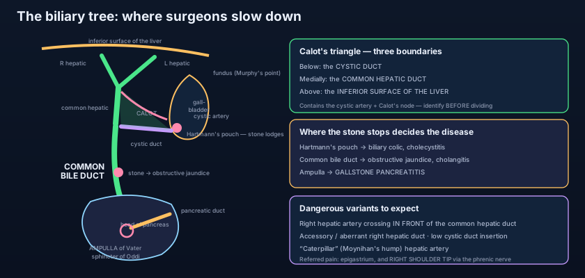
The stone in the neck: a woman finishes a fried supper and within the hour has a severe, constant, gripping pain in the epigastrium boring through to the back and the right shoulder blade. It lasts three hours and then eases as the stone falls back — BILIARY COLIC, a badly named symptom since the pain does not truly come in waves. The stone that stays: the same woman returns days later, this time with fever, right upper quadrant tenderness and a POSITIVE MURPHY'S SIGN — the examiner's fingers held at the tip of the ninth costal cartilage while she breathes in, and the descending inflamed gallbladder strikes them and arrests the breath. The stone in the main road: a man is not in much pain at all, but his wife noticed his eyes were yellow; his urine is dark, his stools are pale and he cannot stop scratching. A stone in the distal bile duct has produced OBSTRUCTIVE JAUNDICE, and if bacteria ascend behind it he develops CHARCOT'S TRIAD — fever with rigors, right upper quadrant pain and jaundice — which is ascending cholangitis, an emergency needing antibiotics and urgent drainage by ERCP. The stone that took a wrong turn: a fourth patient develops severe epigastric pain radiating to the back with a raised serum amylase — a stone impacted at the ampulla has obstructed the pancreatic duct too, and GALLSTONE PANCREATITIS, described in pancreatitis, is under way.
Stones that break the rules
Two syndromes are worth knowing because they behave as if the anatomy had been rearranged. In MIRIZZI SYNDROME a large stone impacted in the cystic duct or Hartmann's pouch compresses the adjacent COMMON HEPATIC DUCT from outside, so the patient becomes jaundiced without any stone ever entering the main duct — and in advanced cases the stone erodes through into it, creating a cholecystocholedochal fistula. Mirizzi is the classic trap of laparoscopic surgery: the inflammation obliterates Calot's triangle, the risk of duct injury rises steeply, and the correct decision is often to abandon the keyhole approach. In GALLSTONE ILEUS a chronically inflamed gallbladder becomes adherent to the duodenum and a large stone ulcerates directly through, creating a CHOLECYSTODUODENAL FISTULA; the stone then travels down the small bowel and impacts, usually at the terminal ileum, causing mechanical obstruction as covered in the small intestine. The plain film shows Rigler's triad: small bowel obstruction, a stone outside the biliary tree, and air in the biliary tree (pneumobilia) — gas that has entered through the fistula. It is a striking reminder that a stone can leave the biliary tree without ever passing through the papilla.
- Order of the ducts: right + left HEPATIC ducts from the porta hepatis → COMMON HEPATIC DUCT → joined by the CYSTIC DUCT → the (common) BILE DUCT, about 6–8 cm long and up to 6 mm wide. "Common hepatic" above the cystic junction, "bile duct" below it.
- The bile duct descends in the FREE EDGE of the lesser omentum — to the RIGHT of the hepatic artery proper and IN FRONT of the portal vein — then behind the first part of the duodenum, then grooving the back of the head of the pancreas.
- It joins the main pancreatic duct at the HEPATOPANCREATIC AMPULLA (of Vater) and opens at the MAJOR DUODENAL PAPILLA in the second part of the duodenum, guarded by the SPHINCTER OF ODDI.
- Gallbladder parts: FUNDUS at the tip of the 9th costal cartilage where the lateral border of rectus crosses the costal margin (Murphy's sign); BODY; INFUNDIBULUM with HARTMANN'S POUCH where stones lodge; NECK continuing as the cystic duct with the SPIRAL VALVE OF HEISTER.
- It stores 30–50 mL and CONCENTRATES bile up to TENFOLD by absorbing water and electrolytes; its mucosa has rugae and NO SUBMUCOSA, which is why gallbladder cancer invades early.
- CALOT'S TRIANGLE (cystohepatic) is bounded by the CYSTIC DUCT, the COMMON HEPATIC DUCT and the INFERIOR SURFACE OF THE LIVER, and contains the CYSTIC ARTERY (usually from the right hepatic artery) and the cystic node.
- The CRITICAL VIEW OF SAFETY: clear the hepatocystic triangle of fat, free the lower third of the gallbladder from the liver bed, and confirm exactly TWO structures entering the gallbladder — and if it cannot be obtained, convert, do a subtotal cholecystectomy, or stop.
- Dangerous variants: a right hepatic artery crossing IN FRONT of the common hepatic duct; an accessory or aberrant right hepatic duct; a low, parallel cystic duct insertion; and Moynihan's hump — the tortuous "caterpillar" right hepatic artery looping into the triangle.
- The supraduodenal bile duct is fed by delicate axial vessels at the 3 O'CLOCK and 9 O'CLOCK positions (from the posterior superior pancreaticoduodenal artery below and the right hepatic artery above) — over-dissection devascularises it and causes a LATE benign stricture.
- Nerve supply is the coeliac plexus (sympathetic, T5–T9) and the vagus; visceral pain is referred to the EPIGASTRIUM, moves to the right upper quadrant when parietal peritoneum is involved, and goes to the RIGHT SHOULDER TIP via phrenic irritation.
- CCK from duodenal I cells after a fatty meal contracts the gallbladder AND relaxes the sphincter of Oddi simultaneously — which is why biliary colic follows a fatty meal by 20–60 minutes.
- Cystic duct block → colic then cholecystitis, NO jaundice. Bile duct block → obstructive jaundice with pale stools and dark urine, and cholangitis with CHARCOT'S TRIAD (fever, right upper quadrant pain, jaundice).
- Thinking a stone in the cystic duct causes jaundice. It does not: liver bile still reaches the duodenum down the common hepatic duct. Jaundice requires obstruction BELOW the cystic junction — or the external compression of Mirizzi syndrome.
- Placing the bile duct behind the portal vein in the free edge of the lesser omentum. The duct is ANTERIOR and to the RIGHT, the hepatic artery anterior and to the LEFT; the portal vein lies BEHIND them both.
- Believing bile duct injury is a rare accident of clumsy hands. It is overwhelmingly a failure of IDENTIFICATION — the common bile duct pulled into line with the cystic duct in an inflamed field — which is why the remedy is a mandatory view, not faster dissection.
During a laparoscopic cholecystectomy the surgeon dissects a triangular window before dividing anything. Which three structures bound that window, and what artery should be found within it?
- Right + left hepatic ducts → common hepatic duct → joined by the cystic duct → the bile duct (6–8 cm long, up to 6 mm wide), which runs in the free edge of the lesser omentum to the right of the hepatic artery and in front of the portal vein, behind the duodenum, through the head of the pancreas, to the ampulla of Vater and the major duodenal papilla guarded by the sphincter of Oddi.
- The gallbladder — fundus at the tip of the 9th costal cartilage (Murphy's sign), body, infundibulum with Hartmann's pouch, neck with the spiral valve of Heister — stores 30–50 mL and concentrates bile up to tenfold; it has rugae but no submucosa.
- Calot's triangle (cystic duct, common hepatic duct, inferior surface of the liver) contains the cystic artery from the right hepatic artery; the critical view of safety and a healthy fear of the variants — anterior right hepatic artery, aberrant right hepatic duct, low cystic insertion, Moynihan's hump — are what prevent the classic bile duct injury.
- Pain is referred to the epigastrium (foregut, T5–T9), to the right upper quadrant once the parietal peritoneum is inflamed, and to the right shoulder tip via the phrenic nerve; block the cystic duct and you get colic then cholecystitis, block the bile duct and you get obstructive jaundice, cholangitis with Charcot's triad, and gallstone pancreatitis.
- Drake RL, Vogl AW, Mitchell AWM. Gray's Anatomy for Students — Abdomen: the gallbladder and the biliary tree.
- Moore KL, Dalley AF, Agur AMR. Clinically Oriented Anatomy — Biliary ducts and gallbladder; the cystohepatic (Calot's) triangle.
- Netter FH. Atlas of Human Anatomy — Gallbladder, extrahepatic bile ducts and pancreatic duct; variations in cystic and hepatic ducts.
- Last RJ. Last's Anatomy: Regional and Applied — The extrahepatic biliary apparatus and its arterial supply.
- Snell RS. Clinical Anatomy by Regions — Gallstones, cholecystectomy and bile duct injury.
- TeachMeAnatomy — The Gallbladder and Biliary Tree; Calot's Triangle.

