
Foregut, Midgut, Hindgut: Why Appendicitis Pain Moves
The abdomen looks, at first, like a bag of unrelated organs — a stomach here, a liver there, yards of intestine coiled between them, and no obvious reason why any of it should be arranged the way it is. Then you learn one idea, and the bag becomes a machine. In the fourth week of life the gut is a single straight tube, and it is cut into three parcels: foregut, midgut, hindgut. Each parcel keeps one artery, one set of sympathetic nerves and one address on the skin for the rest of your life — and it keeps them no matter how far the organ later travels. That is why the pain of an inflamed appendix begins nowhere near the appendix, why a duodenal ulcer hurts in the same place as a heart attack, and why the single most useful question in abdominal pain is not "where does it hurt?" but "where did it start?"
A fifteen-year-old wakes at dawn feeling wrong. There is an ache somewhere around his navel — dull, deep, hard to describe, impossible to point to. He is not hungry, which is unlike him, and he feels faintly sick. He goes back to sleep. By mid-morning the ache has not gone but has changed its character: it is no longer vague and no longer central. It has slid down and to the right, and it is now sharp. When his mother drives over a speed bump he gasps. In the emergency department the doctor asks him to cough, then places a single fingertip low on the right side of his abdomen, two-thirds of the way from the navel to the bony prominence of the hip, and he flinches before she has pressed. Nothing inside him has moved. His appendix is exactly where it was at dawn. What changed is which membrane is being irritated — and the whole story of the abdomen is contained in that sentence.
One tube, cut into three parcels
Everything the abdomen does later is written into a decision made in the fourth week of life. The primitive gut tube runs the length of the embryo, suspended in the midline, and it is divided into three regions not by any visible wall but by the arteries that grow into it from the aorta. Each division receives one ventral branch of the aorta, and that single vessel then defines everything else about the segment: which organs bud from it, which mesentery carries it, which lymph nodes drain it, which autonomic nerves travel along the artery to reach it, and — because those nerves enter the spinal cord at fixed levels — where on the body wall its pain will be felt. The rule is beautifully rigid. An organ can migrate enormously during development, rotate, be plastered against the posterior wall, or end up on the opposite side of the abdomen, and it will still be fed by the artery it was born with and will still refer its pain to the same patch of skin. The four constants of each division — EXTENT, ARTERY, SYMPATHETIC LEVEL, REFERRED SITE — are worth learning as a single unit, because once you know them you can predict the site of pain for any abdominal organ without memorising a list.
The foregut: coeliac trunk, T5–T9, the epigastrium
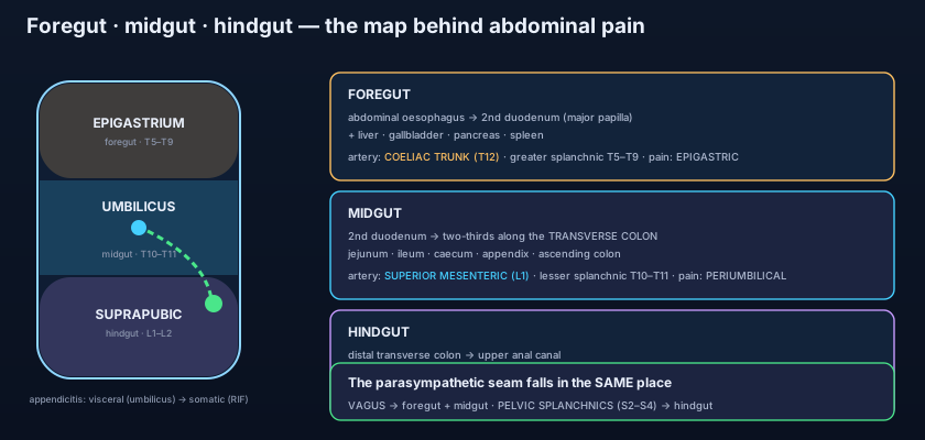
The FOREGUT runs from the abdominal oesophagus to the second part of the duodenum, and its distal limit is precise: the level of the MAJOR DUODENAL PAPILLA, where the bile duct and the main pancreatic duct open together. Every organ that buds off this stretch belongs to it — the stomach, the liver and biliary tree, the pancreas, and the spleen, which is a foregut structure by blood supply even though it is not derived from the gut tube at all. Its artery is the COELIAC TRUNK, which leaves the aorta at the level of T12 and trifurcates almost immediately into the left gastric, splenic and common hepatic arteries. Its sympathetic afferents — the fibres that carry pain — enter the spinal cord between T5 and T9, having travelled up the artery and then through the GREATER SPLANCHNIC nerve, which pierced the crus of the diaphragm to get there. Because T5–T9 dermatomes cover the upper central abdominal wall, foregut pain is felt in the EPIGASTRIUM, roughly at the T7 dermatome. This is why a stomach ulcer, a gallbladder attack and acute pancreatitis all begin in the same place, and why patients so often describe early foregut pain by placing a whole flat hand below the sternum rather than a finger.
The midgut: superior mesenteric artery, T10–T11, the navel
The MIDGUT takes over at the major duodenal papilla and runs all the way to a point roughly TWO-THIRDS ALONG THE TRANSVERSE COLON. That means it owns the distal half of the duodenum, the whole jejunum and ileum, the caecum and appendix, the ascending colon, and most of the transverse colon — by length, by far the largest of the three. Its artery is the SUPERIOR MESENTERIC ARTERY, arising from the aorta at L1, one centimetre below the coeliac trunk, and giving the inferior pancreaticoduodenal, jejunal and ileal, ileocolic, right colic and middle colic branches. Its sympathetic afferents enter the cord at T10 and T11 through the LESSER SPLANCHNIC nerve. The T10 dermatome is the classic landmark of the whole body wall: it runs through the UMBILICUS. So every midgut structure, from the jejunum to the appendix, refers its early pain to a poorly localised ring around the navel — which is exactly where our fifteen-year-old felt it at dawn. It is worth pausing on how strange that is: the appendix sits in the right iliac fossa, and yet its first complaint arrives fifteen centimetres away in the midline, because the appendix is not shouting through the body wall but whispering down an autonomic nerve that was assigned to it in the fourth week of embryonic life.
The hindgut: inferior mesenteric artery, L1–L2, suprapubic
The HINDGUT begins at that same two-thirds point on the transverse colon and continues through the descending colon, the sigmoid, the rectum and down to the upper anal canal — the boundary there being the pectinate line, above which the mucosa is insensitive to pain and below which it is exquisitely sensitive somatic skin. Its artery is the INFERIOR MESENTERIC ARTERY, leaving the abdominal aorta at L3 — three centimetres above the aortic bifurcation at L4 — and branching into the left colic, the sigmoid arteries and the superior rectal artery. Its sympathetic afferents reach the cord at L1 and L2 through the LUMBAR SPLANCHNIC nerves. The L1 dermatome crosses the lower abdominal wall just above the groin, so hindgut pain is referred SUPRAPUBICALLY, in the hypogastric region. That is why the cramping pain of constipation, of an obstructing sigmoid tumour, or of early diverticulitis sits low and central across the pubic hairline before it localises. Line the three up and the pattern is impossible to unsee: T7 epigastrium, T10 umbilicus, L1 suprapubic — three horizontal bands stacked down the front of the abdomen, one for each parcel of the original tube.
Think of the three divisions as three postal districts drawn on a map long before any of the houses were built. Each district was given one road in (its artery) and one telephone exchange (its spinal levels). Later the houses were built, extended and moved — the stomach swung to the left, the liver ballooned to the right, the caecum walked all the way down into the right iliac fossa — but nobody redrew the districts, and nobody rewired the exchanges. So when a house in the midgut district calls for help, the call still comes through the T10 exchange, and the operator still writes down the address the exchange is registered to: the navel. The organ has moved; its paperwork never did. Referred pain is not a mistake the nervous system makes. It is an old address, faithfully honoured.
The vagus stops exactly where the artery stops
The parasympathetic supply obeys the same seam — and that is the proof the seam is real. Sympathetic fibres reduce gut activity and carry its pain; parasympathetic fibres do the opposite, driving secretion and peristalsis, and they carry no pain at all. The remarkable thing is where the parasympathetic supply changes hands. The VAGUS nerve — the tenth cranial nerve, arriving on the oesophagus as anterior and posterior vagal trunks — supplies the foregut AND the midgut, and it stops at precisely the same two-thirds-of-the-transverse-colon line. From there onwards the hindgut is supplied by the PELVIC SPLANCHNIC nerves from the sacral cord, S2, S3 and S4, which run up through the inferior hypogastric plexus in the opposite direction to everything else. Two entirely different nerves, from opposite ends of the neuraxis, meeting at the same point on the transverse colon where the superior mesenteric territory hands over to the inferior mesenteric — because it is not two boundaries, it is one embryological seam seen twice. The relays for all of this sit as ganglia clustered around the origins of the three arteries, the coeliac, superior mesenteric and inferior mesenteric ganglia, and the plexuses that connect them are traced in the autonomic nerves of the abdomen. Practically, the split explains why vagal stimulation empties the stomach and the small bowel but does nothing for a sluggish descending colon, and why sacral nerve damage — a pelvic fracture, a low spinal injury, extensive pelvic surgery — produces constipation and a neurogenic bowel while the stomach behaves perfectly normally.
The loop that left home and came back
The midgut grows faster than the abdomen that must hold it. In week 6 the whole midgut loop, hinged on the superior mesenteric artery and tethered at its apex to the vitellointestinal (vitelline) duct, HERNIATES out of the abdomen into the umbilical cord — a physiological herniation that is normal and universal. While it is outside, it rotates 270 degrees ANTICLOCKWISE around the axis of the superior mesenteric artery, viewed from the front: 90 degrees during herniation and a further 180 degrees as it returns. In week 10 it retracts, small bowel first, and the rotation has by then carried the caecum across the top of the abdomen and down the right side to the right iliac fossa, dragging the ascending colon behind it. Fixation follows: the ascending and descending colon and the duodenum are plastered against the posterior wall and become retroperitoneal, while the jejunum, ileum, transverse colon and sigmoid keep their mesenteries and stay mobile. Every step of this can fail, and each failure has a name. MALROTATION leaves the caecum high and the small bowel suspended on a narrow stalk instead of a broad root, so the whole midgut can twist on its own artery — MIDGUT VOLVULUS, the bilious vomiting of a previously well neonate, a surgical emergency of minutes and hours, not days. Failure of the loop to return leaves an EXOMPHALOS (omphalocele), the bowel still in the cord and covered by a membrane at the umbilicus; a separate defect in the abdominal wall lateral to the cord produces GASTROSCHISIS, bowel floating free in amniotic fluid with no covering at all. And if the vitellointestinal duct fails to close, its remnant persists as a MECKEL'S DIVERTICULUM: the rule of twos — about two per cent of people, two feet from the ileocaecal valve, two inches long, often containing two types of ectopic mucosa (gastric and pancreatic), and the gastric mucosa is why it can bleed painlessly or ulcerate.
Why the three territories talk to each other
Three arteries, but never three separate circulations. If the divisions were truly sealed off, blocking one artery would kill a third of the gut. They are not. Along the whole inner border of the colon runs the MARGINAL ARTERY of Drummond, a continuous vessel formed by the end-to-end anastomosis of every colic branch from the ileocolic to the sigmoid, so blood from the superior mesenteric can travel round to hindgut territory and back again. Deeper and more variable is the ARC OF RIOLAN, a short central connection between the middle colic branch of the superior mesenteric and the left colic branch of the inferior mesenteric, which enlarges dramatically when one of them narrows and can then be seen on angiography as a "meandering artery". At the upper end the PANCREATICODUODENAL ARCADES do the same job for the foregut–midgut junction, linking the superior pancreaticoduodenal artery from the coeliac trunk with the inferior pancreaticoduodenal artery from the superior mesenteric, which is why the duodenum straddling the border between the two divisions has a doubled blood supply from both. But an anastomotic chain is weakest where two supplies meet at their furthest reach, and those WATERSHED points fail first: the SPLENIC FLEXURE, at the superior–inferior mesenteric junction, is the classic site of ischaemic colitis in an elderly patient after a period of low blood pressure; and SUDECK'S POINT at the rectosigmoid, where the last sigmoid branch meets the superior rectal artery, is the reason a low anterior resection anastomosis is watched so anxiously for leakage.
Two membranes, two completely different pains
Now return to the boy. The organs of the abdomen are wrapped in VISCERAL peritoneum, and the body wall is lined by PARIETAL peritoneum, and although the two sheets are continuous — the arrangement laid out in the peritoneum — their nerve supplies could not be more different. Visceral peritoneum and the gut wall itself are supplied only by the autonomic afferents just described. They respond to stretch, distension, ischaemia and spasm, but not to cutting or burning, they are bilateral and few in number, and they enter the cord at the segmental level of the division. The resulting pain is therefore DULL, MIDLINE, poorly localised, often colicky, and accompanied by nausea, sweating and pallor — the patient rubs a whole region and cannot point. Parietal peritoneum, by contrast, is supplied by SOMATIC nerves — the lower intercostal, subcostal and iliohypogastric nerves that also supply the overlying skin and muscle. Their pain is SHARP, precisely LOCALISED to the spot, worse on movement, coughing or a jolt in the car, and it triggers reflex guarding of the muscle above it. So early appendicitis, a midgut structure with an inflamed but untouching tip, produces vague periumbilical visceral pain; and when hours later the inflamed serosa reaches and irritates the PARIETAL peritoneum of the right iliac fossa, the somatic system takes over and the pain migrates, sharpens and can be found with one fingertip at McBurney's point — two-thirds of the way along the line from the umbilicus to the right anterior superior iliac spine. Nothing moved; the message simply changed wires.
"Where did it start, and where is it now?" is the single most informative question in the assessment of abdominal pain, and it costs nothing. A pain that began around the navel and then settled in the right iliac fossa is appendicitis until proved otherwise. A pain that began in the epigastrium and then became a rigid, board-like, agonising pain everywhere is a perforated duodenal ulcer whose acid has spilled onto the whole parietal peritoneum. A pain that began in the epigastrium and moved to the right upper quadrant, worse after fatty food, is the gallbladder. Pain that starts suprapubically and localises to the left iliac fossa in an older patient is diverticulitis. The migration itself is the diagnosis — because the first site tells you which embryological division is involved, and the second tells you that inflammation has now reached the body wall. A patient describing only the second half of that story has already had hours of disease, and the acute abdomen is a race the anatomy lets you enter early.
The dermatome rule works in reverse, too, and that is where it catches people out. Anything that irritates the T10 nerve root or the structures sharing it will produce pain the patient reports as abdominal, whether or not the abdomen is the problem. A child with a right lower lobe PNEUMONIA can present with abdominal pain and be sent to the surgeons, because the lower intercostal nerves supply both the pleura above the diaphragm and the abdominal wall below it. TESTICULAR TORSION frequently presents as central abdominal pain and vomiting with barely a mention of the scrotum, because the testis developed on the posterior abdominal wall at the T10 level and dragged its nerve supply down with it as it descended. Shingles in a thoracic dermatome can mimic an acute abdomen for two days before the rash appears. The lesson is not that referred pain is unreliable — it is that you must examine the chest and the groin of every patient with abdominal pain, precisely because the nervous system files by embryological origin and not by postcode.
The ulcer that announced itself twice: a man with months of gnawing epigastric pain, relieved by food and by the antacids described in peptic ulcer treatment, suddenly doubles over at work. For an hour the pain is epigastric — the duodenum is foregut, T5–T9. Then acid tracks down the right paracolic gutter and inflames the parietal peritoneum of the right iliac fossa, and he is admitted with suspected appendicitis. Free gas under the diaphragm on an erect chest film gives him away. The gallbladder that started in the wrong place: a woman wakes at 2 a.m. with severe epigastric pain after a fried dinner; six hours later it has moved to the right upper quadrant and she cannot take a deep breath while the examiner's hand rests there. The cystic duct stone was foregut visceral pain; the inflamed gallbladder touching the parietal peritoneum under the ribs is somatic. The left-sided appendicitis: a seventy-year-old with two days of vague suprapubic cramping now has sharp, localised tenderness in the left iliac fossa with a fever — sigmoid diverticulitis, hindgut pain that has completed the same journey on the other side. And the neonate who vomited green: a four-day-old feeds poorly and vomits bile-stained milk; he is in theatre within two hours, because bilious vomiting in a newborn is midgut volvulus until an urgent contrast study says otherwise.
- FOREGUT: abdominal oesophagus to the second part of the duodenum at the major duodenal papilla, plus liver, gallbladder, pancreas and spleen. Artery: coeliac trunk (T12). Sympathetics T5–T9 via the greater splanchnic. Pain referred to the EPIGASTRIUM.
- MIDGUT: second part of the duodenum to two-thirds along the transverse colon — jejunum, ileum, caecum, appendix, ascending and most of transverse colon. Artery: superior mesenteric (L1). Sympathetics T10–T11 via the lesser splanchnic. Pain referred PERIUMBILICALLY.
- HINDGUT: distal transverse colon to the upper anal canal — descending colon, sigmoid, rectum. Artery: inferior mesenteric (L3). Sympathetics L1–L2 via the lumbar splanchnics. Pain referred SUPRAPUBICALLY (hypogastrium).
- The referred site is simply the dermatome of those spinal segments: T7 epigastrium, T10 umbilicus, L1 suprapubic. Three horizontal bands stacked down the front of the abdomen.
- Parasympathetic supply splits at the SAME seam: the vagus serves foregut and midgut to the two-thirds point of the transverse colon; pelvic splanchnic nerves (S2–S4) serve the hindgut from there on.
- Midgut development: physiological herniation into the umbilical cord in week 6, 270° anticlockwise rotation about the superior mesenteric artery, retraction in week 10, then fixation of duodenum and ascending/descending colon retroperitoneally.
- Rotation failures: malrotation (high caecum, narrow mesenteric root) predisposing to MIDGUT VOLVULUS — bilious vomiting in a neonate is a surgical emergency; exomphalos (bowel in the cord, membrane-covered) versus gastroschisis (paraumbilical wall defect, no covering).
- Meckel's diverticulum is the persistent vitellointestinal duct — rule of twos: 2% of people, 2 feet from the ileocaecal valve, 2 inches long, 2 types of ectopic mucosa; the gastric mucosa causes painless bleeding or ulceration.
- The territories anastomose: the marginal artery of Drummond along the colon, the arc of Riolan centrally, and the pancreaticoduodenal arcades at the foregut–midgut junction (which is why the duodenum has a double supply).
- Watershed points fail first: the SPLENIC FLEXURE (SMA–IMA junction) is the classic site of ischaemic colitis, and SUDECK'S POINT at the rectosigmoid threatens low anastomoses.
- VISCERAL peritoneum + gut wall = autonomic afferents: dull, midline, poorly localised, colicky pain with nausea, responding to stretch and ischaemia but not to cutting.
- PARIETAL peritoneum = somatic nerves: sharp, precisely localised pain, worse on movement and coughing, with guarding — hence appendicitis pain migrating from the umbilicus to McBurney's point, two-thirds along the umbilicus-to-right-ASIS line.
- Placing the foregut–midgut boundary at the pylorus or at the duodenojejunal flexure. It is at the MAJOR DUODENAL PAPILLA in the second part of the duodenum — which is why the duodenum straddles two divisions and enjoys a double blood supply through the pancreaticoduodenal arcades.
- Assuming the appendix pain "spreads" or that the organ shifts. Nothing moves. The first pain is visceral (autonomic, T10, periumbilical) and the second is somatic (parietal peritoneum of the right iliac fossa) — a change of nerve pathway, not of position.
- Thinking the vagus supplies the whole gut. It stops with the midgut, two-thirds along the transverse colon; the hindgut is parasympathetically supplied by the pelvic splanchnic nerves (S2–S4) travelling upwards from the sacral cord.
A 15-year-old presents with 12 hours of pain. It began as a vague, dull ache around the umbilicus that he could not localise, associated with nausea and anorexia; over the last three hours it has become sharp and is now maximal at a point two-thirds of the way from the umbilicus to the right anterior superior iliac spine, with guarding and pain on coughing. Which single statement best explains the change in the character and site of his pain?
- The gut is divided embryologically into foregut, midgut and hindgut, and each keeps four constants for life: extent, artery, sympathetic level and referred pain site — coeliac trunk / T5–T9 / epigastrium; superior mesenteric artery / T10–T11 / periumbilical; inferior mesenteric artery / L1–L2 / suprapubic.
- The foregut ends at the major duodenal papilla and the midgut ends two-thirds along the transverse colon; the vagus supplies foregut and midgut to that same line, and pelvic splanchnic nerves (S2–S4) supply the hindgut beyond it — one seam seen twice.
- The midgut loop herniates into the umbilical cord in week 6, rotates 270° anticlockwise about the superior mesenteric artery and returns in week 10; failures give malrotation with midgut volvulus, exomphalos, gastroschisis and Meckel's diverticulum.
- Visceral peritoneum (autonomic) gives dull, midline, poorly localised pain; parietal peritoneum (somatic) gives sharp, localised pain with guarding — so appendicitis starts periumbilically and migrates to McBurney's point, and "where did it start?" is the most useful question in abdominal pain.
- Drake RL, Vogl AW, Mitchell AWM. Gray's Anatomy for Students — Abdomen: development of the gut, arterial territories and referred pain.
- Moore KL, Dalley AF, Agur AMR. Clinically Oriented Anatomy — Foregut, midgut and hindgut; visceral versus parietal peritoneal pain.
- Netter FH. Atlas of Human Anatomy — Coeliac trunk, superior and inferior mesenteric arteries; autonomic innervation of the abdomen.
- Last RJ. Last's Anatomy: Regional and Applied — Rotation of the midgut and fixation of the intestines.
- Snell RS. Clinical Anatomy by Regions — Referred abdominal pain, appendicitis and the acute abdomen.
- TeachMeAnatomy — The Gastrointestinal Tract: Embryological Divisions; Referred Pain.

