
The Facial Nerve: Why a Stroke Spares the Forehead
A man's face has fallen on one side. The corner of his mouth droops, his cheek sags, saliva escapes when he drinks. It looks alarming, and it looks like one thing. But a doctor at the bedside will ask him to do something almost trivial before anything else: raise your eyebrows, wrinkle your forehead. If both eyebrows lift and the forehead creases evenly, the danger is in the brain — a stroke — and the emergency clock is running. If the forehead on the drooping side stays smooth and dead and will not move, the lesion is in the nerve itself, out in the face, and the outlook is usually kind. One nerve, one muscle of the forehead, and one quirk of its wiring in the brain decide which of two utterly different diagnoses a face is showing. This is the story of the facial nerve — the nerve that not only moves the face but tastes the front of the tongue, waters the eye, wets the mouth, and dampens sound in the ear — and of the single most useful line in a droop.
A woman of thirty-four wakes, brushes her teeth, and water runs out of the left side of her mouth. In the mirror the left half of her face has gone slack overnight: the forehead is smooth on that side, the eyebrow will not lift, the eye will not close — when she tries, the eyeball rolls up and only white shows — and the corner of her mouth is pulled across to the good side. She thinks she is having a stroke and comes to hospital frightened. The doctor watches her face rather than her scan. He asks her to look up and wrinkle her forehead: the right side furrows, the left stays glassy and still. He asks her to screw her eyes shut: the right lids bury the lashes, the left will not meet. He asks her to bare her teeth: the smile is dragged to the right. Then he asks about her ear — sounds have seemed uncomfortably loud on the left since yesterday — and touches a drop of sugar to the front of her tongue, which she cannot taste on that side. He has not ordered a single test, and he already knows this is not a stroke. It is a lower motor neuron facial palsy — the whole half of the face, forehead and all — and the extra clues have told him almost exactly where along the nerve the fault lies. The one nerve involved, described among its eleven companions in the cranial nerves overview, is doing far more than moving a face.
Can you wrinkle your forehead?
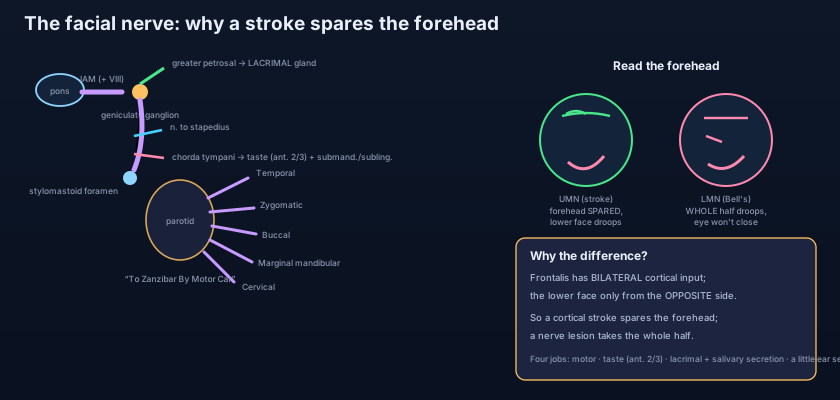
It is the cheapest, fastest and most decisive test in the whole of clinical neurology. A drooping face frightens everyone, and to the untrained eye a Bell's palsy and a stroke look identical. Yet a single question separates them at the bedside, before any scan: can the patient wrinkle the forehead on the weak side? The muscle that raises the eyebrows and corrugates the forehead is FRONTALIS (part of occipitofrontalis), and it holds a secret unique among the muscles of facial expression — it receives cortical commands from BOTH cerebral hemispheres, while every muscle of the lower face is driven by the OPPOSITE hemisphere alone. So a lesion in the brain — a stroke in the motor cortex or the internal capsule, an UPPER MOTOR NEURON lesion — paralyses the contralateral LOWER face but SPARES the forehead, because the surviving hemisphere still reaches frontalis. A lesion of the nerve itself out in the face — a Bell's palsy, a LOWER MOTOR NEURON lesion — paralyses the WHOLE half of the face, forehead included, and the eye on that side will not close. Forehead spared, think brain; forehead paralysed, think nerve. Learn that one line and you have the single most useful sign in a facial droop.
Four jobs crowded into one nerve
Call it "the motor nerve of the face" and you have named barely half of what it does. The facial nerve (CN VII) carries four distinct kinds of fibre. FIRST, and largest, MOTOR (branchial) fibres to all the MUSCLES OF FACIAL EXPRESSION — the mimetic muscles that furrow, smile, grimace and close the eye, laid out in the scalp and face — and, beyond the face, to STAPEDIUS in the middle ear, the POSTERIOR BELLY OF DIGASTRIC and STYLOHYOID: every one of these a derivative of the SECOND pharyngeal arch, whose nerve is VII, as set out in the pharyngeal arches. SECOND, special sensory TASTE from the anterior two-thirds of the tongue, carried by the CHORDA TYMPANI. THIRD, PARASYMPATHETIC (secretomotor) fibres — to the LACRIMAL gland (that keep the eye wet) by way of the greater petrosal nerve and the pterygopalatine ganglion, and to the SUBMANDIBULAR and SUBLINGUAL salivary glands (that wet the mouth) by way of the chorda tympani and the submandibular ganglion, glands mapped in the salivary glands. FOURTH, a little GENERAL SENSATION from the skin of the external ear and the external auditory meatus. Motor, taste, secretomotor, sensory — four jobs, and a lesion at the wrong point knocks out several at once.
Think of the facial nerve not as a single wire but as a bundle of four cables taped together, running down one long, narrow conduit drilled through the hardest bone in the body. Because they share the conduit, the point at which the conduit is crushed decides which cables are cut. Crush it high, near the start, and you sever all four at once — the face falls, the eye runs dry, sounds become painfully loud, taste disappears. Crush it low, after three of the cables have peeled off and left the tunnel, and only the motor cable remains to be cut — the face falls, but the eye still waters, sound is normal, taste is intact. The clinician runs this logic backwards: from which functions survive, he reads how far down the conduit the damage lies. No other cranial nerve gives up its position so precisely, and it is precisely because it is trapped for so long in a bony tube that peels its branches off one by one.
The longest bony course of any nerve
Follow it from the brainstem to the face and every clinical sign falls out of the geography. The facial nerve leaves the brainstem at the PONS as two roots: a larger MOTOR root from the facial motor nucleus, and a smaller NERVUS INTERMEDIUS carrying the sensory (taste and general) and the parasympathetic fibres. Both cross the posterior cranial fossa and enter the INTERNAL ACOUSTIC MEATUS alongside the vestibulocochlear nerve (CN VIII) — which is why an acoustic neuroma growing in that canal, described with the ear, can take the facial nerve with it. From the fundus of the meatus the nerve enters the FACIAL CANAL in the petrous temporal bone, and here it does something no other nerve does: it takes a sharp bend, the GENICULUM, at which sits the GENICULATE GANGLION (the cell bodies of its taste and sensory fibres). At the geniculate ganglion it gives off the GREATER PETROSAL NERVE, which carries parasympathetic fibres forward to the pterygopalatine ganglion and on to the LACRIMAL gland. The nerve then turns and runs down the medial wall of the middle ear, giving two more branches in sequence: the NERVE TO STAPEDIUS, and then the CHORDA TYMPANI, which crosses the eardrum and joins the lingual nerve to carry taste from the anterior two-thirds of the tongue and secretomotor fibres to the submandibular and sublingual glands. Stripped of these branches, the now purely motor nerve exits the skull at the STYLOMASTOID FORAMEN.
Into the parotid, and the five terminal branches
Just after leaving the stylomastoid foramen the nerve gives its POSTERIOR AURICULAR branch (to the occipital belly and the auricular muscles) and twigs to the posterior digastric and stylohyoid. Then it enters the PAROTID GLAND — and this is a point examiners love: the facial nerve runs THROUGH the parotid, dividing it into a superficial and a deep part, but it does NOT supply the gland (the parotid's own secretomotor supply is parasympathetic from the glossopharyngeal nerve, CN IX, via the otic ganglion, a separate pathway detailed in the salivary glands). Within the gland the nerve splits at the "pes anserinus" (goose's foot) into FIVE TERMINAL BRANCHES that fan out across the face: TEMPORAL, ZYGOMATIC, BUCCAL, MARGINAL MANDIBULAR and CERVICAL — remembered as "To Zanzibar By Motor Car" (or "Ten Zebras Bit My Coach"). Each supplies a territory of mimetic muscle: temporal to frontalis and orbicularis oculi above, marginal mandibular to the muscles of the lower lip below, cervical to platysma in the neck. This intimate relationship is the whole reason parotid surgery is dangerous: a tumour, or the knife that removes it, can injure any of the five, and the surgeon works with a nerve stimulator to find and protect them. The marginal mandibular branch, looping down below the mandible, is the one most easily cut in surgery on the submandibular gland — producing an asymmetrical lower lip.
The eye is the emergency, not the smile. When the facial nerve fails and the face droops, the cosmetic asymmetry is what the patient notices — but the danger is the eye that will no longer close. ORBICULARIS OCULI, the sphincter of the eyelids, is facial-nerve muscle; paralyse it and the lid cannot blink or shut, so the cornea is left exposed and dries out. Worse, the loss is often compounded: if the lesion is high enough to take the greater petrosal branch, the LACRIMAL gland stops watering the eye at the same time, so a cornea that cannot be covered also cannot be lubricated. Add BELL'S PHENOMENON — the eyeball rolling upward on attempted closure, so only white sclera shows — and you have the classic picture. Untreated, this leads to exposure keratitis, corneal ulceration and, in the worst case, blindness of an eye whose actual visual apparatus was never touched. This is why the first prescription in any facial palsy is not for the face but for the eye: artificial tears by day, ointment and taping or a watch-glass dressing by night. Save the cornea first; the smile can wait for recovery.
- The facial nerve (CN VII) carries FOUR fibre types: motor to the muscles of facial expression, stapedius, posterior digastric and stylohyoid (all SECOND pharyngeal arch); taste from the anterior two-thirds of the tongue (chorda tympani); parasympathetic secretomotor to the lacrimal, submandibular and sublingual glands; and general sensation from the external ear.
- It leaves the pons as a motor root plus the nervus intermedius (sensory + parasympathetic), enters the INTERNAL ACOUSTIC MEATUS with CN VIII, then runs the longest bony course of any nerve through the FACIAL CANAL.
- Branches in the canal, in order: GREATER PETROSAL (to lacrimal, at the geniculate ganglion) → NERVE TO STAPEDIUS → CHORDA TYMPANI (taste + submandibular/sublingual secretomotor). It then exits at the STYLOMASTOID FORAMEN as a purely motor nerve.
- It passes THROUGH the parotid gland (dividing it) but does NOT supply it — the parotid's secretomotor supply is CN IX via the otic ganglion.
- Five terminal branches from the parotid: TEMPORAL, ZYGOMATIC, BUCCAL, MARGINAL MANDIBULAR, CERVICAL — "To Zanzibar By Motor Car" — plus the posterior auricular branch given off before the gland.
Upper versus lower motor neuron — the forehead decides
The whole distinction rests on how the forehead is wired to the two hemispheres. The facial motor nucleus in the pons is divided in two. The part that drives the LOWER face receives its cortical (upper motor neuron) command from the OPPOSITE hemisphere only. The part that drives the UPPER face — frontalis and orbicularis oculi — receives command from BOTH hemispheres. That single fact generates the two clinical pictures. An UPPER MOTOR NEURON lesion — a stroke, a tumour or a bleed in the contralateral motor cortex or internal capsule, above the nucleus — cuts the crossed pathway to the lower-face part, so the OPPOSITE lower face is paralysed; but the upper-face part still receives fibres from the undamaged (ipsilateral) hemisphere, so the FOREHEAD IS SPARED and the eye still closes. The patient cannot smile symmetrically, yet can wrinkle the brow. A LOWER MOTOR NEURON lesion — anywhere along the nerve itself from the nucleus outward, as in a Bell's palsy or a parotid tumour — destroys the final common path to both halves, so the WHOLE ipsilateral face is paralysed, forehead included, and the eye will not close. Same drooping mouth, opposite meaning: the forehead is the tell.
The commonest cause of a lower motor neuron facial palsy is BELL'S PALSY: an idiopathic, acute, unilateral facial weakness, often preceded by a viral illness (herpes simplex reactivation is implicated), thought to involve swelling of the nerve within its unforgiving bony canal. It comes on over hours to a day or two. The whole half of the face is affected — the forehead does not wrinkle, the eyebrow droops, the eye will not close, the nasolabial fold flattens and the mouth is pulled to the sound side. Because the lesion is usually in the facial canal, it typically takes the branches with it: the patient may have loss of TASTE on the anterior tongue (chorda tympani), HYPERACUSIS — an uncomfortable loudness of sounds — from paralysis of stapedius, and reduced tearing. There may be pain around the ear, and drooling because the lips will not seal. The reassuring part is prognosis: most patients recover substantially, and early corticosteroids improve the odds. But the eye must be protected from day one — lubricating drops by day, ointment and taping by night — because the cornea is exposed and cannot be trusted to look after itself.
Reading the level of the lesion from the extra deficits
Because the nerve peels off its branches in a fixed order down the facial canal, the pattern of associated deficits localises the lesion with striking precision. Work from the top down. A lesion ABOVE the geniculate ganglion takes everything: facial palsy PLUS loss of lacrimation (greater petrosal) PLUS hyperacusis (stapedius) PLUS loss of taste (chorda tympani). A lesion BELOW the geniculate ganglion but above the nerve to stapedius spares lacrimation but still adds hyperacusis and loss of taste. A lesion below the stapedius branch but above the chorda tympani spares hearing but still loses taste. And a lesion at or below the STYLOMASTOID FORAMEN — or out in the parotid — is MOTOR ONLY: the face is paralysed but tearing, hearing and taste are all normal, because the sensory and secretomotor branches have already left the tunnel. So the examiner asks a fixed sequence: is the eye dry (lacrimation)? are sounds too loud (stapedius)? is taste lost on the front of the tongue (chorda tympani)? Each "yes" pushes the lesion higher up the canal. It is one of the most elegant pieces of clinical localisation in the body, and it works precisely because a single nerve sheds its functions one at a time along a known road.
Ramsay Hunt, and telling a palsy from a stroke
Not every lower motor neuron palsy is a Bell's palsy, and one alternative must never be missed. RAMSAY HUNT SYNDROME (herpes zoster oticus) is reactivation of varicella-zoster virus in the GENICULATE GANGLION. It produces a facial palsy that looks like Bell's but comes with a painful vesicular rash in the external ear and auditory canal — sometimes on the palate or anterior tongue — and often with hearing loss, tinnitus and vertigo because CN VIII shares the internal acoustic meatus. It is more severe than Bell's, recovers less well, and needs antivirals as well as steroids, so the ear must always be examined in a facial palsy. Meanwhile the great everyday decision remains stroke versus nerve, and the bedside method is unchanged: FOREHEAD SPARED points to an upper motor neuron (central) cause and demands the stroke pathway; FOREHEAD INVOLVED, with the eye that will not close, points to the nerve. Beyond the forehead, a stroke almost always brings company — arm or leg weakness, dysarthria, a visual field defect — whereas an isolated lower motor neuron palsy is just the face and its own branches. The facial nerve is also monitored intra-operatively during parotidectomy and acoustic neuroma surgery, precisely because its course keeps it in the surgeon's field; and the marginal mandibular branch is the one at particular risk in an operation on the submandibular gland.
- The MOST USEFUL SIGN in a facial droop is the forehead. FOREHEAD SPARED = upper motor neuron (stroke) — frontalis has bilateral cortical input. FOREHEAD PARALYSED with an eye that will not close = lower motor neuron (the nerve itself, e.g. Bell's palsy).
- LEVEL of a lower motor neuron lesion: above the geniculate ganglion loses lacrimation too; above the stapedius branch adds hyperacusis; above the chorda tympani adds loss of taste; at/below the stylomastoid foramen (or in the parotid) is MOTOR ONLY.
- The EYE is the emergency: orbicularis oculi palsy leaves the cornea exposed (worsened if lacrimation is also lost). Protect it — lubricants by day, ointment/taping by night — from day one. Bell's phenomenon (eye rolls up on closure) is characteristic.
- BELL'S PALSY: idiopathic acute LMN palsy, often post-viral, forehead involved, with variable taste loss, hyperacusis and reduced tearing; usually good recovery, helped by early steroids.
- RAMSAY HUNT SYNDROME: zoster of the geniculate ganglion — facial palsy WITH ear vesicles (± hearing loss/vertigo); more severe, needs antivirals + steroids. The marginal mandibular branch is the one at risk in submandibular surgery.
- Calling every facial droop a stroke — or every facial droop a Bell's palsy. The forehead decides: spared = upper motor neuron (stroke, forehead has bilateral input); involved with an eye that won't close = lower motor neuron. Getting this backwards sends a stroke home or admits a Bell's palsy to the stroke unit.
- Thinking the facial nerve supplies the parotid gland because it runs through it. It does not — it merely passes through, dividing the gland; the parotid's secretomotor supply is the glossopharyngeal nerve (CN IX) via the otic ganglion. CN VII supplies the OTHER two large glands (submandibular and sublingual) via the chorda tympani.
- Forgetting the eye in a facial palsy. The cosmetic droop is not the danger — the exposed cornea is. Orbicularis oculi paralysis (worsened by lost lacrimation if the lesion is high) causes exposure keratitis and can blind an otherwise healthy eye. Prescribe lubrication and protection from the first day.
A patient has weakness of the lower left face but can wrinkle both sides of the forehead and close both eyes fully. Which statement is correct?
- The facial nerve (CN VII) does four jobs: MOTOR to the muscles of facial expression, stapedius, posterior digastric and stylohyoid (second pharyngeal arch); TASTE from the anterior two-thirds of the tongue (chorda tympani); PARASYMPATHETIC to the lacrimal, submandibular and sublingual glands; and a little sensation from the external ear.
- It has the longest bony course of any nerve: pons → internal acoustic meatus (with CN VIII) → facial canal → the sharp bend at the GENICULATE GANGLION (greater petrosal to the lacrimal gland) → nerve to stapedius → chorda tympani → stylomastoid foramen → through the parotid (without supplying it) → five terminal branches (temporal, zygomatic, buccal, marginal mandibular, cervical).
- The forehead is the master sign: FRONTALIS has bilateral cortical input, so an upper motor neuron lesion (stroke) spares the forehead and paralyses only the contralateral lower face, while a lower motor neuron lesion (Bell's palsy) paralyses the whole half of the face and the eye will not close.
- Read the LEVEL from the extra deficits (lacrimation → stapedius → taste, top to bottom); protect the exposed EYE first; think Bell's palsy (idiopathic, good recovery, early steroids) but never miss Ramsay Hunt (ear vesicles, zoster of the geniculate ganglion, antivirals).
- Drake RL, Vogl AW, Mitchell AWM. Gray's Anatomy for Students — Head and Neck: the facial nerve, its intracranial course, branches and the parotid gland.
- Moore KL, Dalley AF, Agur AMR. Clinically Oriented Anatomy — Cranial nerves: CN VII; facial nerve lesions and the upper/lower motor neuron distinction.
- Netter FH. Atlas of Human Anatomy — The facial nerve (CN VII): schema, branches in the facial canal and terminal branches in the face.
- Snell RS. Clinical Anatomy by Regions — The facial nerve; Bell's palsy; the geniculate ganglion and branch-level localisation.
- Last RJ. Last's Anatomy: Regional and Applied — The facial nerve, the facial canal and the parotid gland.
- TeachMeAnatomy — The Facial Nerve (CN VII); The Parotid Gland.

