
The Intercostal Space: Where a Needle Must Never Wander
Between every two ribs there is a gap, and there are eleven of them on each side. From the outside it looks like nothing — a soft valley you can sink a fingertip into. From the inside it is one of the most tightly organised spaces in the body: three sheets of muscle laid across each other at deliberate angles, a hidden plane between the inner two carrying an artery, a vein and a nerve in a fixed order, and a bony gutter on the underside of each rib in which those three hide. Every chest drain, every needle decompression, every nerve block in the thorax is an argument with this anatomy. Get the level right and the space lets you in. Get it wrong by a centimetre and you are in a vessel.
Three in the morning, and a young man is brought in after a motorcycle crash. He is breathing fast and shallow, his oxygen falling, and one side of his chest is barely moving. The trachea is nudged away from the injured side and there is no breath sound where there should be one. Someone counts down from the sternal angle — second rib, second space — and slides a cannula in above the third rib, and the hiss of escaping air is audible across the room before the syringe is even removed. Twenty minutes later a formal drain goes in, further out and lower, in a small patch of chest wall that surgeons call the safe triangle: a finger sweeps in, the tube follows, and the lung reinflates on the next film. Nothing in that half-hour was improvised. Every decision — which space, how far out, and which edge of which rib to hug — came from knowing that a nerve and two vessels are lying in a groove under the rib above, and that they will not move out of the way for anybody.
Eleven spaces, three layers of muscle
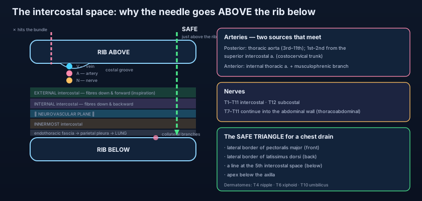
Twelve ribs make eleven gaps — and the twelfth nerve runs below the last rib, not between two. Each intercostal space is named for the rib above it, so the fourth space lies between the fourth and fifth ribs. The bony frame around them is described in the thoracic cage; what fills the gaps is a sandwich of three muscle sheets, and the whole clinical usefulness of the region comes from their order. Outermost is the EXTERNAL intercostal, its fibres running downwards and forwards — the classic memory is that the fibres follow the direction of your hands slipped into your front trouser pockets. It elevates the ribs in inspiration, and it does not reach all the way to the sternum: anteriorly, from the costochondral junctions forwards, the muscle is replaced by the thin external intercostal membrane. Beneath it lies the INTERNAL intercostal, whose fibres cross the external ones at right angles, running downwards and backwards. It depresses the ribs in forced expiration, and it too stops short — posteriorly, from the angles of the ribs backwards, it is replaced by the internal intercostal membrane. Deepest of all is the INNERMOST intercostal, an incomplete layer that is part of a deep group also containing the subcostals (which bridge more than one space at the back) and transversus thoracis (which fans out behind the sternum onto the costal cartilages).
The critical consequence of that stacking is a plane. Between the internal and the innermost layers there is a thin fascial interval, and it is in this interval — the neurovascular plane — that the vessels and nerve of each space travel. The body uses the same design in the abdominal wall: there, the neurovascular plane lies between internal oblique and transversus abdominis, the plane anaesthetists inject when they perform a transversus abdominis plane block. Thorax and abdomen are the same wall built to the same rule, which is unsurprising once you remember that the lower intercostal nerves simply keep going and become the nerves of the abdominal wall. Two layers protect the bundle from the outside; one thin, incomplete layer separates it from the pleura within.
V–A–N: the bundle in the costal groove
Run a finger along the underside of a dried rib and you will feel the gutter that hides them. The inner surface of each rib carries, at its LOWER border, a shallow bony gutter — the costal groove — sheltered under the overhanging inferior margin. The main neurovascular bundle of each space lies in that groove, tucked up against the rib above the space, and the three structures are stacked in a fixed order from top to bottom: vein, artery, nerve — V–A–N. Say it downwards and you have it: the vein is highest and best protected, the nerve lowest and therefore the first thing an ascending needle will meet. This is the single most quoted fact in thoracic anatomy, and it exists because the bundle grew up under the shelter of the rib and has stayed there ever since.
But the groove is only half the story, and the forgotten half is what makes procedures safe or unsafe. Each posterior intercostal artery and each intercostal nerve gives off a COLLATERAL branch that descends and then runs forwards along the UPPER border of the rib BELOW the space. So a space is not empty except at the top — it is a corridor with traffic hugging both its walls: the main bundle under the upper rib, a smaller collateral bundle over the lower rib. The genuinely safe corridor is neither margin but the MIDDLE of the space, and, because the main bundle is by far the larger prize to lose, the classical teaching is to enter just ABOVE the rib below, keeping well away from the lower border of the rib above. In practice the needle is walked onto the top of the lower rib and then slipped just over it, so that bone — not guesswork — guides the depth and the level.
Picture a narrow one-way street with a covered arcade along its northern side. The through traffic — the vein, the artery and the nerve — runs under that arcade, sheltered by the overhang, exactly as the main bundle runs in the costal groove of the rib above. A smaller service lane runs along the southern kerb: the collateral branches. Down the middle of the street there is nothing but air. If you must cross the road blindfolded, you do not walk along the arcade where all the traffic is, and you do not scrape the southern kerb either — you cross in the middle, and you feel for the southern kerb with your foot first so you know exactly where you are. That is the whole doctrine of intercostal needle safety, and it is why the phrase is "just above the rib below" and never "just under the rib above".
Two arterial systems that meet in the middle
Every space is supplied from behind and from in front, and the two supplies shake hands. The POSTERIOR intercostal arteries supply the back and the greater part of each space. The first and second come from the supreme (superior) intercostal artery, a branch of the costocervical trunk off the subclavian artery. The third down to the eleventh arise directly from the descending thoracic aorta, described with the rest of the great vessels in the aorta and great vessels — and because the aorta lies slightly left of the midline, the right posterior intercostals are longer and cross the vertebral bodies to reach their spaces. The ANTERIOR intercostal arteries come from the front: in the upper six spaces directly from the internal thoracic artery, and in the next few from its musculophrenic branch. Below the level where the internal thoracic divides, the lowest spaces have no anterior intercostal artery at all. Each posterior artery meets its anterior partners in the space and anastomoses with them, so that every intercostal space carries a continuous arterial arcade running front to back.
That front-to-back anastomosis is not a curiosity; it is a rescue route waiting to be used. In coarctation of the aorta — a congenital narrowing just distal to the arch — blood cannot easily reach the lower body through the aorta itself, so it goes the long way round: down the subclavian arteries, into the internal thoracic arteries, out along the anterior intercostals, backwards through the anastomosis into the posterior intercostals, and so into the aorta below the narrowing. Those posterior intercostal arteries enlarge and pulsate under the costal grooves for years, and slowly erode the bone above them. On a plain chest radiograph the result is rib notching: scalloped, irregular lower borders of the posterior parts of the ribs, a shadow on a film that is really a map of an anastomosis working overtime. The corresponding veins are simpler — the posterior intercostal veins drain backwards into the azygos system on the right and the hemiazygos and accessory hemiazygos on the left, while the anterior intercostal veins drain forwards into the internal thoracic and musculophrenic veins.
The nerves: twelve segments written on the skin
The thorax is the one region where the body's segmental plan is still openly visible. The intercostal nerves are the anterior (ventral) rami of spinal nerves T1 to T11; the anterior ramus of T12 lies below the twelfth rib and is therefore called the subcostal nerve. Unlike the rami of the limbs, they do not form a plexus — each one keeps its own segment and its own strip of body wall, which is precisely why the thorax is the cleanest place in the body to learn dermatomes. A typical intercostal nerve runs in the neurovascular plane, supplies the intercostal muscles of its own space along with the serratus posterior, levatores costarum and the overlying skin, and gives two named sensory branches: a LATERAL cutaneous branch that pierces the muscles in the mid-axillary line and divides into anterior and posterior twigs, and an ANTERIOR cutaneous branch that emerges near the sternum as the nerve's terminal end. The nerve also supplies the parietal pleura lining its space — which is why parietal pleura hurts sharply and locally, as explained in the pleura and pleural cavity, while visceral pleura feels nothing at all. The wider wiring of the region, including the sympathetic chain and the vagus, belongs to the nerves of the thorax.
Two of the twelve behave differently. The greater part of the first thoracic nerve, T1, leaves its space and climbs to join the brachial plexus, so only a small remnant runs as a true first intercostal nerve and there is no useful T1 dermatome on the chest — its skin territory is on the medial side of the arm. At the other end, the seventh to eleventh nerves do not stop at the costal margin: they leave the intercostal spaces, pass deep to the costal cartilages and continue into the anterior abdominal wall, which is why they are properly called THORACOABDOMINAL nerves. They supply the abdominal muscles and the skin of the abdomen along with T12. This single anatomical fact explains a common bedside puzzle: a patient with a fractured tenth rib who complains bitterly of abdominal pain and tenderness, mimicking an acute abdomen, when the injury is entirely in the chest wall.
Three dermatome landmarks are worth knowing by heart, because they let you place a spinal level on a patient without a single instrument: T4 lies at the nipple line, T6 at the xiphoid process, and T10 at the umbilicus. Ask a patient with a spinal injury where sensation returns, put your finger on the umbilicus, and you have named the segment. Sequence them and they are almost evenly spaced down the trunk — the body wall is still a stack of segments, and the thorax never bothered to disguise it the way the limbs did when they dragged their nerve supply out into plexuses.
Putting a needle through the wall
Every safe procedure in this region is anatomy converted into a rule of thumb. A chest drain goes into the SAFE TRIANGLE, an area bounded in front by the lateral border of pectoralis major, behind by the lateral border of latissimus dorsi, below by a horizontal line at about the level of the fifth intercostal space, and above by the apex below the axilla. The triangle exists to keep the operator away from three dangers at once: too low and the tube enters the abdomen through the diaphragm, striking liver or spleen; too far forward or medial and the muscle is thick and the internal thoracic vessels are near; too far back and the drain lies uncomfortably under the patient and the posterior intercostal vessels are larger. Within the triangle the tube is passed just above the lower rib of the chosen space, using a finger sweep to confirm the pleural cavity is entered and that no lung is adherent to the wall. Needle decompression of a tension pneumothorax classically uses the second intercostal space in the mid-clavicular line — fast, superficial and easy to find in an emergency — though many services now prefer a lateral approach in the fourth or fifth space in the anterior axillary line, because the chest wall there is thinner in adults and the cannula is more likely to reach the pleural cavity at all. The surface landmarks for all of these are laid out in thoracic surface anatomy and procedures.
The same groove that must be avoided by a drain is deliberately sought by a nerve block. In an intercostal nerve block the needle is walked onto the lower border of the rib at the angle, then slipped a few millimetres beneath it into the groove, and a small volume of local anaesthetic is deposited around the nerve. It is superb analgesia for fractured ribs, for a chest drain site, or after a thoracotomy — but the space is richly vascular, so systemic absorption from an intercostal block is faster than from almost any other site in the body, and the maximum safe dose has to be respected strictly. Because each nerve serves only its own segment and there is overlap from the segments above and below, a single space is rarely enough: the block is usually placed at the level of the injury and one space above and one below.
Herpes zoster — shingles — draws the segmental plan on the skin in blisters. The varicella virus that caused chickenpox in childhood retires into a single dorsal root ganglion and sleeps there for decades; when immunity dips it travels back down that one nerve and erupts in exactly its own dermatome, producing a band of pain and vesicles that stops dead at the midline and does not cross to the other side. Nowhere is this more striking than on the trunk, because a thoracic dermatome really is a clean horizontal strip. A rash that respects a single band, burns before it appears, and refuses to cross the midline is anatomy making a diagnosis for you from across the room.
The cracked rib: a man slips on wet stairs and lands on the edge of a step. Nothing is displaced on the film, but for three weeks every laugh, cough and sneeze is a knife, because the fracture ends grate on a nerve lying in the groove millimetres away. He is told to breathe deeply anyway — splinting the chest to avoid pain is how a rib fracture turns into pneumonia. The exhausted breather: a young woman in a severe asthma attack has been using her intercostals and accessory muscles for hours at a rate they were never built to sustain. Her respiratory muscles fatigue exactly as any skeletal muscle does; a rising carbon dioxide and a quietening chest in someone who was noisy an hour ago are the ominous signs, and the emergency management is set out in the acute attack. The rescued lung: an elderly man with a large pleural effusion is drained in the safe triangle, above the lower rib, under ultrasound guidance — and walks out breathing comfortably, having had a needle placed within a centimetre of an artery it never touched.
- There are eleven intercostal spaces on each side, each named for the rib above it; the twelfth thoracic nerve lies below the last rib and is the subcostal nerve.
- External intercostal: fibres downwards and forwards ("hands in front pockets"), elevates ribs in inspiration, replaced ANTERIORLY by the external intercostal membrane.
- Internal intercostal: fibres at right angles, downwards and backwards, depresses ribs in forced expiration, replaced POSTERIORLY by the internal intercostal membrane.
- Innermost intercostal, with the subcostals and transversus thoracis, forms the deepest group; the neurovascular plane lies between the internal and innermost layers — the thoracic equivalent of the TAP plane.
- The main bundle lies in the costal groove at the LOWER border of the rib above, ordered top to bottom vein–artery–nerve (V–A–N).
- Collateral branches run along the UPPER border of the rib below, so the safe entry point is the middle of the space, just above the rib below — never along the lower margin of the rib above.
- Posterior intercostal arteries: spaces 1–2 from the supreme (superior) intercostal artery off the costocervical trunk; spaces 3–11 directly from the thoracic aorta.
- Anterior intercostal arteries come from the internal thoracic artery and its musculophrenic branch; the two systems anastomose front-to-back in every space.
- That anastomosis is the collateral route in coarctation of the aorta — enlarged posterior intercostals erode the ribs and produce rib notching on the chest film.
- Veins drain to the azygos system: posterior intercostal veins into azygos (right) and hemiazygos / accessory hemiazygos (left); anterior ones into the internal thoracic and musculophrenic veins.
- Intercostal nerves = anterior rami T1–T11 (T12 = subcostal); each gives a lateral cutaneous and an anterior cutaneous branch and supplies the parietal pleura of its space. Most of T1 joins the brachial plexus; T7–T11 continue as thoracoabdominal nerves into the abdominal wall.
- Dermatomes to memorise: T4 nipple, T6 xiphoid, T10 umbilicus. Chest drains go into the safe triangle; needle decompression classically in the 2nd space mid-clavicular line, or laterally in the 4th–5th space anterior axillary line.
- Ordering the bundle as artery–vein–nerve. From above downwards it is VEIN, artery, nerve — the vein sits highest, deepest in the shelter of the groove, and the nerve lowest and most exposed.
- Believing the space is safe anywhere except right under the upper rib. Collateral branches hug the UPPER border of the lower rib too — the middle of the space is the target, not either margin.
- Mixing up the two membranes. The EXTERNAL intercostal is replaced ANTERIORLY (by the external intercostal membrane) and the INTERNAL is replaced POSTERIORLY (by the internal intercostal membrane) — each muscle is missing at the end it never needed to reach.
A doctor is inserting a chest drain in the fifth intercostal space. Where should the trocar-free tube be passed, and why?
- Eleven intercostal spaces per side, each named for the rib above, filled by three muscle layers: external (down and forward, inspiratory, replaced anteriorly by a membrane), internal (down and backward, forced expiration, replaced posteriorly by a membrane) and innermost (with subcostals and transversus thoracis).
- The neurovascular plane lies between the internal and innermost layers; the main bundle sits in the costal groove at the lower border of the rib above in the order vein–artery–nerve, with collateral branches along the upper border of the rib below — so needles enter the middle of the space, just above the lower rib.
- Arteries: posterior intercostals (1–2 from the supreme intercostal off the costocervical trunk, 3–11 from the thoracic aorta) anastomose with anterior intercostals from the internal thoracic and musculophrenic arteries — the route that enlarges and notches the ribs in coarctation. Veins drain to the azygos system.
- Nerves are the anterior rami T1–T11 plus the subcostal T12, each with lateral and anterior cutaneous branches; T1 mostly joins the brachial plexus and T7–T11 become thoracoabdominal. Remember T4 nipple, T6 xiphoid, T10 umbilicus — and the safe triangle, the 2nd-space needle decompression, the intercostal block and the shingles band that all follow from them.
- Drake RL, Vogl AW, Mitchell AWM. Gray's Anatomy for Students — Thorax: the thoracic wall and intercostal spaces.
- Moore KL, Dalley AF, Agur AMR. Clinically Oriented Anatomy — Intercostal muscles, nerves and vessels; chest tube insertion.
- Netter FH. Atlas of Human Anatomy — Thoracic wall: intercostal muscles, nerves and arteries.
- Last RJ. Last's Anatomy: Regional and Applied — The thoracic wall and intercostal spaces.
- Snell RS. Clinical Anatomy by Regions — The chest wall, rib fractures and needle thoracostomy.
- TeachMeAnatomy — The Intercostal Muscles; The Intercostal Nerves and Vessels.

