
Hyperkalaemia: The Three-Step Emergency
Potassium is the most abundant ion inside your cells and the most tightly guarded ion in your blood — and for good reason. Let the level in the serum drift up by a couple of millimoles and the heart's electrical membrane begins to fail: the ECG deforms through a predictable, sinister sequence and can end in a flat line. What makes hyperkalaemia a favourite of examiners and a terror on the ward is that the fix is not one drug but a choreographed three-step protocol — protect the heart, shift the potassium, remove it — and getting the order and the logic right is the whole game.
A 68-year-old man with chronic kidney disease is brought to the emergency department feeling "weak and odd." He takes ramipril for his blood pressure, spironolactone for heart failure, and bought ibuprofen last week for his back. A finger-prick shows potassium off the scale, and the monitor tells the real story before the lab does: the T waves have grown tall and tented, the P waves are flattening, and the QRS complexes are starting to smear wide. This is a heart being electrically poisoned in real time — minutes, not hours, from a sine wave and then arrest. Nobody reaches for a drug to lower the potassium first. The first syringe is calcium, to hold the myocardium together while everything else is arranged. In one patient you can see the whole disease: three culprit drugs, a failing kidney, and an ECG counting down.
Why a high potassium stops the heart
The danger is not the number — it is what the number does to a cardiac membrane. Every excitable cell holds a resting voltage across its membrane, and that voltage is set almost entirely by the huge gradient of potassium — high inside the cell, low outside. When extracellular K+ rises, that gradient shrinks and the resting membrane potential drifts upward (less negative), sitting closer to the threshold for firing. At first glance that sounds like it should make cells more excitable, and briefly it does. But a membrane held chronically depolarised cannot reset its fast sodium channels — they stay inactivated — so the cell becomes harder and harder to fire and conduction slows to a crawl. In the heart this shows up as a deteriorating ECG and, ultimately, an electrically silent myocardium. This membrane-potential story is the same physiology taught in the Cardiovascular section, and it is why potassium sits at the centre of cardiac electrophysiology.
The ECG changes march in a recognisable order as potassium climbs, and reading them is a bedside potassium meter. First the T waves become tall, narrow and "tented." Then the P waves flatten and lengthen as atrial conduction fails. Then the QRS complex widens and blurs. Finally the flattened P and the wide QRS merge into a slow, undulating "sine wave" — the pre-terminal pattern that precedes ventricular fibrillation or asystole. The exact number matters less than the trend and the ECG: a patient with sine-wave changes needs treatment before the confirmatory blood result ever returns.
Think of the resting membrane potential as a drawn bowstring, held taut by the potassium gradient. Each heartbeat is a controlled release and re-draw. Raise the extracellular potassium and you let the string go slack — it can no longer be drawn back to full tension, so each shot is weaker and slower, until the bow can't fire at all. IV calcium doesn't restring the bow or remove the excess potassium; it stiffens the frame just enough that the bow keeps firing while you fix the string.
Where the potassium comes from
Four mechanisms, and most real patients have more than one at once. First and biggest: reduced excretion. The kidney is the main exit for potassium, so acute or chronic kidney injury is the commonest serious cause — this ties straight into the renal and nephrotoxic-drugs chapter. Second: drugs, the cause you can actually prevent. Anything that blocks the renin–angiotensin–aldosterone axis retains potassium — ACE inhibitors and ARBs, and the potassium-sparing diuretics spironolactone, eplerenone and amiloride. NSAIDs impair renal perfusion and potassium handling; trimethoprim behaves like a mild amiloride and blocks distal potassium excretion; heparin suppresses aldosterone. The infamous "triple whammy" — an ACE inhibitor or ARB plus a diuretic plus an NSAID, often in a dehydrated older patient — is a classic recipe for a crashing potassium and an acute kidney injury together (see the nephrotoxic-drugs chapter). Third: a shift of potassium out of the cells — metabolic acidosis, insulin deficiency (diabetic ketoacidosis), massive tissue breakdown (rhabdomyolysis, tumour lysis syndrome), and digoxin toxicity, which poisons the Na+/K+-ATPase that normally pumps potassium back in. Fourth: excess intake (usually only dangerous when excretion is already impaired) and pseudohyperkalaemia — a falsely high result from a haemolysed sample or a fist clenched too hard during the draw.
- Hyperkalaemia kills by depolarising the cardiac membrane and inactivating sodium channels — conduction slows to a stop.
- ECG sequence: peaked T waves → flattened P → widened QRS → sine wave → VF/asystole.
- Commonest serious cause is reduced renal excretion (acute or chronic kidney injury).
- Culprit drugs: ACE inhibitors/ARBs, spironolactone/eplerenone/amiloride, NSAIDs, trimethoprim, heparin — the "triple whammy" combines three.
- Cellular shift: acidosis, insulin deficiency/DKA, rhabdomyolysis, tumour lysis, digoxin toxicity.
- Always exclude pseudohyperkalaemia (haemolysed sample) before treating an asymptomatic, normal-ECG result.
Step 1 — Protect the myocardium
The first move buys time; it does not lower the potassium at all. If there are ECG changes (or the potassium is dangerously high), the very first drug is intravenous calcium — calcium gluconate, or calcium chloride via a central line. Calcium raises the threshold potential back away from the depolarised resting potential, restoring the gap the heart needs to fire and conduct. It is cardioprotective and fast, working within minutes, and if the ECG changes persist the dose is simply repeated. The single most important thing to understand is what calcium does not do: it does not lower the serum potassium by a single millimole. It is a holding measure — it keeps the heart electrically stable while the potassium-lowering steps are set up. One important caution links back to the Cardiovascular section: in suspected digoxin toxicity, rapid IV calcium has historically been feared to cause a hypercontracted, non-relaxing "stone heart," so calcium is given slowly and cautiously (or a digoxin-specific antibody is used) rather than pushed.
Step 2 — Shift potassium into the cells
Now actually lower the serum level — fast, but temporarily. The workhorse of step two is insulin given with glucose. Insulin drives potassium into cells by stimulating the Na+/K+-ATPase pump, dropping the serum level within about fifteen minutes. The glucose is co-administered purely to prevent the insulin from causing hypoglycaemia — and the patient's blood sugar must then be monitored, because the classic error is watching the potassium and forgetting the glucose. That insulin drives potassium intracellularly is the same physiology met in the Endocrine section, and it is exactly why insulin deficiency in diabetic ketoacidosis pushes potassium the other way, out of cells. The second agent is nebulised salbutamol — a beta-2 agonist that, like insulin, stimulates the pump and shifts potassium inward, giving an additive effect. Third, if the patient is acidotic, sodium bicarbonate can help shift potassium in as the acidosis is corrected. Like calcium, every step-two treatment is temporary: the potassium has only been hidden inside the cells, not removed from the body, and it will leak back out. Shifting therapies buy an hour or two — they do not end the problem.
Step 3 — Remove potassium from the body
The only definitive step is removing potassium from the body altogether. The modern oral binders are the newer gut agents patiromer and sodium zirconium cyclosilicate, which bind potassium in the gastrointestinal tract and carry it out in the stool; they are better tolerated and faster than the older, slower and much-disliked resins (calcium or sodium polystyrene sulfonate). If the patient is still making urine, a loop diuretic such as furosemide increases renal potassium loss. And for refractory hyperkalaemia — or in established renal failure where none of the above can keep up — the definitive answer is dialysis, which strips potassium out directly. Running through all three steps is the same imperative: treat the cause and stop the culprit drugs. There is little point shifting and binding potassium while an ACE inhibitor, a potassium-sparing diuretic and an NSAID are still on the drug chart driving it back up.
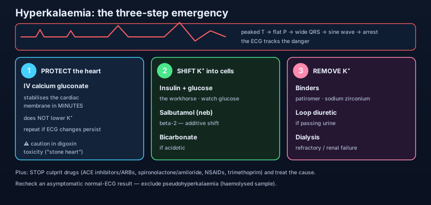
The single cleanest way to hold the whole emergency in your head is the mantra protect, shift, remove — and to know exactly which drug does which. Calcium protects but does not lower the level. Insulin+glucose and salbutamol shift and lower it fast but only temporarily. Binders, diuretics and dialysis remove it for good. Confusing these is the commonest exam trap: give calcium and walk away, and the potassium is still lethal; give a binder alone to a crashing ECG, and the heart arrests before the potassium ever falls.
PROTECT: IV calcium gluconate (or calcium chloride) — onset minutes, no effect on serum K+, repeat if ECG changes persist, give cautiously in digoxin toxicity. SHIFT: insulin (with glucose to prevent hypoglycaemia) — onset ~15 min; nebulised salbutamol — additive; sodium bicarbonate if acidotic. REMOVE: patiromer and sodium zirconium cyclosilicate (gut binders), calcium/sodium polystyrene sulfonate (older resins), furosemide if urine output preserved, and haemodialysis for refractory or renal-failure cases. Throughout: stop the ACE inhibitor/ARB, potassium-sparing diuretic, NSAID and trimethoprim, and treat the underlying cause.
After the crisis: chronic management
Once the acute danger passes, the goal shifts to keeping potassium in a safe range long-term — most often in chronic kidney disease or heart failure. That means dietary potassium restriction, and a careful review of the drug list: many patients are on an ACE inhibitor or ARB and a potassium-sparing diuretic precisely because those drugs protect the kidney and heart. Here the newer binders have quietly changed practice — patiromer and sodium zirconium cyclosilicate can hold potassium down enough to let a patient stay on life-prolonging RAAS therapy that would otherwise have to be stopped. And a final, cheap safeguard sits before every treatment decision: in an asymptomatic patient with a normal ECG and an unexpectedly high potassium, suspect pseudohyperkalaemia from a haemolysed sample and recheck before treating — the danger of over-treating a lab artefact (driving the potassium too low) is real.
- The protocol is ordered: PROTECT (IV calcium) → SHIFT (insulin+glucose, salbutamol, ± bicarbonate) → REMOVE (binders, diuretics, dialysis).
- Calcium protects the heart within minutes but does not lower the potassium at all.
- Insulin+glucose is the workhorse shift; monitor glucose to avoid hypoglycaemia.
- Only removal — binders, diuretics, dialysis — is definitive; shifting is temporary.
- Newer binders (patiromer, sodium zirconium) let patients continue protective RAAS therapy long-term.
- Always stop the culprit drugs and treat the cause — and exclude pseudohyperkalaemia first.
- Believing IV calcium lowers the potassium. It only stabilises the myocardium — without shifting and removal, the potassium is still lethal.
- Giving IV calcium fast in suspected digoxin toxicity — the feared "stone heart." Give slowly and cautiously (or use digoxin-specific antibody).
- Giving insulin without glucose (or without monitoring glucose) — watching the potassium fall while the patient slides into hypoglycaemia.
A patient with acute kidney injury has a potassium of 7.2 mmol/L with peaked T waves and a widening QRS on the monitor. Which is the single most appropriate first step?
- Hyperkalaemia kills by depolarising the cardiac membrane; the ECG marches from peaked T waves through widened QRS to a sine wave and arrest.
- Causes: reduced renal excretion (AKI/CKD), culprit drugs (ACE inhibitors/ARBs, potassium-sparing diuretics, NSAIDs, trimethoprim, the triple whammy), cellular shift (acidosis, DKA, rhabdomyolysis, tumour lysis, digoxin) and pseudohyperkalaemia.
- The emergency is three ordered steps: PROTECT with IV calcium (no effect on the level), SHIFT with insulin+glucose/salbutamol/bicarbonate, REMOVE with binders/diuretics/dialysis.
- Always stop the culprit drugs, treat the cause, give calcium cautiously in digoxin toxicity, and never give potassium as a rapid IV push.
- Rang & Dale's Pharmacology — potassium homeostasis, diuretics and the renin–angiotensin–aldosterone system.
- Katzung's Basic & Clinical Pharmacology — drugs affecting potassium; agents used in hyperkalaemia.
- British National Formulary (BNF) — hyperkalaemia treatment: calcium salts, insulin/glucose, salbutamol, patiromer, sodium zirconium cyclosilicate.
- UK Kidney Association (Renal Association) — Clinical Practice Guidelines: Treatment of Acute Hyperkalaemia in Adults.
- NICE guideline — Acute kidney injury: prevention, detection and management; and Chronic kidney disease.
- Ganong's Review of Medical Physiology — resting membrane potential and the role of the potassium gradient in cardiac excitability.

