
Intravitreal Anti-VEGF: The Drug That Rescued Retinal Vision
A generation ago, wet age-related macular degeneration was a one-way road to legal blindness — you watched the centre of the world dissolve and there was nothing to be done. Then came a class of drugs that does something almost surgical without a scalpel: it reaches into the eye and switches off the single molecule driving the leaky, bleeding new vessels. The result is one of the great therapeutic revolutions of modern medicine — a blinding disease turned into a treatable one. The catch is the delivery: to reach the retina at all, the drug has to be injected through the wall of the eye, again and again, for years.
A 74-year-old man notices that straight lines have started to bend — the door frame kinks, the letters on a page warp, and a grey smudge is growing over the centre of his vision. On examination the macula of his right eye is wet: a membrane of abnormal new vessels has grown up from beneath the retina, leaking fluid and blood into the very spot he reads with. Twenty years ago the ophthalmologist would have had little to offer beyond a magnifier and a sympathetic word. Instead, after topical anaesthetic and a drop of antiseptic, a fine needle passes through the white of his eye and delivers a tiny volume of clear fluid into the vitreous. Over the following weeks the fluid dries up, the distortion recedes, and his reading vision claws back. But the reprieve is conditional: he must return month after month for more injections, because the molecule driving the leak has not gone away — it has only been silenced.
One molecule behind so much blindness
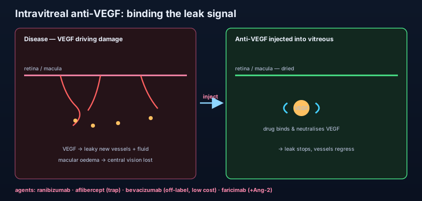
VEGF is a growth signal the retina uses honestly — until disease turns it into a wrecking crew. Vascular endothelial growth factor (VEGF) is the body's master signal for building new blood vessels. In a healthy retina it is kept on a tight leash. But a starved, ischaemic retina — one whose capillaries have been strangled — screams for oxygen by pouring out VEGF. That distress signal does two destructive things at once. First, it drives angiogenesis: the sprouting of fragile, disorganised new vessels (neovascularisation) that bleed easily and grow where they should not. Second, VEGF is a potent permeability factor — it loosens the junctions between endothelial cells, so vessels leak plasma into the retina. In the macula, that leak becomes macular oedema, a waterlogging of the tissue that blurs and destroys central vision. Neutralise VEGF and you attack both halves of the disease at their common root.
This one pathway ties together a whole ward of retinal diseases. Wet (neovascular) age-related macular degeneration (AMD) is the flagship: new vessels invade from the choroid beneath the macula. Diabetic eye disease contributes two of anti-VEGF's biggest indications — diabetic macular oedema (the leading cause of vision loss in working-age diabetics) and proliferative diabetic retinopathy, where VEGF-driven neovascularisation threatens catastrophic bleeding. Add the macular oedema of retinal vein occlusion, the neovascular membranes of pathological myopia, and — in the tiniest patients — retinopathy of prematurity (ROP). Different diseases, one shared engine. That the diabetic story keeps recurring is no accident; it is developed in full in the Endocrine section's diabetic retinopathy chapter.
Think of VEGF as a foreman shouting "build more pipes here!" over a megaphone in a district that is already flooding. The pipes his crew throws up are cheap and leaky, and the more he shouts the worse the flooding gets. Anti-VEGF drugs don't tear down the pipes or drain the water directly — they simply take away the megaphone. With the order silenced, the crew stops laying leaky pipe, the existing shoddy vessels regress, and the flood slowly drains away. But the foreman is still standing there; put the megaphone back (let the drug wear off) and he starts shouting again. That is why the injections must be repeated.
Why it has to go inside the eye
You cannot treat the retina with an eye drop or a tablet. The retina sits at the very back of the eye, behind the vitreous gel, and it is walled off from the bloodstream by the blood–retinal barrier — the same tight, selective barrier logic that governs drug delivery throughout the eye, explained in the Foundations chapter on ocular routes of administration. A topical drop barely penetrates past the cornea; a systemic drug would have to be given in huge, toxic doses to force enough across the barrier, and a large protein antibody would never make it. The only reliable way to bathe the retina in an effective concentration of drug is to place it directly into the vitreous cavity — an intravitreal injection. It is a small, sterile, in-clinic procedure, but it is genuine entry into the interior of the eye, and every risk of the therapy flows from that fact.
The agents: fragments, traps and bispecifics
Every anti-VEGF drug is a protein engineered to grab VEGF and stop it docking on its receptor — but they differ cleverly in form. Ranibizumab is a small antibody fragment (a Fab), engineered specifically for the eye — tiny enough to penetrate the retina well and cleared reasonably fast. Aflibercept takes a different tack: it is a fusion protein, a decoy "VEGF trap" built from pieces of the VEGF receptors themselves, which binds VEGF (and the related placental growth factor) very tightly and tends to last longer between doses. Brolucizumab is an even smaller single-chain fragment, designed to pack more drug into each injection for longer intervals. The newest idea is bispecific: faricimab grips two targets at once — VEGF and angiopoietin-2 (Ang-2), a second molecule that destabilises vessels — a dual mechanism intended to calm the vasculature more completely and stretch treatment intervals further. And then there is bevacizumab, the outlier that reshaped the economics of the whole field.
Bevacizumab is a full-length antibody, and it was never licensed for the eye at all — it is an oncology drug, given intravenously to slow the blood supply of colorectal and other cancers. But ophthalmologists realised it hits exactly the same target, and when a pharmacy splits one systemic vial into many tiny intravitreal doses, the cost per injection collapses to a small fraction of the licensed agents. Used off-label this way, bevacizumab delivers broadly comparable visual results at a tiny fraction of the price — one of the biggest health-economics stories in modern medicine, and the reason it remains a mainstay across much of the world despite never carrying an ocular licence. Its very existence keeps the whole class honest on price.
Ranibizumab (Lucentis) — antibody fragment, eye-specific. Aflibercept (Eylea) — VEGF-trap fusion protein, longer-acting; a higher-dose formulation extends intervals further. Brolucizumab (Beovu) — small single-chain fragment, longer intervals, but carries an intraocular inflammation/vasculitis signal. Faricimab (Vabysmo) — bispecific anti-VEGF + anti-Ang-2, designed for the longest intervals. Bevacizumab (Avastin) — full antibody, licensed for cancer, used off-label intravitreally at a fraction of the cost. Landmark evidence: MARINA and ANCHOR established ranibizumab in wet AMD (the first drugs to improve, not merely stabilise, vision); the VIEW studies showed aflibercept matched ranibizumab with fewer injections.
- VEGF drives both leaky neovascularisation and macular oedema — anti-VEGF hits both at their shared root.
- Indications: wet AMD, diabetic macular oedema, proliferative diabetic retinopathy, RVO oedema, myopic CNV, ROP.
- The retina lies behind the blood–retinal barrier, so the drug must be injected intravitreally — not dropped or swallowed.
- Ranibizumab = antibody fragment; aflibercept = VEGF-trap, longer-acting; brolucizumab/faricimab = longer intervals (faricimab also blocks Ang-2).
- Bevacizumab is an off-label cancer antibody used intravitreally at a fraction of the cost — a major health-economics story.
- MARINA/ANCHOR (ranibizumab) and VIEW (aflibercept) are the landmark trials that turned wet AMD from blinding to treatable.
The regimen — and its relentless burden
Silencing VEGF is easy; keeping it silenced without over-treating is the real art. Because the drug wears off and the disease pushes back, treatment is not a single shot but a schedule. Most regimens open with a loading phase — typically monthly injections for the first three doses — to dry the retina, then move to one of two maintenance strategies. Pro re nata ("as needed", PRN) means the patient is monitored and re-injected only when fluid returns, which minimises injections but demands frequent visits and risks under-treating between them. Treat-and-extend is now the dominant approach: the patient is injected at every visit, but the interval is gradually stretched (say from 4 to 6 to 8 weeks and beyond) as long as the retina stays dry, and pulled back in if fluid recurs. The whole point of the longer-acting agents — aflibercept, brolucizumab, faricimab — is to win the same dryness with fewer trips. Even so, the cumulative burden is real: repeated injections, repeated clinic days, and repeated small procedural risk, often for years, in an elderly population. Non-adherence is itself a leading cause of vision loss slipping back.
Safety, part one: the injection itself
Because every dose is a puncture into a sterile, closed space, the most feared complication is infectious endophthalmitis — bacteria carried into the vitreous, causing a rapidly blinding intraocular infection. It is rare (a fraction of a percent per injection) but devastating, which is why the aseptic ritual — povidone-iodine antisepsis, a sterile lid speculum, a fresh needle — is non-negotiable, and why any patient with a red, painful, blurring eye in the days after an injection is an emergency until proven otherwise. That injection-related infection is exactly the scenario worked through in the Endophthalmitis chapter. Other procedural risks include a transient spike in intraocular pressure from the added volume (usually settling within minutes to an hour, occasionally needing IOP-lowering), subconjunctival haemorrhage (alarming but harmless), a small risk of traumatic cataract or retinal tear/detachment from the needle, and non-infectious intraocular inflammation — the last being a particular signal with brolucizumab, which has been linked to a sight-threatening retinal vasculitis and occlusion that the other agents show far less.
Safety, part two: does blocking VEGF harm the body?
VEGF matters everywhere, so any drug that escapes the eye into the blood raises a theoretical alarm. A tiny amount of intravitreal drug does leak into the systemic circulation, and VEGF is not only a retinal molecule — it maintains blood vessels throughout the body. Systemic VEGF blockade, as used in oncology, is well known to raise blood pressure, impair wound healing, promote bleeding and clotting, and — of most concern for eye patients — increase arterial thromboembolic events (stroke and myocardial infarction). The reassuring news is that the systemic exposure from an intravitreal dose is small — orders of magnitude below an oncology infusion — so in the large trials the excess systemic risk has been low and, for most patients, hard to distinguish from the background rate of an elderly, vascular-disease-prone population. Still, most clinicians exercise caution soon after a recent stroke or myocardial infarction, and weigh the very small systemic risk against the near-certain vision loss of leaving the eye untreated. Agents differ slightly in how much escapes the eye, and the full-antibody bevacizumab has the longest systemic half-life of the group.
The single most illuminating way to understand anti-VEGF is to see it as one drug with two lives. In the eye it is a local, sight-saving therapy given in microgram doses straight to the target, and its dominant risks are those of the needle — infection, pressure, inflammation. In oncology it is the very same molecular idea (bevacizumab and its relatives) given intravenously in far larger doses to starve a tumour's blood supply, and there the risks are systemic and predictable — hypertension, bleeding, impaired healing, clots — as taught in the Oncology anti-angiogenic chapter. Same target, opposite scale. Grasp that contrast and the whole safety profile of intravitreal therapy falls into place: keep the dose local and tiny, and you keep VEGF's body-wide importance largely out of the picture.
- Endophthalmitis is the most feared complication of every injection — rare but blinding; strict antisepsis is mandatory.
- A red, painful, blurring eye in the days after injection = suspected endophthalmitis until proven otherwise (emergency).
- Other ocular risks: transient IOP spike, subconjunctival haemorrhage, cataract/retinal tear, intraocular inflammation.
- Brolucizumab carries a distinct signal of intraocular inflammation and retinal vasculitis/occlusion.
- Systemic exposure is small, so arterial thromboembolic risk is low — but use caution after recent stroke/MI.
- Same molecule, systemic and high-dose in oncology, causes hypertension, bleeding and impaired healing.
- Dismissing a red, aching eye after an injection as "just irritation." Post-injection endophthalmitis presents exactly this way and blinds within days if not treated urgently.
- Believing anti-VEGF cures the disease. It suppresses VEGF, not the underlying process — stop injecting and the oedema and vessels usually return; ongoing monitoring is essential.
- Confusing the local eye dose with an oncology dose. A microgram intravitreal dose is not a systemic infusion — over-fearing stroke risk can wrongly deny a patient sight-saving treatment.
Three days after an intravitreal aflibercept injection for wet AMD, a 78-year-old man returns with a red, painful eye, worsening blurring and floaters. What is the most appropriate immediate action?
- VEGF drives the leaky new vessels and macular oedema of wet AMD, diabetic eye disease, vein occlusion, myopic CNV and ROP; anti-VEGF neutralises it at the shared root.
- The drug must be injected intravitreally to cross the blood–retinal barrier; agents are ranibizumab (fragment), aflibercept (VEGF-trap), brolucizumab, faricimab (also anti-Ang-2), and off-label bevacizumab (cheap cancer antibody).
- Regimens load monthly then maintain by treat-and-extend or PRN, often for years — a heavy but sight-saving burden.
- Main risk is the injection (endophthalmitis, raised IOP, inflammation — brolucizumab vasculitis); systemic exposure is small, so thromboembolic risk is low but warrants caution after recent stroke/MI.
- Kanski's Clinical Ophthalmology: A Systematic Approach — Acquired macular disorders and retinal vascular disease.
- American Academy of Ophthalmology, Basic and Clinical Science Course (BCSC), Section 12: Retina and Vitreous.
- Bartlett & Jaanus, Clinical Ocular Pharmacology — Anti-angiogenic and intravitreal agents.
- Rosenfeld PJ, et al. MARINA Study: Ranibizumab for neovascular age-related macular degeneration. New England Journal of Medicine.
- Heier JS, et al. VIEW 1 and VIEW 2: Intravitreal aflibercept (VEGF Trap-Eye) in wet AMD. Ophthalmology.
- Royal College of Ophthalmologists / NICE guidance on age-related macular degeneration and diabetic retinopathy.

