
The Abdominal Aorta: Nine Levels, One Vessel
It is about as wide as a garden hose and about as long as your forearm, and it lies flat against the front of your lumbar spine, roughly a hand's breadth behind your navel. In the thirteen centimetres between the diaphragm and the fourth lumbar vertebra, this single tube hands out everything: the blood for your stomach and liver, both kidneys, both adrenal glands, the ovaries or testes, nine metres of intestine, and the muscular wall that holds it all in. Every branch leaves at a level you can name, and the levels are not random — they are the fossilised memory of how the gut was built. And because the aorta's wall is elastic tissue that the body cannot replace once it is lost, this same vessel can widen silently for twenty years and then, in a single afternoon, kill a man who never knew it was there.
A retired carpenter, sixty-nine years old, a smoker since he was fifteen, is called in for a screening scan he nearly ignored. He feels perfectly well. He has never had chest pain, never been short of breath, and mows his own lawn. The sonographer presses the probe just above his umbilicus, slightly to the left, and measures a black circle on the screen: five point eight centimetres across. A normal abdominal aorta at that point measures about two. Nothing about him has changed in the ten minutes since he walked in — but a number has, and that number will now decide his year. Somewhere between his renal arteries and his bifurcation, the elastic sheets in his aortic wall have been thinning for two decades, and the pressure of every heartbeat has slowly pushed the weakened segment outwards, a millimetre at a time, in complete silence. He is one of the lucky ones. The unlucky ones arrive by ambulance with sudden back pain, a falling blood pressure and a pulsating mass, and most of them do not reach an operating theatre.
Thirteen centimetres on the spine
The abdominal aorta begins where the thoracic aorta ends — at a gap the diaphragm arches over rather than pierces. The vessel enters the abdomen through the AORTIC HIATUS at the level of the twelfth thoracic vertebra, passing behind the median arcuate ligament between the two crura described in the diaphragm — which is why the aorta is never squeezed by breathing. From T12 it descends on the bodies of the lumbar vertebrae, lying slightly to the LEFT of the midline, so that a surgeon opening the retroperitoneum finds it left of centre and an ultrasound probe is angled a little to the left of the umbilicus. It narrows steadily as it gives off branch after branch, from about two and a half centimetres at the hiatus to under two at its end. At the level of the FOURTH LUMBAR VERTEBRA it BIFURCATES into the right and left COMMON ILIAC arteries, which diverge to supply the pelvis and the lower limbs. The aorta does not simply stop there: a thin twig, the MEDIAN SACRAL ARTERY, continues straight down over the fifth lumbar vertebra, the sacrum and the coccyx — the shrunken remnant of the tail artery that in other vertebrates is the main continuation of the trunk. In the human body it is the vestige of an axis that once ran the whole length of the animal.
Its neighbours matter as much as its branches, because every one of them is something a surgeon can injure. On its RIGHT lies the INFERIOR VENA CAVA, running in parallel, the two great vessels lying side by side like a pipe and its return — a relationship traced in the portal system and the inferior vena cava. Crossing IN FRONT of the aorta, just below the origin of the superior mesenteric artery, is the LEFT RENAL VEIN, squeezed in the fork between the two arteries: when that fork narrows, the vein can be compressed — the "nutcracker" phenomenon. BEHIND the aorta lie the lumbar vertebral bodies and the anterior longitudinal ligament, together with the CISTERNA CHYLI, the sac-like origin of the thoracic duct, and the beginnings of the thoracic duct and the azygos system. IN FRONT, from above downwards, lie the coeliac plexus, the body of the pancreas, the splenic vein, the left renal vein and the THIRD (horizontal) PART OF THE DUODENUM, which is caught between the aorta behind and the superior mesenteric artery in front — the anatomical basis of superior mesenteric artery syndrome, in which severe weight loss removes the fat pad and the duodenum is pinched between two arteries. On its LEFT lie the sympathetic trunk, the left crus and the fourth part of the duodenum; on both sides, the lumbar lymph nodes chain along it like beads.
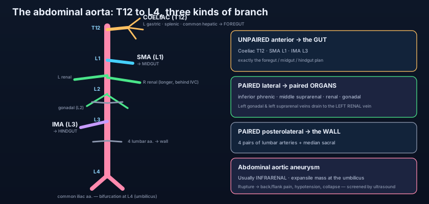
Think of a motorway running north to south down the middle of a country, with three completely different kinds of exit. The first kind are the great single junctions — three of them, spaced far apart, each feeding an entire province: these are the unpaired anterior branches to the gut, and there is only one of each because the gut itself is a single midline tube. The second kind are the matched pairs of slip roads, one to the left and one to the right at the same milepost, each serving a town that exists twice: these are the lateral branches to the kidneys, the adrenals and the gonads, and they are paired because those organs are paired. The third kind are the little unnamed service roads leaving at regular intervals along the whole route to maintain the road itself: these are the lumbar arteries to the body wall. Once you see the motorway this way, you never have to memorise a list again — you ask what an organ IS, and the answer tells you which kind of exit brings its blood.
The unpaired anterior branches: three arteries for one gut
The gut developed as three segments with three blood supplies, and it never changed its mind. The COELIAC TRUNK leaves the front of the aorta at T12, immediately below the hiatus, as a short, wide stump barely a centimetre and a half long. It TRIFURCATES into three vessels that between them supply the entire FOREGUT: the LEFT GASTRIC artery, which runs to the lesser curvature of the stomach and gives oesophageal branches upwards; the SPLENIC artery, which takes a famously tortuous course along the upper border of the pancreas to the spleen, giving pancreatic branches, the short gastric arteries and the left gastro-omental artery; and the COMMON HEPATIC artery, which runs right to become the hepatic artery proper — supplying the liver and, through the cystic artery, the gallbladder — after giving off the gastroduodenal artery and the right gastric artery. Foregut territory ends halfway down the second part of the duodenum, exactly where the bile duct enters. That is not a coincidence: the boundary of a blood supply is the boundary of an embryological segment, a plan set out in foregut, midgut and hindgut.
The SUPERIOR MESENTERIC ARTERY leaves the aorta at L1, one centimetre below the coeliac trunk, and is the artery of the MIDGUT. It passes behind the neck of the pancreas, crosses in front of the third part of the duodenum and the left renal vein, and enters the root of the mesentery, where it runs downwards and to the right in a great curve. From its concave right side come the INFERIOR PANCREATICODUODENAL artery — first branch, sharing the duodenum with the foregut supply — then the MIDDLE COLIC, the RIGHT COLIC and the ILEOCOLIC arteries; from its convex left side come twelve to fifteen JEJUNAL and ILEAL branches, which form arcades in the mesentery: one or two tiers near the jejunum with long straight vessels, four or five tiers near the ileum with short ones, a difference a surgeon uses to tell the two ends of the small bowel apart at operation. Its territory runs from the middle of the second part of the duodenum to the junction of the proximal two thirds and distal third of the TRANSVERSE COLON.
The INFERIOR MESENTERIC ARTERY leaves the aorta at L3, three or four centimetres above the bifurcation, and is a noticeably smaller vessel — it is the artery of the HINDGUT. It passes downwards and to the left behind the peritoneum and gives the LEFT COLIC artery to the descending colon, two to four SIGMOID arteries to the sigmoid, and then continues over the pelvic brim as the SUPERIOR RECTAL artery to the upper rectum. Its territory therefore runs from the distal third of the transverse colon to the upper part of the anal canal, the hindgut of the large intestine. Note the descending order of size — coeliac, then SMA, then IMA — and the descending order of level — T12, L1, L3. The three great gut arteries leave the aorta in the same sequence, and at the same relative distances, as the three parts of the gut lie along its length. The map of the branches IS the map of the gut.
The paired lateral branches: one for each of the twins
Every organ that exists twice is fed twice, at the same level, from the sides of the aorta. The INFERIOR PHRENIC arteries arise first, at T12, immediately after the aorta clears the hiatus. They run upwards and laterally over the undersurface of the diaphragm and each gives off the SUPERIOR SUPRARENAL arteries to the adrenal gland on its side. Next, at L1, come the small MIDDLE SUPRARENAL arteries, running straight out to the adrenals; this triple supply from three different sources — inferior phrenic above, aorta directly, and renal artery below — is peculiar to the adrenal glands and is explained in the adrenal glands. At L1 to L2, just below the neck of the pancreas, come the large RENAL arteries, which carry a fifth of the cardiac output to organs weighing three hundred grams between them; the RIGHT renal artery is the LONGER of the two, and it passes BEHIND the inferior vena cava to reach a kidney that sits slightly lower because of the liver, as covered in the kidneys and ureters. Accessory renal arteries are common — present in perhaps a quarter of people — and enter the kidney at its poles rather than its hilum, a fact of considerable importance in transplant surgery.
Last of the paired lateral branches, at L2, are the GONADAL arteries — TESTICULAR in the male, OVARIAN in the female. They are strikingly long, thin and delicate, and they descend almost vertically over psoas major, crossing the ureter and, in the male, running the whole way to the deep inguinal ring and through the inguinal canal to the scrotum. Their absurd length is a fossil of development: the gonad forms high on the posterior abdominal wall, level with the second lumbar vertebra, and then migrates downwards — to the pelvis in the female, out of the body altogether in the male — dragging its artery behind it like a lifeline paid out from a fixed anchor. The vein tells the same story in reverse and with an asymmetry: the RIGHT gonadal vein drains directly into the inferior vena cava, while the LEFT drains into the LEFT RENAL VEIN at a right angle, which is why a varicocele — a bag of dilated veins in the scrotum — is far commoner on the left, and why a new left varicocele in an older man raises the question of a renal tumour obstructing that vein.
Three organs, three completely different reasons for the length of their artery — and all three are stories about movement. The adrenal gland never moved, so it is fed straight out of the aorta by a short stub at L1. The kidney climbed only a little, from the pelvis to L1–L2, so its artery is short and horizontal but the left renal vein must cross the entire midline in front of the aorta to reach the cava. The gonad travelled furthest of all, from L2 to the scrotum, and its artery is a thin thread the length of a shoelace. Arterial length is a record of embryonic migration: an artery is recruited at the organ's birthplace and never released, so measuring the vessel tells you how far its organ walked.
The posterolateral branches: paying for the wall
Four pairs of LUMBAR ARTERIES leave the back of the aorta, one pair opposite each of the first four lumbar vertebrae. They are the abdominal equivalents of the posterior intercostal arteries — the same segmental plan, one pair per body segment — and they pass backwards around the vertebral bodies, deep to the psoas major and the tendinous arches of that muscle, to supply the muscles of the posterior abdominal wall, the vertebral column and its contents, and, through their dorsal branches, the deep muscles of the back. A fifth pair, when present, arises from the median sacral artery. Because the branches to the spinal canal come from the lumbar arteries, aortic surgery near this level carries a real risk of spinal cord ischaemia. The MEDIAN SACRAL ARTERY completes the posterior group as the true midline continuation of the aorta. Together with the segmental supply of the lower intercostals and the subcostal, they clothe and feed the structures described in the posterior abdominal wall — the plainest possible demonstration that the aorta serves the container as faithfully as it serves the contents.
The safety nets: how the gut survives a blocked artery
Three great arteries would be a fragile design if they were truly independent. They are not. Between the coeliac trunk and the superior mesenteric artery run the PANCREATICODUODENAL ARCADES: the superior pancreaticoduodenal artery, a branch of the gastroduodenal from the coeliac side, meets the inferior pancreaticoduodenal artery from the SMA in anterior and posterior loops around the head of the pancreas. Occlude the coeliac trunk slowly and the whole foregut can be perfused backwards through these arcades — which is why a patient can have a completely blocked coeliac origin and no symptoms at all. Between the superior and inferior mesenteric arteries runs the MARGINAL ARTERY OF DRUMMOND, a continuous vessel that parallels the whole length of the colon just inside its mesenteric border, formed by the terminal loops of the ileocolic, right colic, middle colic, left colic and sigmoid arteries. Deeper and less constant is the ARC OF RIOLAN, a short direct communication between the middle colic and the left colic — the "meandering mesenteric artery", which enlarges dramatically when one of the two mesenteric trunks is occluded. Finally, at the bottom, the SUPERIOR RECTAL artery from the IMA anastomoses with the middle and inferior rectal arteries from the internal iliac and internal pudendal — a junction between two different arterial territories AND, on the venous side, between the portal and systemic circulations.
The widening that no one feels
An ABDOMINAL AORTIC ANEURYSM is a permanent dilatation of the vessel to more than one and a half times its normal diameter — in practice, three centimetres or more. Around ninety per cent are INFRARENAL, arising below the renal arteries and above the bifurcation, and the reason is structural: the number of elastic lamellae in the aortic wall falls steadily from the chest downwards, the infrarenal segment has no vasa vasorum in its media, and the reflected pressure wave from the bifurcation batters that exact spot with every beat. Smoking, age, male sex, hypertension and a family history push the same wall further. Most are found by accident or by screening, because a slowly enlarging aorta produces no symptoms at all. On examination the classic sign is not a pulsatile mass — a thin person's normal aorta is pulsatile — but an EXPANSILE one: fingers placed on either side of it are pushed APART, not merely upwards, with each systole. Ultrasound is the screening tool, cheap and definitive, and in many countries men are offered a single scan at sixty-five.
Risk of rupture rises steeply with diameter, which is why repair is usually offered at around five and a half centimetres, or earlier if the aneurysm is growing fast or the patient is symptomatic. RUPTURE announces itself as sudden severe back, flank or abdominal pain, hypotension and collapse, often with a pulsatile abdominal mass — and the pain is frequently mistaken for renal colic in a patient who has never had a stone in his life. Bleeding into the retroperitoneum on the left is sometimes temporarily contained, buying an hour; free rupture into the peritoneal cavity is usually fatal within minutes. Repair is either OPEN — clamping the aorta and sewing in a synthetic graft — or ENDOVASCULAR (EVAR), in which a stent-graft is threaded up from the femoral arteries and deployed inside the sac, excluding it from the circulation. EVAR has far lower early mortality and a much shorter recovery, but needs a suitable "neck" of normal aorta below the renal arteries and lifelong surveillance for endoleak; open repair is more durable. Neither is a small undertaking, which is precisely why the screening scan on a well man matters so much.
Angina of the gut
The same atherosclerosis that widens one aorta narrows another's branch origins, and the gut behaves exactly like the heart. CHRONIC MESENTERIC ISCHAEMIA — mesenteric angina — is dull central abdominal pain beginning fifteen to thirty minutes after eating, when blood flow to the gut should rise and cannot, and easing an hour or two later. Patients learn to eat less to avoid it, and the classic presentation is unexplained weight loss with "food fear" in an arteriopath who has smoked for forty years. Because of the collateral arcades, symptoms usually require at least two of the three gut arteries to be significantly stenosed. ACUTE MESENTERIC ISCHAEMIA is a different and far more lethal event: an embolus, most often from a heart in atrial fibrillation, lodges in the SUPERIOR MESENTERIC ARTERY, typically just beyond the origin of the middle colic, and the midgut dies. Its signature is PAIN OUT OF PROPORTION TO THE SIGNS — an agonised patient with a soft, almost normal abdomen — followed hours later by peritonitis, a rising lactate and shock once the bowel wall is dead. The window in which anything can be saved is measured in hours, and the diagnosis is made by the doctor who thinks of it early rather than the one who examines carefully.
The dissection that came down: a tall man with sudden tearing chest pain radiating to his back arrives with unequal arm pressures, and by the time the CT is done the intimal flap has propagated the whole length of the aorta — his left kidney is not enhancing because the false lumen has sheared across the origin of its artery, and his left leg is cold. The pain after lunch: a thin seventy-two-year-old woman who has lost eleven kilograms in a year admits she stopped eating because of the pain, and CT angiography shows tight stenoses at the origins of both the coeliac trunk and the superior mesenteric artery. The pain no one can explain: a man with pancreatic cancer whose pain is untouched by morphine is offered a COELIAC PLEXUS BLOCK, in which alcohol is injected around the origin of the coeliac trunk on the front of the aorta at T12 to destroy the nerve plexus that carries the pain — an intervention described further in upper GI and pancreatic cancer. The finger on the navel: a medical student learns to find the bifurcation by placing a finger at the umbilicus and a thumb on the highest point of each iliac crest, because both landmarks fall at L4 — an approximation, but the one that lets you feel an aorta in seconds at the bedside.
- The abdominal aorta enters at the AORTIC HIATUS at T12 (behind the median arcuate ligament, not through muscle), descends slightly LEFT of the midline on the lumbar vertebral bodies, and BIFURCATES at L4 into the two common iliac arteries, continuing as the thin median sacral artery.
- Relations: inferior vena cava on the RIGHT; left renal vein crossing IN FRONT below the SMA; cisterna chyli and thoracic duct BEHIND; pancreas, splenic vein and the third part of the duodenum IN FRONT.
- UNPAIRED ANTERIOR branches supply the GUT: coeliac trunk at T12 (left gastric, splenic, common hepatic — FOREGUT); superior mesenteric at L1 (inferior pancreaticoduodenal, jejunal and ileal, ileocolic, right and middle colic — MIDGUT); inferior mesenteric at L3 (left colic, sigmoid, superior rectal — HINDGUT).
- PAIRED LATERAL branches supply the PAIRED ORGANS: inferior phrenic at T12 (giving the superior suprarenal), middle suprarenal at L1, renal at L1–L2 (right longer, passing BEHIND the IVC), gonadal at L2.
- PAIRED POSTEROLATERAL branches supply the WALL: four pairs of lumbar arteries opposite L1–L4, plus the median sacral — the abdominal equivalent of the segmental posterior intercostal plan.
- The gonadal artery is long and slender because the gonad descended from L2; the LEFT gonadal VEIN drains into the left renal vein at a right angle, which is why varicocele is commoner on the left.
- COLLATERALS between coeliac and SMA are the anterior and posterior PANCREATICODUODENAL ARCADES — enough to keep the foregut alive through a completely occluded coeliac origin.
- COLLATERALS between SMA and IMA are the MARGINAL ARTERY OF DRUMMOND, running the whole length of the colon inside its mesenteric border, and the less constant ARC OF RIOLAN (the meandering mesenteric artery).
- The superior, middle and inferior rectal anastomosis links the IMA with the internal iliac territory — and on the venous side links the portal and systemic circulations.
- ABDOMINAL AORTIC ANEURYSM: dilatation ≥3 cm, about 90% INFRARENAL (fewest elastic lamellae, no medial vasa vasorum, reflected pressure wave from the bifurcation); silent until it ruptures; screened by ultrasound; repaired at about 5.5 cm by open graft or EVAR.
- The sign is an EXPANSILE mass (fingers pushed apart), not merely a pulsatile one; rupture presents with back or flank pain, hypotension and collapse, and is often mistaken for renal colic.
- Chronic mesenteric ischaemia = post-prandial pain, food fear and weight loss (usually ≥2 of 3 gut arteries stenosed); acute mesenteric ischaemia = SMA embolus in atrial fibrillation with PAIN OUT OF PROPORTION to the signs.
- Saying the aorta bifurcates at the umbilicus. The umbilicus is only a rough SURFACE marker for L4 in a slim adult; it drifts caudally with age and obesity, and the reliable bony landmark is the line joining the highest points of the iliac crests.
- Calling any pulsation in a thin abdomen an aneurysm. A normal aorta is easily palpable and pulsatile in a slim person; what defines an aneurysm on examination is EXPANSILE pulsation that pushes the fingers apart.
- Assuming the paired branches are symmetrical. The RIGHT renal artery is longer and passes behind the inferior vena cava, the RIGHT kidney sits lower, and the LEFT gonadal vein drains into the left renal vein rather than the cava — asymmetries that matter in surgery and in explaining varicocele.
A 68-year-old man in atrial fibrillation develops sudden, severe, generalised abdominal pain. His abdomen is soft and only mildly tender, and bowel sounds are present — the pain seems far worse than the examination suggests. Which vessel is most likely occluded, and at what vertebral level does it arise from the aorta?
- The abdominal aorta runs from the aortic hiatus at T12 to its bifurcation at L4, slightly left of the midline on the lumbar vertebrae, with the IVC on its right, the left renal vein crossing in front below the SMA, and the cisterna chyli behind; it continues as the median sacral artery.
- Three UNPAIRED anterior branches feed the gut in embryological order: coeliac trunk T12 (left gastric, splenic, common hepatic — foregut), SMA L1 (midgut), IMA L3 (hindgut).
- PAIRED lateral branches feed the paired organs — inferior phrenic T12, middle suprarenal L1, renal L1–L2, gonadal L2 — and paired posterolateral lumbar arteries feed the wall.
- Collaterals (pancreaticoduodenal arcades, marginal artery of Drummond, arc of Riolan, rectal anastomosis) let the gut survive slow occlusion but not sudden embolism; and the infrarenal segment's weak wall makes it the home of the abdominal aortic aneurysm — silent, expansile on examination, screened by ultrasound, lethal on rupture.
- Drake RL, Vogl AW, Mitchell AWM. Gray's Anatomy for Students — Abdomen: the abdominal aorta and its branches.
- Moore KL, Dalley AF, Agur AMR. Clinically Oriented Anatomy — Posterior abdominal wall: aorta, its branches and the lumbar vessels.
- Netter FH. Atlas of Human Anatomy — Arteries of the abdomen; coeliac, superior and inferior mesenteric distributions.
- Last RJ. Last's Anatomy: Regional and Applied — The abdominal aorta and the collateral circulation of the gut.
- Snell RS. Clinical Anatomy by Regions — Abdominal aortic aneurysm and mesenteric ischaemia.
- TeachMeAnatomy — The Abdominal Aorta; The Coeliac Trunk; The Superior and Inferior Mesenteric Arteries.