
The Abdominal Wall: Nine Layers and One Hidden Line
Run your hand down your own chest and you feel bone under every fingertip — ribs, cartilage, sternum. Keep going past the costal margin and the bone simply stops. For the next twenty centimetres, between the ribs above and the pelvis below, there is nothing hard at all: your liver, your stomach, your spleen, your intestines and the aorta itself are held inside your body by sheets of muscle and tendon a few millimetres thick. It looks like a design flaw until you try to bend forward, cough, sing, vomit, laugh or give birth — none of which a bony cage would permit. The abdomen traded armour for movement, and the price of that trade is written all over the wall: in the way its layers are stacked, in the way three muscles cross each other at three different angles, and in one line, halfway between the navel and the pubis, where the whole architecture quietly changes.
A man in his seventies is admitted with a chest infection and a cough that shakes the bed. He is on warfarin for an irregular heartbeat. On the third night the cough finally tears something, and by morning there is a tender lump in his lower abdomen and his blood pressure is drifting down. The surgical registrar puts two fingers on the mass and asks him to lift his head off the pillow: the lump stays exactly where it is and becomes, if anything, easier to feel. That single manoeuvre tells her the bleeding is not inside the abdominal cavity but inside the wall itself — trapped within the rectus sheath. The scan confirms it: a torn inferior epigastric artery, and because the tear lies below a line most people have never heard of, the blood is not confined in a neat compartment. It is spreading freely across the front of the bladder, tracking downwards where nothing stands in its way. Everything about that night — the mass, the sign, the free spread, the volume of blood lost — is decided by the architecture of a wall no thicker than a paperback.
A wall where a cage should be
The thorax protects its organs with bone. The abdomen protects its organs with tension. The boundaries of the anterior abdominal wall are easy to draw on a living body: above, the xiphoid process and the costal margins of the seventh to tenth cartilages, continuous with the bony ring described in the thoracic cage; below, the iliac crests, the anterior superior iliac spines, the inguinal ligaments and the pubic bones. Between those landmarks lies a laminated sheet of muscle whose job is not only containment. It compresses the abdominal viscera to generate the pressure needed for coughing, sneezing, vomiting, defecation, micturition and childbirth; it flexes and rotates the trunk; it works with the diaphragm above and the pelvic floor below as the walls of a single pressurised cylinder; and it stabilises the spine before you lift anything at all, contracting a fraction of a second before the arm that does the lifting even moves. A bony cage would do the first job magnificently and every other job not at all. So the body used muscle — and then made that muscle strong enough by stacking it, the way plywood is made strong: not by thickness, but by direction.
Nine layers, from skin to peritoneum
A surgeon's knife crosses the same nine layers in the same order every single time. From outside in: SKIN; SUPERFICIAL FASCIA; DEEP FASCIA, a barely perceptible investing film over the outermost muscle; the three FLAT MUSCLES — external oblique, internal oblique and transversus abdominis, each with its own aponeurosis; the TRANSVERSALIS FASCIA, a distinct membranous layer lining the whole inner surface of the abdominal muscles and continuous with the fascia lining the diaphragm, the psoas and the pelvis; the EXTRAPERITONEAL FAT, a variable layer of loose areolar tissue that lets the surgeon strip the peritoneum away from the wall without tearing it; and finally the PARIETAL PERITONEUM, the glistening serous lining discussed in the peritoneum. The relationship of these layers to the skin and its coverings elsewhere in the body is set out in skin and fascia. The one layer that refuses to be simple is the superficial fascia, and it refuses only below the level of the umbilicus.
Below the umbilicus the superficial fascia splits into two named laminae. The outer is CAMPER'S FASCIA, the fatty layer, continuous with the general subcutaneous fat of the body and responsible for most of the contour of the abdomen. Deep to it lies SCARPA'S FASCIA, a thin, tough, membranous layer with almost no fat in it — and the whole of its clinical importance lies in where it goes. Laterally and above it fades into the fat. Below, it fuses firmly with the fascia lata of the thigh about a centimetre below the inguinal ligament, so nothing beneath it can descend into the thigh. Medially it continues over the pubis into the penis or labia and into the scrotum as the dartos fascia, and then into the perineum as COLLES' FASCIA, which is attached at the back to the posterior border of the perineal membrane and at the sides to the ischiopubic rami. Now imagine a young man who falls astride a scaffolding bar and ruptures the bulbar urethra. Urine escapes into the superficial perineal pouch and is guided by those attachments: it cannot go backwards, it cannot go into the thighs — so it swells the scrotum, tracks along the penis and climbs the front of the abdominal wall beneath Scarpa's fascia. The pattern of the swelling is not random; it is a map of a membrane.
Plywood is stronger than a plank of the same thickness for one reason: its thin sheets are glued together with the grain of each running across the grain of the last, so a split that starts in one layer runs straight into fibres it cannot follow. The abdominal wall is built on exactly that principle. External oblique runs downwards and forwards, internal oblique runs upwards and forwards at right angles to it, and transversus abdominis runs horizontally around the body — three grains, three directions, one laminate. A tear in any single sheet is stopped by the next. It is also why the safest way into the abdomen through the flank is to SPLIT each muscle along its own fibres rather than cut across them: the plywood is left intact, and when the retractors come out the layers close over each other like shutters.
Three flat muscles at three angles
Put your hands in your front pockets and your fingers lie along the fibres of external oblique. EXTERNAL OBLIQUE is the outermost and largest. It arises by fleshy slips from the outer surfaces of the lower eight ribs (five to twelve), interdigitating with serratus anterior above and latissimus dorsi behind, and its fibres sweep DOWNWARDS AND FORWARDS to insert into the anterior half of the iliac crest and, through a broad aponeurosis, into the linea alba and the pubic crest. Its lower border is free between the anterior superior iliac spine and the pubic tubercle, and it rolls backwards on itself to form the INGUINAL LIGAMENT — the floor of the canal described in the inguinal canal — while a triangular gap in the aponeurosis just above the pubic tubercle forms the superficial inguinal ring. INTERNAL OBLIQUE lies deep to it and runs the other way: from the thoracolumbar fascia, the anterior two-thirds of the iliac crest and the lateral two-thirds of the inguinal ligament, its fibres fan UPWARDS AND FORWARDS, at right angles to the layer above, to reach the lower three or four costal cartilages and the linea alba. Its lowest fibres arch over the spermatic cord and join those of transversus to form the conjoint tendon on the pubic crest and pecten pubis, and they also send the cremaster down into the scrotum. TRANSVERSUS ABDOMINIS is the deepest — from the inner surfaces of the lower six costal cartilages (interdigitating there with the diaphragm), the thoracolumbar fascia, the iliac crest and the lateral third of the inguinal ligament — and its fibres run HORIZONTALLY forwards, the deep corset of the body. Acting together the three compress the abdomen; acting on one side the two obliques rotate and side-bend the trunk, external oblique turning it to the OPPOSITE side and internal oblique to the SAME side.
In front of all three, on either side of the midline, lies RECTUS ABDOMINIS: a long strap arising from the pubic crest, pubic tubercle and the front of the symphysis, and inserting above into the fifth, sixth and seventh costal cartilages and the xiphoid process. It is the great flexor of the trunk and it is crossed by three or four TENDINOUS INTERSECTIONS — fibrous bands lying roughly at the xiphoid, at the umbilicus and midway between, with an inconstant fourth below the umbilicus. These are the lines of the so-called six-pack, and their crucial anatomical property is that they are fused to the ANTERIOR wall of the rectus sheath and NOT to the posterior wall. Blood or pus in front of the muscle is therefore trapped in small compartments, while the space behind the muscle is uninterrupted from the costal margin to the pubis. Finally, in about eight out of ten people, a small triangular PYRAMIDALIS sits in front of the lowest part of rectus, running from the pubic crest to the linea alba; it tenses the linea alba, it is supplied by the subcostal nerve (T12) rather than by the thoracoabdominal nerves, and it is entirely absent in the rest of the population without anyone noticing.
The rectus sheath — and the line that changes it
The three aponeuroses do not simply pass in front of rectus. Above one level they wrap it; below that level they abandon its back. ABOVE THE ARCUATE LINE the arrangement is a genuine sheath. The aponeurosis of internal oblique reaches the lateral border of rectus and SPLITS into two laminae: one passes in front of the muscle and one behind it. The aponeurosis of external oblique joins the anterior lamina, and the aponeurosis of transversus abdominis joins the posterior one. So the ANTERIOR wall is external oblique plus half of internal oblique, and the POSTERIOR wall is the other half of internal oblique plus transversus. Both walls fuse again in the midline at the linea alba, so the sheaths of the two sides are continuous with each other. (In the uppermost quarter, above the costal margin, even this simplification breaks down: there is no posterior wall at all, because there is no transversus or internal oblique that high — rectus lies directly on the fifth, sixth and seventh costal cartilages.) The sheath also contains the superior and inferior epigastric vessels, the terminal parts of the lower six thoracoabdominal nerves, and lymphatics; it holds rectus in place, prevents it from bowstringing forwards when the trunk flexes, and gives the surgeon a tough, well-vascularised layer that holds sutures.
BELOW THE ARCUATE LINE everything changes. The arcuate line (linea arcuata, the semicircular line of Douglas) lies about midway between the umbilicus and the pubic symphysis — roughly a third of the way down from the umbilicus in many people — and it marks the lower free edge of the posterior wall. Below it, ALL THREE aponeuroses — external oblique, internal oblique and transversus abdominis — pass IN FRONT of rectus abdominis. Nothing aponeurotic passes behind it. The posterior surface of the muscle is separated from the extraperitoneal fat and peritoneum by transversalis fascia alone, a membrane you can see light through. Three consequences follow immediately and all of them matter. First, the anterior wall down here is very strong and the back of the muscle is very weak. Second, the INFERIOR EPIGASTRIC VESSELS, which enter the sheath by curving around the arcuate line from below, lie directly against the posterior surface of the muscle with only fascia between them and the peritoneum — which is why a laparoscopic port placed carelessly in the lower abdomen can spear an artery the surgeon never saw, and why those same vessels are the landmark that separates a direct from an indirect inguinal hernia. Third, a rectus sheath haematoma arising above the arcuate line is contained by the tendinous intersections and the intact posterior wall, whereas one arising below it has no posterior boundary at all and spreads freely into the retropubic space, which is why the same torn artery can cause a far larger bleed depending only on where it tore.
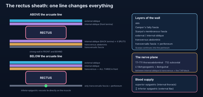
The arcuate line is the single most profitable line in abdominal anatomy, because four different exam answers fall out of one fact. Above it the posterior sheath exists, so the inferior epigastric vessels are separated from rectus by aponeurosis; below it the posterior sheath is gone, so those vessels lie directly on the muscle. Above it a haematoma is compartmentalised by the tendinous intersections; below it, it is not. Above it the surgeon closing a laparotomy has two aponeurotic layers to bite; below it, only one. And the weak point where the linea semilunaris crosses the arcuate line is precisely where the rare Spigelian hernia pushes through — a hernia that hides beneath an intact external oblique aponeurosis and is famously missed on examination. Learn one line; collect four consequences.
The linea alba and the linea semilunaris
Where the aponeuroses of the two sides meet in the midline they interlace into the LINEA ALBA, a tendinous raphe running from the xiphoid process to the pubic symphysis. It is wide above the umbilicus and narrows to a few millimetres below it, and it is essentially avascular — which is exactly why the midline incision is the fastest way into the abdomen, crosses no muscle bellies and cuts no nerves, and can be extended from xiphoid to pubis in seconds when a patient is bleeding. Its price is a slower, less certain heal: an avascular line is a poorly perfused line, and incisional hernia through a midline scar is far commoner than through a muscle-splitting one. The umbilicus is a puckered scar in this raphe, the sealed remnant of the cord, and it is the meeting point of the round ligament of the liver above and the obliterated umbilical arteries and urachus below. The LINEA SEMILUNARIS is the curved lateral border of rectus, palpable in a thin person as a groove running from the tip of the ninth costal cartilage to the pubic tubercle; it marks where the three aponeuroses split or rearrange themselves, and where the sheath is entered from the side. In pregnancy or long-standing obesity the linea alba stretches and thins until the two recti separate — DIVARICATION OF RECTI, which produces a ridge that bulges alarmingly when the patient sits up but is not a true hernia at all, because there is no defect ring and nothing can strangulate in it.
Two arteries that meet in the middle
The wall is supplied from above and from below, and the two supplies shake hands inside the rectus sheath. The SUPERIOR EPIGASTRIC ARTERY is a terminal branch of the internal thoracic artery. It enters the abdomen between the sternal and costal origins of the muscle described in the diaphragm, slips into the upper rectus sheath and descends behind the muscle. The INFERIOR EPIGASTRIC ARTERY arises from the external iliac artery just above the inguinal ligament, runs upwards and medially in the extraperitoneal tissue raising a peritoneal fold, hooks around the arcuate line and ascends behind rectus to meet its partner. The anastomosis between them is usually modest — but it is a true anastomosis between the subclavian and iliac systems, and in COARCTATION OF THE AORTA it becomes a lifeline: blood from the subclavian passes down the internal thoracic, into the superior epigastric, down into the inferior epigastric and out into the external iliac, bypassing the narrowed segment altogether to reach the legs. Alongside these run the deep circumflex iliac artery from the external iliac, the musculophrenic artery, the lower posterior intercostal arteries (T7–T11), the subcostal and the lumbar arteries laterally. Superficially, three small branches of the femoral artery — the superficial epigastric, superficial circumflex iliac and superficial external pudendal — supply the skin and fat, and it is the deep inferior epigastric perforators piercing rectus to reach that skin which allow the whole lower abdominal wall to be transferred as a DIEP flap to reconstruct a breast. Venous drainage mirrors the arteries above and below the umbilicus, and the small paraumbilical veins running in the falciform ligament connect the wall to the portal system — the channel that becomes the caput medusae of portal hypertension.
Nerves in a plane between two muscles
Every nerve of this wall travels in the same corridor — and an anaesthetist can flood that corridor with a needle. The anterior abdominal wall is supplied segmentally, exactly like the chest wall above it. The THORACOABDOMINAL NERVES are the anterior rami of T7 to T11 — the lower intercostal nerves, which simply leave their intercostal spaces at the costal margin and continue into the abdomen. The SUBCOSTAL NERVE is T12, and the ILIOHYPOGASTRIC and ILIOINGUINAL nerves are branches of L1 from the plexus described in the lumbar plexus. All of them run forwards in the same NEUROVASCULAR PLANE, between internal oblique and transversus abdominis, before piercing the rectus sheath to supply rectus and end as anterior cutaneous branches; each also gives a lateral cutaneous branch on the way. Because they are segmental, the dermatomes are beautifully orderly: T7 over the xiphoid, T10 exactly at the umbilicus, T12 above the pubis and L1 over the groin and the root of the scrotum or the mons. That plane is the target of the TAP BLOCK (transversus abdominis plane block), in which local anaesthetic is deposited under ultrasound guidance between the two muscles and spreads along the corridor to numb the whole wall on that side — a technique that has transformed pain control after caesarean section and open appendicectomy. The segmental arrangement also explains the reflexes: stroking the skin of a quadrant makes the umbilicus twitch towards it, upper abdominal reflexes testing T7–T9 and lower ones T10–T12, so a lost reflex localises a spinal level.
The MIDLINE incision runs through the linea alba: bloodless, nerve-free, extensible in seconds, the incision of the bleeding patient and the unknown diagnosis — but the slowest to heal and the likeliest to herniate. The PARAMEDIAN incision, now largely historical, ran through the anterior sheath, retracted rectus laterally and cut the posterior sheath, so the muscle came to lie as a living buttress over the repair; it was strong but slow, and it denervated the muscle medial to it if placed too far out. The GRIDIRON (McBurney) incision for appendicectomy is placed at right angles to a line from the umbilicus to the right anterior superior iliac spine, at the junction of its outer and middle thirds, over the appendix as located in the large intestine and appendix; each of the three muscles is SPLIT along its own fibres and none is cut, so the layers close like a valve and almost no nerve is divided — the Lanz variant simply lays the skin part of it in a transverse crease for cosmesis. The PFANNENSTIEL incision, the standard caesarean approach, is a curved transverse cut two fingers above the pubis: skin and anterior rectus sheath are divided transversely, the sheath is peeled off the muscle, and the recti are retracted sideways rather than cut — strong, cosmetic and comfortable, but the lateral ends of the incision are exactly where the iliohypogastric and ilioinguinal nerves can be caught in a suture, producing the chronic burning groin pain that outlasts everything else about the operation. The KOCHER subcostal incision, running parallel to and two centimetres below the right costal margin for open gallbladder or liver surgery, must divide rectus and will cut across some thoracoabdominal nerves; keeping it short and not extending it too far medially is what preserves the rest.
- Layers from outside in: skin, superficial fascia, deep fascia, external oblique, internal oblique, transversus abdominis, transversalis fascia, extraperitoneal fat, parietal peritoneum.
- BELOW the umbilicus the superficial fascia is in TWO layers: fatty CAMPER'S outside and membranous SCARPA'S deep to it; Scarpa's fuses with fascia lata below the inguinal ligament and continues into the perineum as Colles' fascia — hence extravasated urine from a ruptured urethra fills the scrotum and abdominal wall but never the thighs.
- External oblique: fibres downwards and forwards ("hands in pockets"); its rolled lower border is the INGUINAL LIGAMENT between the ASIS and the pubic tubercle, and its aponeurotic gap is the superficial inguinal ring.
- Internal oblique: fibres upwards and forwards, at right angles to external oblique; its lowest fibres form the conjoint tendon with transversus and give off the cremaster. Transversus abdominis: horizontal fibres, the deep corset.
- Rectus abdominis runs from the pubic crest to the 5th–7th costal cartilages and xiphoid, crossed by three or four TENDINOUS INTERSECTIONS fused to the ANTERIOR sheath only; pyramidalis (absent in ~20%) tenses the linea alba and is supplied by T12.
- ABOVE the arcuate line: internal oblique aponeurosis SPLITS — external oblique + half of internal oblique in front, half of internal oblique + transversus behind.
- BELOW the arcuate line (about midway between umbilicus and pubis): ALL THREE aponeuroses pass IN FRONT, leaving only transversalis fascia behind rectus — so the inferior epigastric vessels lie directly on the muscle and a haematoma there spreads freely.
- The LINEA ALBA is the avascular midline raphe from xiphoid to symphysis (the midline incision, and the umbilicus within it); the LINEA SEMILUNARIS is the curved lateral border of rectus, and a Spigelian hernia occurs where it crosses the arcuate line.
- Arteries: SUPERIOR EPIGASTRIC (from the internal thoracic) meets INFERIOR EPIGASTRIC (from the external iliac) inside the rectus sheath — a vital collateral in coarctation of the aorta; plus the deep circumflex iliac, lower posterior intercostals, subcostal and lumbar arteries, and three superficial branches of the femoral.
- Nerves: thoracoabdominal T7–T11, subcostal T12, iliohypogastric and ilioinguinal from L1 — all running in the plane between internal oblique and transversus (the TAP block plane). T10 supplies the umbilicus.
- Incisions and their price: midline (fast, avascular, herniates); gridiron/McBurney (muscle-splitting, nerve-sparing); Pfannenstiel (cosmetic, but risks trapping iliohypogastric/ilioinguinal); Kocher subcostal (divides rectus and some thoracoabdominal nerves).
- Wall pathology: divarication of recti (a stretched linea alba, not a true hernia), umbilical and paraumbilical hernia, incisional hernia, rectus sheath haematoma — and the retroperitoneal bruising of Cullen's (periumbilical) and Grey Turner's (flank) signs.
- Saying the external oblique aponeurosis passes behind rectus below the arcuate line. Below the line ALL THREE aponeuroses pass in FRONT; behind the muscle there is only transversalis fascia — which is the whole reason the inferior epigastric vessels are exposed there.
- Assuming the tendinous intersections cross the whole muscle. They are fused to the ANTERIOR sheath only, so the space behind rectus is one continuous corridor — which is why blood collects in compartments in front and spreads freely behind.
- Calling divarication of recti a hernia. There is no defect and no ring, so nothing can obstruct or strangulate; it is a stretched linea alba, managed quite differently from a true umbilical or incisional hernia.
During laparoscopic surgery a port is inserted 4 cm lateral to the midline and 4 cm above the pubic symphysis, and brisk bleeding follows from a vessel lying immediately behind the rectus abdominis muscle. Which anatomical fact best explains the injury?
- The abdomen has no bony cage: nine layers — skin, superficial fascia (fatty Camper's and membranous Scarpa's below the umbilicus), deep fascia, the three flat muscles, transversalis fascia, extraperitoneal fat and parietal peritoneum — trade armour for the movement needed to breathe, bend, cough, vomit and give birth.
- The three flat muscles cross at three angles — external oblique downwards and forwards (its rolled border being the inguinal ligament), internal oblique upwards and forwards, transversus horizontally — with rectus abdominis in front, crossed by tendinous intersections fused to the anterior sheath only, and the small pyramidalis below.
- The rectus sheath changes at the arcuate line: above it the internal oblique aponeurosis splits around rectus; below it all three aponeuroses pass in front, leaving only transversalis fascia behind — so the inferior epigastric vessels lie bare on the muscle and a haematoma there spreads freely.
- Supply and clinics: superior epigastric meets inferior epigastric inside the sheath (a lifeline in coarctation), and T7–T11, T12 and L1 nerves run between internal oblique and transversus (the TAP plane, with T10 at the umbilicus) — which together explain the choice of incision, the herniae, the TAP block and the cough impulse.
- Drake RL, Vogl AW, Mitchell AWM. Gray's Anatomy for Students — Abdomen: the anterior abdominal wall, rectus sheath and arcuate line.
- Moore KL, Dalley AF, Agur AMR. Clinically Oriented Anatomy — Anterolateral abdominal wall: fascia, muscles, neurovasculature and surgical incisions.
- Netter FH. Atlas of Human Anatomy — Anterior abdominal wall: superficial and intermediate dissections; rectus sheath cross-sections.
- Last RJ. Last's Anatomy: Regional and Applied — The anterior abdominal wall and the inguinal region.
- Snell RS. Clinical Anatomy by Regions — Abdominal wall incisions, rectus sheath haematoma and abdominal herniae.
- TeachMeAnatomy — The Anterolateral Abdominal Wall; The Rectus Sheath; Transversus Abdominis Plane Block.

