
The Knee: The Largest, Most Exposed Joint in the Body
Look at the knee honestly and it should not work at all. Two of the longest bones in the body meet almost end-to-end, a rounded pair of condyles balanced on a nearly flat tabletop, with barely any bony grip to hold them together. Compare that with the hip, where a deep socket swallows the head of the femur and makes dislocation a genuine event. The knee has no socket. What holds it is soft tissue: two crossed ligaments inside the joint, two more on the sides, two crescents of cartilage that deepen the surface, and the pull of the muscles crossing it. That design buys enormous range and enormous power — and it is exactly why the knee is the most commonly injured major joint in the human body.
A footballer plants her left foot to change direction. The boot's studs bite the turf and the foot stops dead; her body keeps turning. For a fraction of a second the whole rotational force of a sprinting adult passes through one joint that cannot rotate — and something inside it gives way with a sound she will describe afterwards as a pop she heard rather than felt. She goes down untouched, without contact from any opponent. Within an hour the knee is tense and swollen, hot and tight, because the torn ligament she has just ruptured has its own blood supply and has bled directly into the joint cavity. On the sideline a physiotherapist grips her tibia and pulls it forward; it slides further than it should. The diagnosis is made at the pitch, before any scanner is switched on, by nothing but a knowledge of what lives inside the knee and what each structure is there to stop.
Three joints in one cavity
The knee is not one articulation but three, wrapped in a single synovial envelope. Formally the knee is a modified hinge — a bicondylar synovial joint — and it contains three articulations inside one continuous cavity: the medial tibiofemoral joint, the lateral tibiofemoral joint, and the patellofemoral joint between the back of the patella and the trochlear groove on the front of the femur. The two condyles of the femur, described in the femur and patella, roll and glide on the tibial plateau of the tibia and fibula; the fibula takes no part at all in the knee, which surprises students every year. Calling it a "hinge" is a useful simplification rather than the truth: a pure hinge only flexes and extends, whereas the knee also permits a modest amount of rotation once it is flexed, and this extra freedom is precisely where it is vulnerable. The synovial cavity that wraps all three is the largest in the body, and it is continuous — which is why blood or pus anywhere inside it distends the whole joint.
Because the capsule is huge and the joint is crossed by heavy tendons, the knee is padded with more bursae than any other joint — small fluid-filled sacs that let tendon and skin slide over bone. The suprapatellar bursa sits above the patella beneath quadriceps femoris and communicates freely with the joint cavity, so a knee effusion balloons upwards into it. The prepatellar bursa lies between the skin and the front of the patella; inflame it by kneeling upright on a hard floor for hours and you get "housemaid's knee". The infrapatellar bursae lie around the patellar ligament, and inflaming them by kneeling more upright still — leaning back on the heels, as at prayer — gives "clergyman's knee". Medially, the pes anserine bursa cushions the conjoined tendons of sartorius, gracilis and semitendinosus, a classic site of medial knee pain in runners. Posteriorly, the semimembranosus bursa can swell into the popliteal fossa as a Baker's cyst.
The menisci: making a socket out of nothing
If the tibia will not give you a socket, build one out of cartilage. The menisci are two crescents of fibrocartilage sitting on the tibial plateau, thick at their outer rim and tapering to a thin free inner edge, so that in cross-section each is a wedge. That wedge shape turns a nearly flat tabletop into a shallow dish that cradles the femoral condyles. They do four jobs: they deepen the articular surface and improve congruence, they spread load over a far wider area (halving the pressure the cartilage must bear), they absorb shock, and they help distribute synovial fluid for lubrication. The medial meniscus is the larger of the two, a wide open C, and — the single most consequential fact about it — it is firmly attached to the deep fibres of the tibial (medial) collateral ligament and to the capsule, which tethers it and leaves it relatively immobile. The lateral meniscus is smaller and more nearly a closed circle, and it is NOT attached to the fibular (lateral) collateral ligament; the popliteus tendon separates the two. So the lateral meniscus is free to slide out of the way as the joint moves, while the medial one is dragged along by whatever happens to the ligament it is bound to.
The menisci also carry one of the most clinically useful blood-supply facts in the body. Only the outer third of each meniscus is vascular, fed from the capsule by branches of the genicular arteries — the so-called red zone. The middle third is a red–white transition, and the inner third is entirely avascular, nourished only by diffusion from synovial fluid — the white zone. The consequence is blunt: a tear in the peripheral red zone can heal, and is worth repairing with sutures; a tear in the inner white zone will not heal no matter what you do, and is trimmed away instead. Every surgical decision about a torn meniscus starts with asking where in that vascular gradient the tear sits.
The cruciates: two ligaments crossing inside the joint
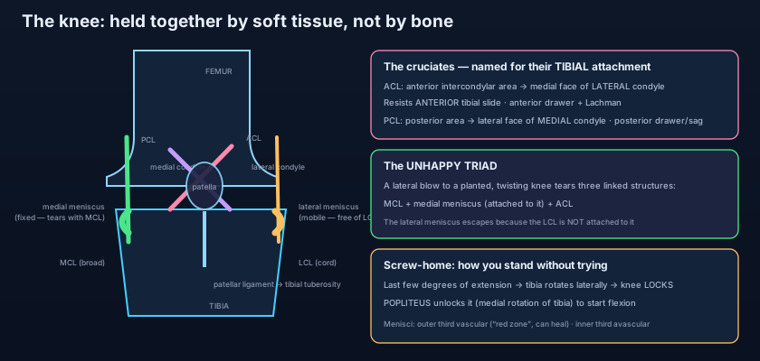
They are named for where they attach on the tibia, and they cross like the strokes of an X. The anterior cruciate ligament (ACL) runs from the anterior intercondylar area of the tibia upwards, backwards and laterally to the medial surface of the LATERAL femoral condyle. Its job is to resist anterior translation of the tibia on the femur — it stops the shin sliding forwards out from under the thigh — and it is also the main restraint against hyperextension and excessive internal rotation of the tibia. Two bedside tests interrogate it: the anterior drawer test, with the knee flexed to ninety degrees and the tibia pulled forward, and Lachman's test, the same pull at about twenty to thirty degrees of flexion, which is the more sensitive of the two because the hamstrings cannot guard against it.
The posterior cruciate ligament (PCL) runs from the posterior intercondylar area of the tibia upwards, forwards and medially to the lateral surface of the MEDIAL femoral condyle. It is the stronger and thicker of the two, and it resists posterior translation of the tibia — it stops the shin sliding backwards. It is tested by the posterior drawer test, and by the sag sign: lay the patient supine with both hips and knees flexed, and the injured tibia visibly drops back compared with the other side. A useful memory: each cruciate resists the movement its own name describes, and each attaches to the femoral condyle on the OPPOSITE side from its tibial name — anterior to lateral, posterior to medial.
Outside the joint, the collaterals guard the sides — and one of them holds a meniscus. The tibial (medial) collateral ligament, or MCL, is a broad flat band running from the medial femoral epicondyle to the medial condyle and shaft of the tibia. It resists valgus force — a blow to the outside of the knee that would splay the leg outwards — and, critically, its deep fibres are fused to the medial meniscus. The fibular (lateral) collateral ligament, or LCL, is completely different in character: a rounded cord running from the lateral femoral epicondyle to the head of the fibula, standing free of the capsule and free of the lateral meniscus, resisting varus force. Around them sit the rest of the supporting cast: the patellar ligament, which is the continuation of the quadriceps tendon from the patella to the tibial tuberosity; the oblique popliteal ligament, an expansion of the semimembranosus tendon reinforcing the back of the capsule; and the arcuate popliteal ligament arching over the popliteus tendon. The muscles that cross the knee, covered in the muscles of the thigh, are the dynamic half of this system — a well-trained quadriceps is functionally part of the knee's stability.
The screw-home mechanism: how the knee locks itself
Standing still should be exhausting. It isn't, and here is why. The medial femoral condyle has a longer articular surface than the lateral one. As the knee approaches full extension the lateral condyle runs out of surface first and stops moving, while the medial condyle keeps gliding — and because one side is still travelling while the other has halted, the tibia is forced to rotate laterally through the last ten to fifteen degrees. This is the screw-home mechanism, and its effect is to twist all the ligaments taut at once: the cruciates wind on each other, the collaterals tighten, and the joint jams into its close-packed position. The knee is now locked. A person standing at ease can switch off the quadriceps almost completely and hang on ligaments alone. Watch a queue of people waiting for a bus and you are watching a row of locked knees doing free work.
But a lock that cannot be opened is a trap, so the body keeps a key. Popliteus is a small flat triangular muscle in the floor of the popliteal fossa, running from the lateral femoral condyle downwards and medially to the back of the tibia. When the foot is off the ground it rotates the tibia medially; when the foot is planted it rotates the femur laterally on the fixed tibia. Either way the effect is the same — it undoes the screw-home rotation and unlocks the knee so that flexion can begin. Every single time you sit down, popliteus fires first. It is one of the most elegant small details in the whole limb: a muscle whose entire career is turning a key.
Think of the knee as a tent pole standing on a dinner plate. The pole has enormous leverage and the plate has no rim to hold it — nothing in the shape of the two parts keeps them together. What keeps the tent standing is guy ropes: two running diagonally through the middle of the structure (the cruciates), two down the sides (the collaterals), and a pair of rubber wedges under the pole to stop it skidding on the china (the menisci). Cut a guy rope and the pole does not fall immediately, but every gust now moves it further than it should. Compare that with the hip, which is a ball dropped deep into a cup: you could cut its ligaments and it would still not come apart easily. The hip is built by its bones; the knee is built by its ropes.
When the ropes give way
The classic injury is the unhappy triad — sometimes called O'Donoghue's triad — and it follows directly from the anatomy. A force from the outside of the knee while the foot is planted and the body twists drives the joint into valgus with external rotation. The MCL, guarding against valgus, tears first. Because the MCL's deep fibres are welded to the medial meniscus, the meniscus is dragged and torn with it. And with the medial restraint gone, the rotational force falls on the ACL, which fails last. Three structures, one mechanism, and the link between them is a single anatomical attachment. An isolated ACL rupture behaves differently again: the audible pop, then rapid swelling within one or two hours because the ligament bleeds into the joint — a haemarthrosis, tense and painful, quite unlike the slower effusion of a meniscal tear that appears the next morning.
Meniscal tears produce their own signature. A longitudinal tear whose inner fragment flips into the intercondylar notch is a bucket-handle tear, and it physically blocks extension: the patient presents with a locked knee, held in slight flexion, unable to straighten it however hard they try, often with a history of clicking and giving way. Posteriorly, a swollen semimembranosus bursa becomes a Baker's cyst, a soft fullness behind the knee that can rupture and mimic a deep vein thrombosis in the calf. The PCL has its own mechanism entirely: a dashboard injury, where a front-seat passenger's flexed knee strikes the dashboard in a collision and the tibia is driven backwards. And the joint has a dangerous neighbour — the common fibular nerve winds subcutaneously around the neck of the fibula just below the lateral side of the knee, so a lateral blow, a varus injury, or a tight plaster there can cause foot drop, as described in the sciatic, tibial and fibular nerves.
Two ligaments with almost identical names behave in opposite ways, and the difference is one attachment. The medial collateral ligament is broad, flat, fused to the capsule and welded to the medial meniscus — so it tears together with the meniscus, and so a medial meniscal tear is far commoner than a lateral one. The lateral collateral ligament is a free round cord that never touches the lateral meniscus — so lateral injuries stay isolated. When you are asked why the medial meniscus is injured three to five times more often than the lateral, the answer is not that it is bigger or weaker. It is that it is tied down and cannot get out of the way.
The pivot: a basketball player lands from a jump on a slightly extended knee and turns at the same moment. No contact, an audible pop, a knee swollen tight within the hour — a torn ACL with haemarthrosis, confirmed with Lachman's test. The locked knee: a young man squats to lift a box, feels something shift, and stands up unable to fully straighten his leg; the knee is held at fifteen degrees of flexion — a bucket-handle medial meniscal tear jammed in the notch. The dashboard: a passenger in a head-on collision, knees flexed against the dash, arrives with a posterior sag sign — a PCL rupture. The worn joint: a woman of seventy with years of gradually worsening medial knee pain, stiffness after sitting, crepitus on movement and a bow-legged (varus) stance as the medial compartment cartilage thins — osteoarthritis, managed with weight loss, quadriceps strengthening and anti-inflammatory analgesia, and eventually with a total knee replacement that resurfaces the femoral condyles and the tibial plateau in metal and polyethylene.
- The knee is a modified hinge (bicondylar synovial) with three articulations in one continuous cavity: medial tibiofemoral, lateral tibiofemoral, and patellofemoral. The fibula takes no part.
- It has the largest synovial cavity in the body and many bursae: suprapatellar (communicates with the joint), prepatellar ("housemaid's knee"), infrapatellar ("clergyman's knee"), pes anserine and semimembranosus (Baker's cyst).
- The medial meniscus is a larger C, relatively immobile, and ATTACHED to the MCL; the lateral meniscus is more circular, mobile, and free of the LCL.
- The menisci deepen the surface, spread load, absorb shock and aid lubrication; the outer third is vascular (red zone, can heal), the inner third avascular (white zone, cannot).
- ACL: anterior intercondylar area → medial surface of the LATERAL femoral condyle; resists ANTERIOR tibial translation (anterior drawer, Lachman).
- PCL: posterior intercondylar area → lateral surface of the MEDIAL femoral condyle; the stronger ligament, resisting POSTERIOR translation (posterior drawer, sag sign).
- Tibial (medial) collateral ligament: broad and flat, resists valgus, deep fibres fused to the medial meniscus. Fibular (lateral): a free cord, resists varus, never touches the lateral meniscus.
- Other reinforcements: the patellar ligament (quadriceps tendon → tibial tuberosity), the oblique popliteal ligament (from semimembranosus) and the arcuate popliteal ligament.
- Screw-home: the longer medial condyle forces the tibia to rotate LATERALLY in the last degrees of extension, tightening every ligament and locking the knee for effortless standing.
- Popliteus UNLOCKS the knee — rotating the tibia medially (or the femur laterally on a fixed tibia) to begin flexion.
- Unhappy triad = ACL + MCL + medial meniscus, from a valgus twisting force on a planted foot; the medial meniscus is dragged in because it is bound to the MCL.
- Rapid tense swelling after a pop = haemarthrosis (ACL); a knee that cannot straighten = bucket-handle meniscal tear; the common fibular nerve at the fibular neck is at risk → foot drop.
- Thinking the fibula forms part of the knee joint. It does not — the knee is femur, tibia and patella only; the fibula meets the tibia at a separate, small proximal tibiofibular joint.
- Mixing up the cruciate attachments. The ANTERIOR cruciate attaches to the LATERAL femoral condyle and the POSTERIOR to the MEDIAL — each crosses to the opposite side, which is exactly why they form an X.
- Assuming any torn meniscus can be stitched. Only the vascular outer third (the red zone) can heal; a tear in the avascular inner third is trimmed, not repaired.
A footballer is struck on the lateral side of the knee while his foot is planted, forcing the joint into valgus with external rotation. Which combination of structures is most likely injured, and why are they injured together?
- The knee is a modified hinge (bicondylar synovial joint) with three articulations — medial and lateral tibiofemoral and patellofemoral — in the largest synovial cavity in the body, surrounded by numerous bursae; it has almost no bony stability and depends on soft tissue.
- The menisci deepen the surface, spread load and lubricate; the medial is larger, less mobile and attached to the MCL (hence torn with it), the lateral is more circular, mobile and free — and only the outer third of either is vascular enough to heal.
- The ACL resists anterior tibial translation (anterior drawer, Lachman) and the PCL posterior translation (posterior drawer, sag sign), while the broad MCL resists valgus and the cord-like LCL resists varus.
- The screw-home mechanism laterally rotates the tibia to lock the knee in full extension for effortless standing, and popliteus unlocks it to begin flexion; the classic failures are the unhappy triad, the ACL pop with haemarthrosis, the locked knee of a bucket-handle tear, the dashboard PCL injury and, in age, osteoarthritis.
- Drake RL, Vogl AW, Mitchell AWM. Gray's Anatomy for Students — Lower limb: the knee joint.
- Moore KL, Dalley AF, Agur AMR. Clinically Oriented Anatomy — The knee joint, menisci and cruciate ligaments.
- Netter FH. Atlas of Human Anatomy — Knee: ligaments, menisci and bursae.
- Last RJ. Last's Anatomy: Regional and Applied — The knee and popliteal fossa.
- Snell RS. Clinical Anatomy by Regions — Knee injuries, the unhappy triad and meniscal tears.
- TeachMeAnatomy — The Knee Joint; The Menisci of the Knee.

