
The Pelvic Floor: The Muscle Sheet Holding Everything Up
In a four-legged animal the outlet of the pelvis is a back wall: the viscera rest on the abdominal wall, and the muscle closing the outlet has almost nothing to carry. Stand that animal upright and the geometry turns through ninety degrees. The wall becomes a FLOOR, and a sheet of striated muscle a few millimetres thick is suddenly the last thing between the entire weight of the abdominal contents and the outside world. It must hold that column of pressure through every cough, every lift, every step — and then, on command and only on command, open to let urine and faeces out, and once or twice in a lifetime stretch far enough to let a whole child through and close again afterwards. No other muscle in the body is asked to be both a floor and a door.
A thirty-four-year-old woman comes to the physiotherapy room a year after the birth of her first child — a long second stage that ended with forceps. Her complaint is small and constant: a leak of urine when she coughs, when she laughs, when she lifts her toddler out of the car. She has been doing her exercises faithfully, she says, and they have changed nothing. The physiotherapist asks her to squeeze, and watches. The patient's buttocks tighten, her thighs draw together and her breath is held — and the one muscle that matters does not move at all. She has been training the gluteal muscles for a year. So the instruction is changed to a sensation instead of a name: draw up as though you were stopping yourself from passing wind, and lift, without squeezing your buttocks, without holding your breath. On the fourth attempt something small and deep contracts. It is the first time in her life she has knowingly used a muscle that has been holding her organs off the ground since the day she learned to stand.
What standing upright cost us
Anatomy is often a record of a compromise, and the pelvic floor is the clearest one in the body. The bony pelvis is a ring open below: the outlet described in the walls of the pelvis is a wide aperture bounded by the pubic arch in front, the ischial tuberosities at the sides and the coccyx behind, and it is closed by soft tissue alone. In a quadruped that aperture faces backwards and the muscle closing it is little more than a tail-wagging sheet. In a biped it faces DOWNWARDS, and the entire hydrostatic column of the abdomen — liver, gut, bladder, uterus, and every rise in intra-abdominal pressure they transmit — presses on it. The body's answer was to convert that sheet into a funnel-shaped, tonically active, striated muscular diaphragm which is unusual in two ways: it is skeletal muscle that never entirely relaxes, contracting quietly all day and even in sleep; and it is a load-bearing structure that must nevertheless be perforated in the midline by three tubes. Every disorder in this article follows from that one design conflict — the sheet has to hold, and the sheet has holes.
The pelvic diaphragm: a funnel slung between three attachments
The muscular floor proper is the PELVIC DIAPHRAGM (الحجاب الحوضي), formed by two muscles on each side: LEVATOR ANI in front and laterally, and COCCYGEUS behind. Together they make not a flat plate but a shallow FUNNEL, sloping downwards and medially towards the midline hiatuses, with its convexity facing the perineum below and its concavity facing the pelvic cavity above. The line of origin is a continuous horseshoe running from the body of the PUBIS in front, backwards along the TENDINOUS ARCH OF LEVATOR ANI, to the ISCHIAL SPINE behind. That tendinous arch — the arcus tendineus levator ani — is not a ligament in its own right but a thickened white band in the fascia covering OBTURATOR INTERNUS on the side wall, and it is the anatomical trick that solves an engineering problem: the pelvic side wall has no bone at the right height for a muscle to arise from, so the floor takes its origin from a tendon painted onto the fascia of another muscle. Above the diaphragm lies pelvic fascia and the peritoneum; below it lie the ischioanal fat and the muscles of the perineum. The pelvic diaphragm is the SUPPORT layer; the perineal membrane and the deep perineal pouch below it are a separate, more anterior sheet, and confusing the two is the commonest error students make here.
Levator ani in three parts
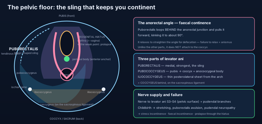
One muscle, three names, three completely different jobs — and only one of them is a true sling. PUBORECTALIS is the medial, thickest and strongest part. Its fibres arise from the back of the body of the pubis on each side, run backwards almost horizontally alongside the urethra, vagina and anal canal without attaching to any of them, and — this is the whole point — meet their fellows of the opposite side BEHIND the anorectal junction to form a continuous U-shaped SLING. Puborectalis has no bony attachment posteriorly at all: it does not reach the coccyx. Because both ends are anchored to the pubis in front, its resting tone pulls the anorectal junction FORWARDS, kinking the gut tube and producing the ANORECTAL ANGLE of roughly 90 degrees between the axis of the rectum and that of the anal canal. That angle, not any sphincter, is the principal mechanism of gross faecal continence, as detailed in the anal canal. PUBOCOCCYGEUS is the main intermediate part, arising from the pubis lateral to puborectalis and from the front of the tendinous arch, sweeping backwards and medially to insert into the coccyx and into the ANOCOCCYGEAL BODY (the levator plate) in the midline; its medial fibres are named after the structures they pass and grip — PUBOPERINEALIS to the perineal body, PUBOVAGINALIS in the female, PUBOANALIS to the intersphincteric groove, and in the male the slip called LEVATOR PROSTATAE, which supports the prostate and is a functional urethral elevator. ILIOCOCCYGEUS is the thin, flat, posterolateral part, arising from the tendinous arch and the ischial spine and passing backwards and medially to the coccyx and the anococcygeal body; in most adults it is more aponeurotic than muscular, a translucent sheet rather than a working belly, and it is the part most easily torn or thinned.
Coccygeus, and the back of the floor
COCCYGEUS, also called ISCHIOCOCCYGEUS, completes the diaphragm behind. It is a small triangular muscle arising from the tip of the ISCHIAL SPINE and inserting into the lateral margin of the COCCYX and the lowest piece of the SACRUM. It lies directly on the pelvic surface of the SACROSPINOUS LIGAMENT and shares its shape so exactly that the muscle and the ligament are best regarded as two faces of a single structure — degenerate muscle in front, ligament behind. Functionally it does very little lifting: it supports the posterior part of the floor, pulls the coccyx forwards after defecation and childbirth, and in humans is largely a vestige of the muscle that wagged a tail. Its practical importance is topographical: because the ischial spine and the sacrospinous ligament are its landmarks, coccygeus marks the plane at which the pudendal nerve hooks round the spine, and it is the muscle the surgeon works through when the sacrospinous ligament is used to suspend a prolapsed vault.
The hiatuses, and the plate behind them
A floor with holes in it fails at the holes. In the midline, between the medial borders of the two levator muscles, is a gap — the LEVATOR HIATUS — divided into two functional parts. In front is the UROGENITAL HIATUS, transmitting the urethra and, in the female, the vagina; behind it is the ANAL HIATUS, transmitting the anal canal. The urogenital hiatus is the weak point of the human pelvis. It is bounded by muscle on each side and closed below only by the perineal membrane and the perineal body, and it is through this aperture, when the levator muscles are torn or denervated and the hiatus widens, that pelvic organs descend — the whole subject of continence and pelvic organ prolapse. Behind the anorectal junction the two pubococcygeus muscles meet and fuse in the midline as a firm horizontal shelf, the LEVATOR PLATE (anococcygeal raphe), running from the anorectal junction back to the coccyx. Its orientation is the quiet hero of pelvic support: while the plate stays HORIZONTAL, the vagina and rectum lie flattened upon it, and any rise in abdominal pressure presses them DOWN ONTO the shelf, closing them like a flap valve against a table top. When the plate is damaged and sags into a more vertical slope, the same rise in pressure now pushes the organs along the plate towards the widened hiatus instead of onto it. The identical force that maintained continence yesterday drives descent today, and nothing has changed except an angle.
At the front edge of the anal hiatus, in the midline between the anal canal behind and the vaginal opening (or the bulb of the penis) in front, lies the PERINEAL BODY — a small pyramidal fibromuscular node into which an extraordinary number of structures converge: the two levator ani muscles through their puboperinealis slips, the external anal sphincter, the superficial and deep transverse perineal muscles, the bulbospongiosus, and the fibres of the perineal membrane. It is the central anchoring post of the anterior floor. Its thickness in the female — the tissue between the vagina and the anal canal — is only a couple of centimetres, and it is exactly the tissue that tears, or is deliberately cut in an episiotomy, during delivery. Destroy that node and the two halves of the floor lose their midline anchor: the levator hiatus gapes, the vaginal opening no longer closes, and the anal sphincter loses the fixed point it pulls against.
Picture a trampoline stretched across a circular frame, with the household's furniture standing on it. To be any use it must do two contradictory things. It must be TAUT — sag it, and everything on it slides towards the middle and eventually through. But it must also YIELD on demand: three chutes pass through the fabric in the centre, and twice a day something has to be let down them and then the fabric must close again as if nothing had passed. A well-strung trampoline does both: it takes the sudden load of a jump without stretching (the cough, the lift), and it can be opened at the middle when you choose (micturition, defecation). Now cut some of the springs at one edge, as a difficult delivery does, and the whole sheet slopes towards the hole. It is still a trampoline; it simply cannot hold anything up any more, and everything on it drifts, slowly and permanently, towards the one part that was never meant to bear weight.
Nerves: two surfaces, two supplies
Levator ani is supplied from ABOVE by the NERVE TO LEVATOR ANI, a direct somatic branch of the sacral plexus arising from the anterior rami of S3 and S4, which runs on the PELVIC (superior) surface of the muscle. From BELOW, its perineal surface receives contributions from the PUDENDAL NERVE (S2, S3, S4) — chiefly the inferior rectal and perineal branches — described in full in the pudendal nerve. Coccygeus is supplied by branches of S4 and S5. Three consequences follow. First, the floor is SOMATIC muscle under voluntary control, unlike the smooth muscle of the bladder or gut, which is why it can be trained at all. Second, because the innervation is segmental and paired, an injury on one side leaves the other side working — asymmetrical weakness, and an asymmetrically descending hiatus, is common. Third, and most important clinically, the pudendal nerve is stretched over the ischial spine during the descent of the fetal head, so a prolonged second stage can denervate the very muscles it is exhausting: an injury that is mechanical and neurological at once, and the reason continence sometimes fails years after a delivery in a woman with no visible tear at all.
What the floor actually does, minute by minute
Four things, and they are best learned as four separate behaviours. ONE — TONIC SUPPORT: the muscle maintains a continuous low-grade contraction against gravity, holding the viscera up and keeping the hiatus narrow; this is the baseline that never switches off. TWO — REFLEX PRE-CONTRACTION: milliseconds BEFORE a cough, a sneeze, a laugh or a lift, the floor contracts harder, anticipating the surge in intra-abdominal pressure rather than reacting to it. This feed-forward reflex is the single mechanism whose failure produces stress incontinence, and it is precisely what a trained floor recovers. THREE — SPHINCTERIC ACTION: the puborectalis sling maintains the anorectal angle and, with the pubovaginalis and levator prostatae slips, compresses and elevates the urethra, adding to the closure pressure generated by the urethral sphincters themselves, as set out in the urinary bladder. FOUR — CONTROLLED RELAXATION: during micturition and defecation the floor must RELAX and descend, the puborectalis in particular releasing so that the anorectal angle straightens from about 90 degrees to nearly 140 and the tube becomes passable. Note the symmetry that makes this subject clinically rich: a floor that cannot contract leaks, and a floor that cannot relax obstructs. Both are diseases of the same muscle.
Ask a student what keeps faeces in and the answer is almost always the anal sphincters. It is only half true, and the smaller half. The internal anal sphincter, smooth muscle in tonic contraction, contributes most of the RESTING pressure but is involuntary and easily overcome; the external sphincter is voluntary but fatigues within a minute. What holds continence hour after hour, without attention and without effort, is a bend: the puborectalis sling pulling the anorectal junction forwards to make the ANORECTAL ANGLE, so that rising rectal pressure presses the anterior rectal wall down over the top of the anal canal like a flap valve — the harder you push, the tighter the seal. This explains three clinical facts at once. A patient whose puborectalis is avulsed from the pubis can have perfectly normal sphincter pressures on manometry and still be incontinent to solid stool, because the angle has straightened. A patient in whom the puborectalis fails to RELAX on straining — anismus, or paradoxical contraction — has a permanently kinked outlet and cannot evacuate, no matter how hard they push. And the reason squatting eases defecation is not folklore: hip flexion beyond ninety degrees opens the anorectal angle mechanically, doing for the patient what the muscle is meant to do for itself.
- The PELVIC DIAPHRAGM is a funnel of LEVATOR ANI plus COCCYGEUS, arising along a continuous horseshoe: the body of the pubis in front, the TENDINOUS ARCH OF LEVATOR ANI (a thickening in the obturator internus fascia) at the side, and the ISCHIAL SPINE behind.
- LEVATOR ANI has three parts: PUBORECTALIS (medial, strongest, a U-shaped sling behind the anorectal junction with NO posterior bony attachment), PUBOCOCCYGEUS (with the puboperinealis, pubovaginalis, puboanalis and levator prostatae slips), and ILIOCOCCYGEUS (thin, posterolateral, largely aponeurotic).
- COCCYGEUS (ischiococcygeus) runs from the ischial spine to the coccyx and lowest sacrum, lying directly on the SACROSPINOUS LIGAMENT — muscle and ligament are two faces of one structure.
- The midline LEVATOR HIATUS has a UROGENITAL part in front (urethra, and vagina in the female — the weak point through which prolapse occurs) and an ANAL part behind; the LEVATOR PLATE behind the anorectal junction must stay HORIZONTAL for pressure to close the organs onto it rather than push them through the hiatus.
- The PERINEAL BODY is the anterior anchoring node where levator ani, external anal sphincter, transverse perineal muscles, bulbospongiosus and the perineal membrane converge — the tissue torn or cut in delivery.
- NERVES: nerve to levator ani from S3–S4 on the pelvic surface, pudendal branches (inferior rectal and perineal) on the perineal surface; coccygeus from S4–S5. It is SOMATIC, voluntary muscle — which is why it can be trained.
When the floor fails
Almost every pelvic floor disorder is one of three injuries: the muscle is stretched, the muscle is detached, or the nerve is damaged. Vaginal delivery is the dominant risk factor, and it injures in all three ways at once, as the anatomy of childbirth describes. The levator hiatus must distend to several times its resting area to admit the fetal head, and the medial fibres of puborectalis undergo the greatest stretch of any skeletal muscle in the body — a lengthening of two- to threefold, far beyond the point at which muscle normally tears. In up to a fifth of first vaginal deliveries the puborectalis is AVULSED from its origin on the pubic bone on one or both sides, a lesion invisible on inspection and detectable on transperineal ultrasound as a widened, asymmetrical hiatus. Add the stretch of the pudendal nerve over the ischial spine during a long second stage, and a perineal tear extending into the perineal body or the external anal sphincter, and one delivery can produce mechanical, structural and neurological injury together. Risk rises with instrumental delivery — forceps above all — a prolonged second stage, a large baby and a mid-line episiotomy that extends. The consequences declare themselves at different times: stress incontinence often within months, prolapse and faecal incontinence often decades later, when the reserve of an already-thinned muscle is finally exhausted by ageing and oestrogen withdrawal.
Pelvic floor muscle training works — it is first-line treatment for stress urinary incontinence and for mild prolapse — but only if the patient contracts levator ani and nothing else. The commonest failure is substitution: the patient squeezes the gluteal muscles, adducts the thighs, or holds the breath and bears DOWN, which is the exact opposite of the intended movement and loads the hiatus rather than closing it. The instruction that works is a sensation, not an anatomical name: draw up and in as though preventing the passage of wind, and hold, while breathing normally and keeping the buttocks and thighs still. The correct action is a lift and an inward draw, felt deep and in the midline; nothing visible should happen outside. Effective programmes teach both fast contractions (for the pre-cough reflex) and sustained ones (for tonic support), continue for at least three months, and are supervised at first, because a substantial proportion of women taught only by leaflet perform the manoeuvre incorrectly. The same training is given to men before and after radical prostatectomy, where removal of the prostate takes away the passive urethral support of the levator prostatae slip and leaves continence dependent on the external sphincter and the surviving floor — which is why a man who has trained his pelvic floor before the operation recovers continence sooner than one who begins afterwards.
- FUNCTION is four behaviours: tonic support against gravity; a feed-forward REFLEX contraction milliseconds before a cough or lift; sphincteric action (anorectal angle plus urethral elevation and compression); and controlled RELAXATION for micturition and defecation.
- A floor that cannot CONTRACT leaks (stress incontinence, faecal incontinence when the anorectal angle straightens); a floor that cannot RELAX obstructs (anismus, obstructed defecation). Same muscle, opposite failures.
- Childbirth injures in three ways at once: extreme STRETCH of the hiatus and of puborectalis, AVULSION of puborectalis from the pubis (in up to a fifth of first vaginal deliveries), and PUDENDAL NEUROPATHY from stretch over the ischial spine.
- The LEVATOR HIATUS is measured on transperineal ultrasound: a widened, asymmetrical hiatus is the signature of avulsion, and hiatal area correlates with prolapse.
- Chronic cough, obesity, constipation with chronic straining and repeated heavy lifting all raise intra-abdominal pressure day after day — chronic overload of a sheet designed for intermittent load, and every one of them is a modifiable risk factor.
- In the male, radical prostatectomy removes the passive urethral support provided by the levator prostatae slip, leaving continence dependent on the external sphincter and the remaining floor — hence pre- and post-operative pelvic floor training.
- Calling the pelvic floor and the urogenital diaphragm the same thing. The PELVIC DIAPHRAGM (levator ani and coccygeus) is the support layer, slung from pubis to ischial spine. The perineal membrane and deep perineal pouch are a separate, smaller, more anterior sheet BELOW it, closing the urogenital triangle. Damage to each produces a different clinical picture.
- Teaching a pelvic floor contraction as a squeeze of the buttocks or a bearing-down effort. Gluteal squeezing, thigh adduction and breath-holding are the classic substitutions, and bearing down loads the hiatus rather than closing it. The correct action is an inward LIFT, felt deep in the midline, with nothing moving on the outside.
- Assuming faecal continence is purely sphincteric. Manometry may be normal while a patient is incontinent to solid stool, because the anorectal angle — maintained by the puborectalis sling, not by any sphincter — has been lost.
A woman is incontinent to solid stool one year after a forceps delivery. Anorectal manometry shows normal resting and squeeze pressures, and endoanal ultrasound shows both anal sphincters to be intact. Transperineal ultrasound shows a widened, asymmetrical levator hiatus. Which single structure best explains her incontinence?
- Standing upright turned the pelvic outlet from a back wall into a floor, and the PELVIC DIAPHRAGM — levator ani plus coccygeus — is the funnel-shaped, tonically contracting muscular sheet that now carries the abdominal viscera, slung from the pubis in front, the tendinous arch of levator ani (in the obturator internus fascia) at the side, and the ischial spine behind.
- Levator ani has three parts: PUBORECTALIS — a U-shaped sling passing behind the anorectal junction with no posterior attachment, creating the ~90° ANORECTAL ANGLE that is the main mechanism of faecal continence; PUBOCOCCYGEUS with its puboperinealis, pubovaginalis, puboanalis and levator prostatae slips; and the thin, aponeurotic ILIOCOCCYGEUS. Coccygeus runs from the ischial spine to the coccyx on the sacrospinous ligament.
- The midline UROGENITAL HIATUS (urethra, vagina) is the designed weak point through which prolapse occurs; the LEVATOR PLATE behind must stay horizontal so pressure closes the organs onto it, and the PERINEAL BODY anchors the front. Nerve supply: nerve to levator ani S3–S4 above, pudendal branches below; coccygeus S4–S5.
- The floor supports tonically, pre-contracts by reflex before a cough or lift, maintains the anorectal angle and urethral closure, and relaxes on command — so failure to contract gives stress and faecal incontinence, and failure to relax gives anismus and obstructed defecation. Childbirth injures it by stretch, puborectalis avulsion and pudendal neuropathy; training works only when the correct muscle is found (the sensation of stopping wind, not squeezing the buttocks).
- Drake RL, Vogl AW, Mitchell AWM. Gray's Anatomy for Students — Pelvis and Perineum: the pelvic floor, levator ani and coccygeus.
- Moore KL, Dalley AF, Agur AMR. Clinically Oriented Anatomy — Pelvic diaphragm; puborectalis and the anorectal angle; the perineal body.
- Netter FH. Atlas of Human Anatomy — Pelvic diaphragm from above and below; levator ani and its named parts.
- Last RJ. Last's Anatomy: Regional and Applied — The pelvic floor and its fascial attachments; the tendinous arch of levator ani.
- Standring S (ed). Gray's Anatomy: The Anatomical Basis of Clinical Practice — True pelvis: levator ani, levator hiatus and levator plate.
- TeachMeAnatomy — The Pelvic Floor; Levator Ani; The Perineal Body.

