
The Perineum: Two Triangles and the Spaces Between Them
It is one of the smallest regions in the body and one of the most feared in the examination hall — a diamond of tissue you could cover with the palm of one hand, containing the outlets of two systems, three sets of muscles, two closed fatty spaces and a membrane that decides where a leak will and will not go. Students skip it because the names are unfamiliar and the diagrams look like nothing they have seen. Then a young man arrives with a swollen, bruised scrotum and blue-black staining creeping up his lower abdomen, and stops abruptly, as if drawn with a ruler, at the crease of each groin. Nobody drew that line. A sheet of fascia did — and once you can explain why the bruising stopped there, you understand the perineum.
A twenty-four-year-old scaffolder slips on a wet plank and lands astride a steel bar. He is winded, embarrassed, and walks it off. Two hours later he cannot pass urine; there is a bead of blood at the tip of the penis and he is desperate to void but nothing comes. By the time he reaches the emergency department his scrotum is the size of a grapefruit and the colour of a storm cloud, the shaft of the penis is swollen, and the discolouration has climbed across the pubis and is spreading over the lower abdominal wall in the shape of a butterfly. The junior doctor examining him notices something odd, and it is the finding that makes the diagnosis: the bruising has not gone anywhere near the thighs. On both sides it stops dead in the groin crease. He has torn the spongy part of his urethra against the bar, urine and blood have poured into a closed fascial pouch in the perineum, and that pouch has walls — open at the front, sealed at the sides, sealed behind. The swelling has not spread randomly. It has simply gone everywhere the fascia allowed it to go, and stopped precisely where the fascia said stop.
A diamond with four bony corners
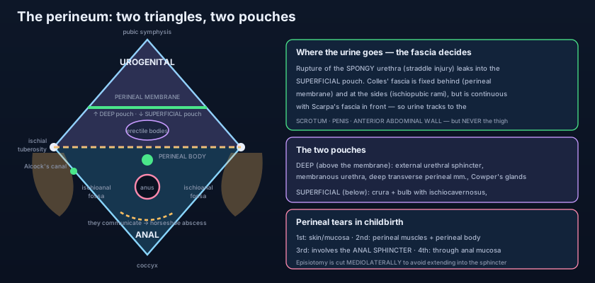
The perineum is everything below the pelvic floor — the floor is its roof. Anatomically the perineum is the diamond-shaped region lying BELOW the pelvic diaphragm, between the thighs. Its roof is the levator ani and coccygeus described in the pelvic floor; its floor is the skin. Its outline is drawn on bone and ligament, and the four corners are easy to find on a skeleton or on a living patient in the lithotomy position: ANTERIORLY the pubic symphysis; ANTEROLATERALLY the ischiopubic rami running back on each side to the ISCHIAL TUBEROSITIES, the two prominences you sit on; POSTEROLATERALLY the SACROTUBEROUS LIGAMENTS sweeping back from the tuberosities to the sacrum; and POSTERIORLY the tip of the COCCYX. Those landmarks are laid out in the hip bone and the bony pelvis, and every one of them can be palpated — which is why the perineum, alone among the deep regions, is a place the examining finger can reach directly. The diamond is not flat: it is pitched like a shallow tent, sloping upward from the skin towards the pelvic floor above, and its two halves lie in slightly different planes.
One line, two triangles
Draw a line transversely between the two ischial tuberosities and the diamond splits into two triangles, and this single line organises the entire region. In front of it lies the UROGENITAL TRIANGLE, containing the external genitalia, the outlet of the urinary tract and, in the female, the vaginal opening. Behind it lies the ANAL TRIANGLE, containing the anal canal and its sphincters. The division is not a textbook convenience — the two triangles are built differently. The anal triangle is a simple space: a canal in the middle and fat on either side, with no fascial floor beneath it. The urogenital triangle is closed off by a tough sheet, the perineal membrane, which stretches between the ischiopubic rami and turns the front half of the perineum into a sandwich of two distinct compartments. Everything clinical about the perineum comes from that structural asymmetry: infection behaves one way behind the line and a completely different way in front of it.
The anal triangle and its two fatty wedges
In the middle, a canal; on each side, a wedge of fat doing an unglamorous but essential job. The centre of the anal triangle is occupied by the anal canal and the EXTERNAL ANAL SPHINCTER wrapped around it, a somatic, voluntary muscle described with the rest of that canal in the anal canal. On either side lies the ISCHIOANAL FOSSA — still widely called the ischiorectal fossa — a wedge-shaped space packed with coarse fat and crossed by fibrous strands. Its boundaries are worth memorising as a shape rather than a list: MEDIALLY the sloping undersurface of levator ani and the external anal sphincter, which form the roof and the medial wall together, meeting the lateral wall at a sharp apex above; LATERALLY the vertical fascia over obturator internus and the ischial tuberosity below it; POSTERIORLY the sacrotuberous ligament and the gluteus maximus; ANTERIORLY the posterior edge of the perineal membrane; and INFERIORLY the perineal skin. Duplicated in the fascia of the lateral wall is the PUDENDAL CANAL, ALCOCK'S CANAL, a sleeve carrying the internal pudendal artery and vein and the pudendal nerve forwards along the ischial ramus. The fat is not padding for its own sake: it is soft, mobile, compressible tissue that allows the anal canal to distend hugely during defaecation and allows the whole perineum to be flattened by the descending fetal head without tearing the levator plate.
Two features of these fossae generate almost all the surgery done in the region. The first is that the two fossae COMMUNICATE WITH EACH OTHER posteriorly, behind the anal canal and deep to the anococcygeal body — so they are not two sealed compartments but one horseshoe-shaped space with a narrow bridge at the back. The second is that each fossa sends an ANTERIOR RECESS forwards, above the perineal membrane and below levator ani, into the urogenital triangle, where pus can hide out of reach of a finger placed in the anal triangle. Add the fact that fat is poorly vascularised and therefore poorly defended, and the consequences write themselves: infection of an anal gland at the level of the pectinate line tracks into the fat and forms a PERIANAL or ISCHIOANAL ABSCESS; if it is neglected it crosses the posterior bridge and becomes a HORSESHOE ABSCESS involving both sides, which cannot be cured by draining one side alone; and once the abscess has discharged, the epithelialised track that remains between the anal canal and the perineal skin is a FISTULA-IN-ANO, a problem measured in years rather than days. Whenever a perianal abscess recurs after apparently adequate drainage, the anatomy is telling you that the other limb of the horseshoe, or an anterior recess, was never opened.
Think of the anal triangle as two identical storerooms on either side of a lift shaft, each packed floor to ceiling with soft crates. What the plans do not show, and what the caretaker only discovers when water gets in, is that the two storerooms share a low crawl-space behind the shaft, and that each has a small annexe running forward under the floor of the next department. Flood one room and the water does not politely stay there: it seeps through the crawl-space into its twin and forwards into the annexe, and a survey that opens only one door will always report the leak as fixed and always be wrong. That crawl-space is the posterior communication behind the anal canal, the annexe is the anterior recess above the perineal membrane, and the surveyor who forgets them is the surgeon whose patient comes back.
The urogenital triangle: one membrane, two pouches
Everything in the front half of the perineum is described as being either above the membrane or below it. The PERINEAL MEMBRANE is a tough triangular sheet of dense fibrous tissue stretched horizontally between the two ischiopubic rami, with its apex at the pubic arch and its free posterior border running between the ischial tuberosities. It is perforated by the urethra in both sexes and additionally by the vagina in the female. Above it lies the DEEP PERINEAL POUCH, a thin compartment between the membrane below and the fascia of levator ani above. Its contents are the ones that matter most functionally: the EXTERNAL URETHRAL SPHINCTER, a circular skeletal muscle under voluntary control that grips the membranous urethra and is the muscle a patient uses to interrupt the stream; the MEMBRANOUS URETHRA itself, the narrowest and least forgiving segment of the male urethra as set out in the urethra; the DEEP TRANSVERSE PERINEAL muscles running transversely to steady the perineal body; the BULBOURETHRAL (COWPER'S) GLANDS in the male, sitting within the sphincter and sending their long ducts down into the spongy urethra below; and the internal pudendal vessels with the dorsal nerve and artery of the penis or clitoris travelling forwards to reach the erectile tissue. In the female the corresponding pouch contains the sphincter urethrae complex and the vessels, but the greater vestibular glands lie below the membrane, not above it.
Below the membrane lies the SUPERFICIAL PERINEAL POUCH, and this is the compartment that produces the classic clinical picture. Its roof is the perineal membrane; its floor is COLLES' FASCIA, the membranous layer of superficial perineal fascia. Its contents are the erectile bodies and the muscles that squeeze them. The two CRURA of the penis or clitoris are anchored to the ischiopubic rami and covered by ISCHIOCAVERNOSUS on each side; the BULB of the penis, or the paired vestibular bulbs in the female, lies in the midline covered by BULBOSPONGIOSUS; and the SUPERFICIAL TRANSVERSE PERINEAL muscles run from each ischial tuberosity to meet in the midline behind. Also in this pouch in the female are the GREATER VESTIBULAR (BARTHOLIN'S) GLANDS, whose obstruction produces the familiar Bartholin's cyst or abscess at the posterior part of the vaginal opening. Functionally these muscles are a vascular pump: ischiocavernosus compresses the crus to raise pressure inside the corpus cavernosum and maintain rigidity, and bulbospongiosus compresses the bulb to empty the urethra of its last drops of urine and to expel semen at ejaculation. They are supplied, like everything else here, by the pudendal nerve.
Colles' fascia is the whole trick, and it is defined entirely by where it sticks and where it does not. Behind, it is firmly attached to the free posterior border of the perineal membrane. At the sides, it is firmly attached to the ischiopubic rami. In front, it is attached to nothing at all — because in front it simply CONTINUES, as the dartos fascia of the scrotum and penis, and then upwards over the pubis as SCARPA'S FASCIA, the membranous layer of superficial fascia of the anterior abdominal wall described in the anterior abdominal wall. So the superficial perineal pouch is a bag sealed at the back and along both sides, with its only opening pointing forwards and upwards. Anything that fills it can only travel one way. And the reason it never reaches the thigh is a second attachment further up: Scarpa's fascia fuses with the FASCIA LATA about a fingerbreadth below the inguinal ligament. That fusion line is the ruler that drew the edge of the bruise.
When the urethra tears: two ruptures, two destinations
The male urethra can be torn at two places, and the level of the tear determines where the urine goes with complete reliability. A STRADDLE INJURY — falling astride a bar, a bicycle crossbar, the edge of a manhole — crushes the BULB of the penis and the SPONGY (bulbar) URETHRA within it against the inferior surface of the pubic arch. The tear is therefore below the perineal membrane, inside the superficial perineal pouch, and urine and blood extravasate into that pouch. Constrained by Colles' fascia, they distend the SCROTUM, track along the shaft of the PENIS deep to the dartos, and pass upwards over the pubis deep to Scarpa's fascia onto the ANTERIOR ABDOMINAL WALL — and, because of the fusion of Scarpa's fascia with the fascia lata, they never enter the THIGH. This pattern of swelling is so characteristic that it is a diagnosis by inspection. Contrast the second injury: a PELVIC FRACTURE, typically from a road traffic collision, shears the prostate upwards and ruptures the MEMBRANOUS URETHRA at the prostatomembranous junction. That tear lies ABOVE the perineal membrane, so urine collects in the deep perineal pouch and extraperitoneally around the bladder base, producing no scrotal swelling at all — instead a high-riding prostate on rectal examination, blood at the meatus, and a patient who cannot void. In both cases the rule is the same: blood at the meatus means do NOT pass a urethral catheter until the urethra has been imaged, because a blind catheter converts a partial tear into a complete one.
Return to the scaffolder. Every element of his presentation is a fascial attachment made visible. Blood at the meatus and inability to void: the urethra is torn. Butterfly bruising of the perineum: blood filling the superficial pouch and taking the shape of the pouch itself, whose posterior edge is the free border of the perineal membrane. Massive scrotal and penile swelling: the pouch is continuous with dartos. Discolouration climbing the lower abdominal wall: the pouch is continuous with the plane deep to Scarpa's fascia. Thighs entirely spared: Scarpa's fascia fuses with the fascia lata below the inguinal ligament. Management follows directly — no blind catheter, a retrograde urethrogram to define the level of the tear, and a SUPRAPUBIC catheter placed above the pubis to divert urine while the injury is assessed. The most useful anatomical fact in the whole encounter is a negative one: the sparing of the thigh, which excludes a leak into the deep pouch or a rupture into the thigh compartments and tells you exactly which segment of urethra has failed.
- The perineum is the diamond BELOW the pelvic floor: pubic symphysis in front, ischiopubic rami and ischial tuberosities anterolaterally, sacrotuberous ligaments posterolaterally, coccyx behind. Its roof is levator ani; its floor is skin.
- A transverse line between the ISCHIAL TUBEROSITIES divides it into the anterior UROGENITAL triangle and the posterior ANAL triangle — and the two are built completely differently.
- The ISCHIOANAL FOSSAE flank the anal canal: medial wall = levator ani and external anal sphincter; lateral wall = obturator internus fascia carrying the PUDENDAL (ALCOCK'S) CANAL with the internal pudendal vessels and pudendal nerve; roof = the apex where the two meet; floor = skin.
- The fossae COMMUNICATE posteriorly behind the anal canal and send ANTERIOR RECESSES into the urogenital triangle — hence the horseshoe abscess and the abscess that recurs after one-sided drainage.
- The PERINEAL MEMBRANE spans the ischiopubic rami and divides the urogenital triangle into a DEEP pouch above (external urethral sphincter, membranous urethra, deep transverse perineal muscles, bulbourethral glands, dorsal neurovascular bundle) and a SUPERFICIAL pouch below.
- The SUPERFICIAL pouch, floored by COLLES' FASCIA, contains the crura and bulb with ISCHIOCAVERNOSUS, BULBOSPONGIOSUS and the SUPERFICIAL TRANSVERSE PERINEAL muscles, plus the greater vestibular (Bartholin's) glands in the female.
The perineal body: the keystone in the middle
One small fibromuscular node holds the whole floor together, and childbirth is what threatens it. In the midline, exactly on the intertuberous line, sits the PERINEAL BODY — a pyramidal fibromuscular mass between the anal canal behind and the vaginal vestibule (or the bulb of the penis) in front. It is where an extraordinary number of muscles converge and interlace their fibres: BULBOSPONGIOSUS from in front, the SUPERFICIAL and DEEP TRANSVERSE PERINEAL muscles from each side, the EXTERNAL ANAL SPHINCTER from behind, and fibres of LEVATOR ANI, especially puborectalis and pubococcygeus, from above. It is the mechanical keystone of the perineum: the point through which the pull of every one of those muscles is transmitted, and therefore the point whose failure loosens all of them at once. In the female it is larger, sits directly between vagina and anal canal, and is only about three to four centimetres from the anal margin — which is why it is so exposed during the second stage of labour. A perineal body torn and badly repaired is followed years later by a widened vaginal opening, a descending pelvic floor, and prolapse of the rectum or the posterior vaginal wall, because the anchor has gone.
One artery and one nerve for the whole region
The perineum is unusually simple in its supply: essentially everything here is fed by the INTERNAL PUDENDAL ARTERY and innervated by the PUDENDAL NERVE (S2, S3, S4), described in detail in the pudendal nerve. Both leave the pelvis through the greater sciatic foramen below piriformis, hook forwards around the sacrospinous ligament at the ISCHIAL SPINE, re-enter through the lesser sciatic foramen, and run forwards in the pudendal canal on the lateral wall of the ischioanal fossa. The branches follow the anatomy exactly: the INFERIOR RECTAL nerve and artery cross the fossa to supply the external anal sphincter and the skin around the anus; the PERINEAL branch supplies the muscles of both pouches and the posterior scrotal or labial skin; and the DORSAL NERVE and artery of the penis or clitoris run forward in the deep pouch to reach the erectile tissue. Two practical consequences. First, because the ischial spine is palpable through the vagina or the rectum, a needle placed onto it delivers a PUDENDAL NERVE BLOCK that anaesthetises the whole perineum for an instrumental delivery, a perineal repair or a minor perineal operation. Second, the pudendal supply stops at a line inside the anal canal: above the pectinate line the innervation is autonomic and painless, below it somatic and exquisitely painful — the reason an internal haemorrhoid can be banded in the clinic without anaesthesia while an anal fissure a centimetre lower is one of the most severe pains in surgery.
Learn the mnemonic "S2, S3, S4 keep the pelvis off the floor" and it will pay for itself three times over. Those roots supply the pudendal nerve and therefore the external anal sphincter, the external urethral sphincter and the perineal muscles — so a cauda equina lesion, a sacral fracture or an epidural that spreads too far shows itself as saddle anaesthesia, urinary retention or overflow, and loss of anal tone. The same roots carry the parasympathetic pelvic splanchnic nerves that produce erection. And the same roots supply the sensory skin over the perineum, which is why saddle anaesthesia and sphincter failure are inseparable red flags in a patient with back pain: they mark the level, and they demand imaging today, not next week.
In the second stage of labour the perineum thins from a thick pad to a translucent sheet as the fetal head distends it, and the midwife's hands do two things at once: one supports the perineal body and flexes the head to present its smallest diameter, the other slows the crowning so the tissues have time to stretch rather than split. When they split, the tear is graded by what it involves: FIRST degree, skin and vaginal mucosa only; SECOND degree, into the perineal muscles and the perineal body — this is the level of a standard episiotomy; THIRD degree, into the EXTERNAL ANAL SPHINCTER, subdivided by how much of the sphincter complex is torn; FOURTH degree, through the sphincter and into the anal canal mucosa. The third and fourth degrees are the ones that change a life, because an unrecognised or poorly repaired sphincter tear means faecal urgency or frank incontinence years later, and every delivery is therefore followed by a deliberate examination for one. This is also the reason an episiotomy, when it is needed, is cut MEDIOLATERALLY rather than in the midline: a midline cut runs directly along the fibres of the perineal body straight towards the anal sphincter and extends into it with alarming ease, whereas an oblique cut angled towards the ischial tuberosity heads away from the sphincter into the fat of the ischioanal fossa. It bleeds more and hurts more, and it is chosen anyway. The obstetric mechanics behind all of this are set out in the anatomy of childbirth.
- The PERINEAL BODY is the fibromuscular node in the midline between the anal canal and the vaginal vestibule (or the bulb) where bulbospongiosus, the superficial and deep transverse perineal muscles, the external anal sphincter and levator ani all converge — the keystone of perineal support.
- COLLES' FASCIA is attached to the perineal membrane behind and the ischiopubic rami at the sides, but is continuous in front with dartos and with SCARPA'S FASCIA of the abdominal wall — so the superficial pouch opens forwards and upwards only.
- Rupture of the SPONGY urethra (straddle injury) → urine into the superficial pouch → SCROTUM, PENIS and ANTERIOR ABDOMINAL WALL, but NEVER the thigh, because Scarpa's fascia fuses with the fascia lata. Rupture of the MEMBRANOUS urethra (pelvic fracture) → urine ABOVE the membrane, no scrotal swelling, high-riding prostate.
- Blood at the external urethral meatus after pelvic or perineal trauma is an absolute contraindication to a blind urethral catheter — image first, divert with a suprapubic catheter.
- The INTERNAL PUDENDAL ARTERY and the PUDENDAL NERVE (S2–S4) supply essentially the whole perineum, reaching it through the pudendal canal after hooking around the ISCHIAL SPINE — the landmark for a pudendal block.
- Obstetric tears: 1st = skin/mucosa; 2nd = perineal muscles and perineal body; 3rd = external anal sphincter; 4th = through into anal mucosa. Episiotomy is cut MEDIOLATERALLY to steer the tear away from the sphincter.
- Believing the two ischioanal fossae are separate closed spaces. They communicate behind the anal canal and each sends an anterior recess above the perineal membrane — which is precisely why a horseshoe abscess exists and why draining one side only guarantees recurrence.
- Confusing the two urethral ruptures. Scrotal, penile and abdominal wall swelling with a butterfly perineal bruise means the SPONGY urethra and the SUPERFICIAL pouch; a high-riding prostate with no scrotal swelling after a pelvic fracture means the MEMBRANOUS urethra, above the perineal membrane.
- Placing the external urethral sphincter in the superficial pouch. The voluntary sphincter and the membranous urethra lie in the DEEP pouch, ABOVE the perineal membrane; the superficial pouch below contains the erectile bodies and their three muscles.
A 30-year-old man falls astride a scaffolding bar. He cannot pass urine and there is blood at the external urethral meatus. Examination shows a butterfly-shaped perineal bruise, a grossly swollen scrotum and penis, and discolouration spreading over the lower anterior abdominal wall — but both thighs are entirely normal. Which structure is responsible for the thighs being spared?
- The perineum is the diamond below the pelvic floor — pubic symphysis, ischiopubic rami and ischial tuberosities, sacrotuberous ligaments, coccyx — divided by a line between the ischial tuberosities into an anterior urogenital triangle and a posterior anal triangle.
- The anal triangle holds the anal canal and external sphincter flanked by the fat-filled ischioanal fossae — medial wall levator ani and sphincter, lateral wall obturator internus fascia carrying the pudendal (Alcock's) canal — which communicate behind the anal canal (horseshoe abscess) and send anterior recesses forwards; poor vascularity plus fat makes them the classic site of abscess and fistula-in-ano.
- The perineal membrane splits the urogenital triangle into a deep pouch above (external urethral sphincter, membranous urethra, deep transverse perineal muscles, bulbourethral glands, dorsal neurovascular bundle) and a superficial pouch below, floored by Colles' fascia and containing the crura and bulb with ischiocavernosus, bulbospongiosus and superficial transverse perineal muscles.
- Rupture of the spongy urethra sends urine into the superficial pouch and thence to scrotum, penis and anterior abdominal wall but never the thigh (Scarpa fuses with fascia lata), unlike membranous rupture above the membrane; the perineal body is the muscular keystone torn in childbirth; and one artery and one nerve — internal pudendal and pudendal (S2–S4) — serve the entire region.
- Drake RL, Vogl AW, Mitchell AWM. Gray's Anatomy for Students — Pelvis and Perineum: the perineal triangles, perineal membrane and the deep and superficial perineal pouches.
- Moore KL, Dalley AF, Agur AMR. Clinically Oriented Anatomy — The perineum; ischioanal fossae; extravasation of urine and the attachments of the perineal fascia.
- Netter FH. Atlas of Human Anatomy — Perineum and external genitalia (male and female); coronal sections of the ischioanal fossa.
- Last RJ. Last's Anatomy: Regional and Applied — The perineum, perineal membrane and pudendal canal.
- Snell RS. Clinical Anatomy by Regions — Perineal body, ischiorectal abscess, fistula-in-ano and rupture of the urethra.
- TeachMeAnatomy — The Perineum; The Ischioanal Fossa; The Perineal Pouches.

