
The Prostate: Two Diseases, Two Different Zones
Nature made one decision about this gland that determines almost everything that goes wrong with it: she wrapped it around the urethra. A walnut of glandular tissue, sitting under the bladder, with the only outflow tract of the urinary system running straight through its middle. Anything that enlarges it can enlarge in only one direction that matters — inwards, onto the tube. And then a second fact doubles the story: the two commonest diseases of the prostate do not begin in the same part of it. Benign enlargement starts in the small ring of tissue hugging the urethra, so it obstructs early and loudly. Cancer starts in the outer shell at the back, so it is felt by a finger through the rectal wall long before it ever narrows anything. One organ, two zones, two entirely different clinical stories — and the whole of prostatic medicine falls out of that single anatomical fact.
A man of sixty-eight has stopped drinking water after seven in the evening. He does not mention it as a symptom; he mentions it as a household arrangement, the way one mentions the route to work. He gets up three or four times a night. When he stands at the toilet he waits — a few seconds at first, then longer — before anything comes, and when it does the stream is thin and stops and starts, and he finishes, walks away, and finds a few drops arrive afterwards anyway. He has learned where every public toilet on his usual streets is. Two floors up in the same hospital, another man of the same age has no urinary symptoms at all. He came in because of low back pain that woke him at night and did not settle with rest, and the X-ray showed bone that looked too white rather than too thin. Both men have a diseased prostate. The first has a benign growth in the small collar of tissue that surrounds the urethra. The second has a cancer that began in the outer shell at the back of the gland, far from the urethra, and has already travelled up a network of valveless veins into his lumbar spine. The gland is the same. The zone is not.
A walnut in a very awkward place
Everything the prostate does to a patient begins with what it is touching. The prostate is a fibromuscular glandular organ about 3 cm long, roughly the size and shape of a walnut, weighing about 20 g in a healthy young adult. Its BASE is fused to the neck of the bladder above, so there is no plane between them and the muscle of the internal urethral sphincter blends directly into the gland. Its APEX rests below on the external urethral sphincter and the perineal membrane, which is why the surgeon dissecting the apex is working millimetres from the muscle of continence. In FRONT lies the pubic symphysis, separated by the fat and veins of the retropubic space (the cave of Retzius) and anchored forwards by the PUBOPROSTATIC LIGAMENTS. BEHIND lies the rectum, and here the whole clinical importance of the organ is decided: the prostate and the rectum are separated only by the thin sheet of DENONVILLIERS' FASCIA — the rectoprostatic fascia — so that a finger in the rectum lies, for practical purposes, on the back of the gland. That single relation is why the prostate is one of the very few internal organs a clinician can palpate directly, and why a rectal examination has been the front line of prostate medicine for a century.
Described as a solid, the gland has a base above, an apex below, and three surfaces between them: an ANTERIOR surface behind the symphysis, a POSTERIOR surface against the rectum, and two INFEROLATERAL surfaces resting on the levator ani of the pelvic floor. The older anatomical description divides it into five LOBES: an ANTERIOR lobe or isthmus in front of the urethra, which is almost entirely fibromuscular and contains little glandular tissue; a MIDDLE lobe between the urethra and the ejaculatory ducts; a POSTERIOR lobe behind them; and two LATERAL lobes forming the bulk of the gland on either side. This lobar language survives in the operating theatre and in older textbooks — a middle lobe growing upwards into the bladder like a ball valve is a real and troublesome finding — but it does not map onto the way disease actually behaves. For that, anatomy had to be redrawn.
McNeal's zones — the spine of the subject
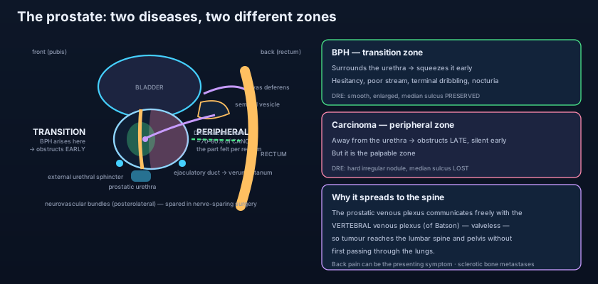
Divide the gland the way disease divides it, and the clinic becomes predictable. John McNeal's zonal anatomy replaced the lobes because it explains what the lobes never could. The TRANSITION ZONE is a small pair of lobules surrounding the prostatic urethra, making up only about 5% of the glandular tissue in a young man — and it is the exclusive site of BENIGN PROSTATIC HYPERPLASIA. The CENTRAL ZONE forms about 25% of the glandular tissue, a cone of tissue surrounding the ejaculatory ducts as they run from the base to the verumontanum; it is relatively resistant to both diseases. The PERIPHERAL ZONE is the largest, about 70% of the glandular tissue, forming the posterior and lateral outer shell of the gland — the part that lies against Denonvilliers' fascia and the rectum, and therefore the part a finger reaches — and it is where roughly 70–80% of PROSTATE CARCINOMA arises. In front of all three lies the ANTERIOR FIBROMUSCULAR STROMA, a thick non-glandular apron that gives the gland its front wall and contains essentially no acini at all.
State the consequence plainly, because it is the single most useful sentence in the subject. BPH = TRANSITION ZONE = EARLY OBSTRUCTION: the growth begins in the very tissue that hugs the urethra, so even a modest enlargement compresses the outflow tract and the patient presents with symptoms while the gland is still small. CANCER = PERIPHERAL ZONE = PALPABLE NODULE, LATE OBSTRUCTION: the growth begins in the outer shell at the back, far from the urethra, so it is felt as a hard nodule through the rectal wall — or found by a raised PSA — while the man is still passing urine perfectly well. Two corollaries follow immediately and are worth carrying to any examination. First, the SIZE of a prostate correlates poorly with symptoms, because what matters is where the tissue grew, not how much of it there is. Second, urinary symptoms are a BAD screening test for cancer: by the time a peripheral-zone tumour obstructs the urethra it has usually crossed most of the gland, and the men who present with obstruction overwhelmingly have benign disease.
Picture a small block of flats built around a single central staircase. The transition zone is the ring of storerooms opening directly onto that staircase; the peripheral zone is the row of outer flats along the back wall, facing the street. Now let two different builders start work. The first extends the storerooms — and because they open onto the stairwell, the very first thing anyone notices is that the staircase has narrowed and the residents are climbing it slowly, one at a time. The second builder converts the back flats, and for a long time nobody inside the building notices anything at all, because the staircase is untouched; but a neighbour walking down the street can see the change through the rear wall. Benign hyperplasia is the storeroom builder — it blocks the stairs at once. Carcinoma is the back-flat builder — it is visible from outside long before it interferes with the traffic in the middle.
The urethra running through the middle
The PROSTATIC URETHRA is the widest and most dilatable part of the male urethra, about 3 cm long, entering at the base and leaving at the apex, and bent forwards through roughly 35 degrees at its midpoint. Along its posterior wall runs a longitudinal ridge, the URETHRAL CREST, and midway along the crest is a rounded eminence — the SEMINAL COLLICULUS, or verumontanum. On its summit opens the blind PROSTATIC UTRICLE, a small midline diverticulum a few millimetres deep that is the male vestigial remnant of the paramesonephric (Müllerian) ducts. Flanking the utricle, one on each side, are the two slit-like openings of the EJACULATORY DUCTS. On either side of the crest lies a shallow groove, the PROSTATIC SINUS, and into the floor of these two sinuses drain the 20–30 PROSTATIC DUCTS that carry the gland's secretion into the stream. The arrangement is deliberate: the exit of the seminal tract and the exit of the glandular tract meet on the same short stretch of tube, and that stretch is bracketed above by the internal sphincter at the bladder neck and below by the external sphincter — an anatomy set out in full in the urethra.
Two capsules, and what lies between them
Histologically the prostate is 30–50 branched tubuloalveolar glands embedded in an abundant FIBROMUSCULAR STROMA — so much smooth muscle that the gland is, functionally, a muscular organ as much as a secretory one. That smooth muscle contracts at emission to expel the prostatic secretion, and it is the same muscle whose tone is relaxed by the alpha-1 blockers used for benign obstruction. Around the glandular substance is a thin TRUE CAPSULE of dense fibrous tissue. Outside that, derived from the pelvic fascia, is a FALSE CAPSULE or PROSTATIC SHEATH, and in the loose space between the two lies the PROSTATIC VENOUS PLEXUS — a nest of thin-walled, valveless veins that is the single most important structure to know about when anyone puts an instrument near this gland. The prostatic secretion itself is thin, milky and slightly acidic, rich in citric acid, zinc, acid phosphatase and the proteolytic enzyme prostate-specific antigen (PSA), whose job is to liquefy coagulated semen — and which, leaking into the blood when the glandular architecture is disturbed, became the most used tumour marker in medicine.
The seminal vesicles and the ejaculatory ducts
Their name has misled generations of students: they are not stores, they are factories. Each SEMINAL VESICLE is a single coiled tube about 5 cm long when unravelled, packed into a lobulated sac 3–5 cm long, lying obliquely on the BASE OF THE BLADDER, lateral to the ampulla of the vas deferens on its own side, above the prostate and in front of the rectum — close enough to the rectal wall to be reached on rectal examination when they are enlarged or invaded. They do NOT store spermatozoa. They SECRETE: a viscid, alkaline, FRUCTOSE-RICH fluid, together with prostaglandins and clotting proteins, that makes up the majority of the ejaculate by volume. The fructose is the energy substrate for the sperm; the alkalinity buffers the acidity of the vagina and of the prostatic fluid. At its lower end each vesicle narrows into a duct that joins the VAS DEFERENS of its own side to form the EJACULATORY DUCT — a short tube, about 2 cm long, that pierces the posterior surface of the prostate, runs forwards and downwards through the central zone, and opens on the verumontanum beside the utricle. Downstream of the prostate, in the deep perineal pouch, sit the pea-sized BULBOURETHRAL (COWPER'S) GLANDS, whose ducts pierce the perineal membrane to open into the bulbous part of the spongy urethra, delivering a clear alkaline lubricating secretion ahead of the ejaculate.
- The prostate is a walnut-sized fibromuscular gland, about 3 cm long and 20 g in a young adult, fused above to the bladder neck, resting below on the external urethral sphincter and perineal membrane, behind the pubic symphysis (retropubic space, puboprostatic ligaments) and directly in front of the rectum across DENONVILLIERS' FASCIA — hence palpable per rectum.
- The five ANATOMICAL lobes (anterior/isthmus, middle, posterior, two lateral) are the older description; McNEAL'S ZONES are the description that explains disease.
- TRANSITION zone ≈ 5% of glandular tissue, surrounds the prostatic urethra → the site of BENIGN PROSTATIC HYPERPLASIA → obstructs EARLY.
- PERIPHERAL zone ≈ 70%, posterior and lateral, the part felt on rectal examination → the site of about 70–80% of PROSTATE CARCINOMA → palpable but obstructs LATE.
- CENTRAL zone ≈ 25% surrounds the ejaculatory ducts and is relatively disease-resistant; the ANTERIOR FIBROMUSCULAR STROMA is non-glandular.
- The prostatic urethra bears the urethral crest, the VERUMONTANUM (seminal colliculus) with the prostatic utricle and the two ejaculatory duct openings, and the prostatic sinuses receiving 20–30 prostatic ducts.
Blood, lymph and the road to the spine
The arterial supply comes from three branches of the internal iliac system: the INFERIOR VESICAL artery (the main supply, running to the base), the MIDDLE RECTAL artery, and the INTERNAL PUDENDAL artery — all traced in the internal iliac artery. The venous drainage is where the anatomy turns clinical. The PROSTATIC VENOUS PLEXUS, lying between the true and false capsules, receives the deep dorsal vein of the penis, drains into the internal iliac veins, and — crucially — communicates freely and without valves with the internal vertebral venous plexus, BATSON'S PLEXUS, running the length of the vertebral canal. Because those veins have no valves and because pressure in them rises with every cough, strain and lift, tumour cells shed from the prostate can travel directly backwards into the vertebral and pelvic veins WITHOUT first passing through the inferior vena cava and the lungs. That is the anatomical reason prostate cancer metastasises characteristically to the LUMBAR SPINE and PELVIS — and often to bone before lung. The deposits are typically OSTEOBLASTIC (sclerotic), showing as dense white areas on a radiograph rather than the punched-out lucencies of most other tumours, and back pain in an older man is therefore a presentation of prostate cancer until proved otherwise. Lymphatic drainage is to the INTERNAL ILIAC and SACRAL nodes, with some flow to the external iliac group.
The nerves that matter here are not the ones that make the gland work. Innervation is from the INFERIOR HYPOGASTRIC (PELVIC) PLEXUS, whose prostatic extension carries sympathetic fibres from L1–L2 — which drive the smooth muscle of the gland and the internal sphincter at emission — and parasympathetic fibres from the pelvic splanchnic nerves S2–S4, which drive glandular secretion, as set out in the autonomic nerves of the pelvis. But the nerves that dominate operative practice are the cavernous nerves travelling in the NEUROVASCULAR BUNDLES described by Patrick Walsh, which run POSTEROLATERALLY on each side of the prostate between the layers of fascia at roughly the five- and seven-o'clock positions, carrying the parasympathetic fibres onward to the erectile tissue. They lie so close to the capsule that the classic radical prostatectomy sacrificed them and left almost every patient with erectile dysfunction. Walsh's demonstration of exactly where they run made NERVE-SPARING prostatectomy possible: the surgeon dissects between the prostate and the bundle, preserving the nerves when the tumour allows it. The trade-off is unavoidable and honest — sparing the bundle preserves potency, but leaving it means leaving a plane closer to the cancer.
Everything the examiner wants can be recovered from one sentence: the disease of the INSIDE obstructs, the disease of the OUTSIDE is felt. Benign hyperplasia grows in the transition zone against the urethra, so a gland that is only modestly enlarged already produces hesitancy, a poor stream, terminal dribbling and nocturia — and on rectal examination it feels SMOOTH, symmetrically enlarged, rubbery, with the MEDIAN SULCUS between the lateral lobes still preserved. Carcinoma grows in the peripheral zone against Denonvilliers' fascia, so it produces no symptoms for years — and on rectal examination it feels HARD, IRREGULAR and NODULAR, asymmetric, with LOSS of the median sulcus and, when it has spread beyond the capsule, fixity to surrounding tissue. Smooth and symmetrical with a sulcus versus hard and craggy without one: two fingers' worth of information, and it is entirely a statement about which zone the disease started in.
The first man from the opening scene is examined: a smooth, symmetrically enlarged, rubbery gland with the median sulcus intact. His flow rate is low, there is residual urine on ultrasound, and one night — after a cold remedy containing a sympathomimetic — he cannot pass urine at all and arrives with a painfully distended bladder that a catheter relieves in seconds. He is started on an alpha-1 blocker to relax the prostatic smooth muscle, the pharmacology of which is set out in alpha-blockers for BPH, with a 5-alpha-reductase inhibitor added to shrink the gland; when medicine fails he has a TURP, in which the obstructing transition-zone tissue is resected from inside the urethra. He is warned beforehand of the characteristic consequence: because the resection destroys the internal sphincter at the bladder neck, semen at emission takes the path of least resistance backwards into the bladder — RETROGRADE EJACULATION, harmless but permanent, and the commonest reason a man regrets not having been told. The second man is examined and the finger finds a hard irregular nodule with no median sulcus; his PSA is markedly raised; biopsy — transrectal, or increasingly transperineal to avoid seeding rectal organisms into the gland — confirms adenocarcinoma of the peripheral zone, and a bone scan lights up the lumbar spine and pelvis exactly where Batson's plexus predicted. His options, including the androgen-deprivation strategies covered in prostate cancer and androgen deprivation, are decided in a clinic where the two great surgical trade-offs — incontinence from the sphincter at the apex, impotence from the bundles at the sides — are named out loud before anything is signed.
- Blood supply: inferior vesical, middle rectal and internal pudendal arteries. Venous drainage: the PROSTATIC VENOUS PLEXUS between true and false capsules → internal iliac veins.
- That plexus communicates freely and WITHOUT VALVES with the vertebral venous plexus of BATSON — so prostate cancer reaches the lumbar spine and pelvis without passing through the lungs, and the deposits are typically OSTEOBLASTIC (sclerotic).
- Lymphatics → internal iliac and sacral nodes. Nerves → inferior hypogastric (prostatic) plexus: sympathetic L1–L2 for emission, parasympathetic S2–S4 for secretion.
- The NEUROVASCULAR BUNDLES of Walsh run POSTEROLATERALLY beside the gland (about five and seven o'clock) — spared in nerve-sparing prostatectomy to preserve erectile function.
- SEMINAL VESICLES: paired coiled tubes about 5 cm long on the base of the bladder, lateral to the vasa deferentia and in front of the rectum. They do NOT store sperm — they secrete the fructose-rich alkaline bulk of semen; each joins the vas deferens to form the EJACULATORY DUCT, which pierces the prostate and opens at the verumontanum.
- DRE: BPH = smooth, symmetrically enlarged, median sulcus PRESERVED. Carcinoma = hard, irregular nodule, sulcus LOST, ± fixity. TURP destroys the internal sphincter → RETROGRADE EJACULATION; radical prostatectomy risks incontinence (apex/external sphincter) and impotence (bundles).
- Assuming a big prostate means cancer and a small one excludes it. Size reflects transition-zone hyperplasia; carcinoma begins in the peripheral zone and a lethal tumour can sit in a gland of entirely normal size. Equally, a very large benign gland can be free of cancer altogether.
- Treating urinary symptoms as a screen for prostate cancer. Peripheral-zone tumours obstruct late, so the classical presentations of cancer are a nodule found on examination, a raised PSA, or bone pain — while the man with a poor stream almost always has benign disease.
- Believing the seminal vesicles store sperm. They are secretory glands: sperm are stored in the epididymis and the ampulla of the vas deferens, while the vesicles contribute the fructose-rich alkaline fluid that makes up most of the ejaculate volume.
A 66-year-old man is found on rectal examination to have a hard, irregular nodule on the right side of the prostate with loss of the median sulcus; he has no urinary symptoms. In which zone of the prostate did this lesion almost certainly arise, and why does it obstruct so late?
- The prostate is a walnut-sized fibromuscular gland (~3 cm, ~20 g) below the bladder neck, above the external sphincter and perineal membrane, behind the pubic symphysis and directly in front of the rectum across Denonvilliers' fascia — which is why it is palpable per rectum.
- McNeal's zones explain the two diseases: TRANSITION zone (~5%, around the urethra) → benign prostatic hyperplasia → EARLY obstruction; PERIPHERAL zone (~70%, posterior, the part felt per rectum) → 70–80% of carcinoma → palpable nodule, LATE obstruction; plus the central zone (~25%, around the ejaculatory ducts) and the anterior fibromuscular stroma.
- The prostatic urethra carries the urethral crest and the verumontanum bearing the prostatic utricle and the two ejaculatory duct openings, with 20–30 prostatic ducts draining into the prostatic sinuses; the seminal vesicles do not store sperm but secrete the fructose-rich alkaline bulk of semen and join each vas deferens to form the ejaculatory duct.
- The prostatic venous plexus communicates valvelessly with Batson's vertebral plexus — the route to sclerotic lumbar and pelvic bone metastases; the neurovascular bundles run posterolaterally and are spared to preserve potency; and the clinic follows the zones: smooth gland with a preserved sulcus in BPH versus a hard nodule with a lost sulcus in cancer, TURP causing retrograde ejaculation, radical prostatectomy risking incontinence and impotence.
- Drake RL, Vogl AW, Mitchell AWM. Gray's Anatomy for Students — Pelvis: the prostate, seminal vesicles and ejaculatory ducts.
- Moore KL, Dalley AF, Agur AMR. Clinically Oriented Anatomy — Male internal genital organs; prostatic urethra and the prostatic venous plexus.
- Netter FH. Atlas of Human Anatomy — Male pelvis: bladder, prostate and seminal vesicles; zonal anatomy of the prostate.
- Last RJ. Last's Anatomy: Regional and Applied — The prostate: capsules, relations and lobes.
- Snell RS. Clinical Anatomy by Regions — Digital rectal examination; benign prostatic hyperplasia and carcinoma of the prostate.
- TeachMeAnatomy — The Prostate Gland; The Seminal Vesicles and Vas Deferens.

