
The Mediastinum: The Crowded Corridor Between the Lungs
The two lungs occupy almost the whole chest, and between them they leave a single narrow slot — a corridor no wider than your hand, running from the root of the neck to the diaphragm. Every structure that has business on both sides of that line has to pass through it: the heart, the aorta and every great vein, the trachea, the oesophagus, the thoracic duct, the nerves that drive breathing and slow the heart. Nothing is spare and nothing has room. That crowding is the reason a mass here is dangerous out of all proportion to its size — and it is also the reason radiologists can name that mass, before any biopsy, from one question alone: which compartment is it in?
A 24-year-old comes to the clinic because his shirt collars have stopped fitting. For three weeks he has had a dry cough, some night sweats, and a face that looks puffy in the mornings. The chest X-ray is the first clue: the central shadow — the mediastinum — is wider than it should be, its border bulging out over the left lung field. On the CT the radiologist does not begin by measuring the mass. She begins by locating it: it sits in front of the great vessels, packed into the narrow space behind the sternum, with the heart pushed backwards and the great veins flattened against it. Before a single cell has been examined, the anatomy has already narrowed the diagnosis to a short list, and the swollen face has explained itself — the mass is pressing on the thinnest-walled vessel in the chest. The compartment made the diagnosis. The biopsy only confirmed it.
A corridor, not a room
The mediastinum is defined entirely by what surrounds it. The mediastinum is the median partition of the thorax: everything that lies between the two pleural sacs, from the thoracic inlet above to the diaphragm below. Its walls are borrowed from its neighbours. In front is the sternum with the costal cartilages, described in the thoracic cage. Behind are the bodies of the twelve thoracic vertebrae. Above, the superior thoracic aperture — the thoracic inlet — opens the mediastinum directly into the root of the neck, which is why an infection or a haemorrhage can travel between the two without crossing any barrier. Below, the diaphragm closes it, pierced by three openings that let the aorta, the oesophagus and the inferior vena cava continue into the abdomen. On each side stands the mediastinal pleura, the medial wall of each pleural sac, covered in the pleura and the pleural cavity — a membrane, not a wall, and therefore the reason mediastinal disease so readily produces a pleural effusion.
Two properties of this corridor matter clinically before any of its contents are named. First, it is mobile: apart from the vertebrae behind it, the mediastinum is soft tissue slung between two elastic lungs, so a pressure difference between the two pleural cavities will physically push it sideways. Second, it is continuous — with the neck above and, through the diaphragmatic openings, with the abdomen below — and it is filled with loose areolar tissue. Loose tissue offers no resistance to spreading pus, and no resistance to air. That single fact explains both surgical mediastinitis and the crackling subcutaneous emphysema that can appear at a patient's neck after an oesophageal tear.
The plane of the sternal angle
Run a finger down the front of your chest and you will find the single most useful landmark in the body. About five centimetres below the suprasternal notch the sternum changes angle: the manubrium meets the body at a palpable transverse ridge, the sternal angle of Louis. Take a horizontal plane through it — backwards to the disc between the fourth and fifth thoracic vertebrae, T4/T5 — and that plane is a roll-call of events. It passes through the second costal cartilage, the rib you count from. It cuts the trachea exactly at its bifurcation into the two main bronchi. It marks both the beginning and the end of the arch of the aorta, so that the whole arch lies above it. It is where the arch of the azygos vein turns forwards to join the superior vena cava, and roughly where the thoracic duct crosses the midline from right to left. And, for our purposes, it is the line that divides the SUPERIOR mediastinum above from the INFERIOR mediastinum below.
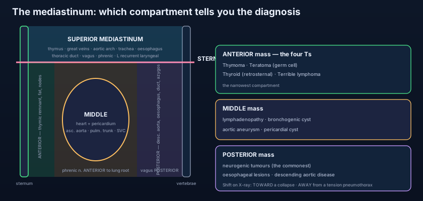
The inferior mediastinum is then subdivided not by a bony plane but by an organ — the fibrous pericardium. Whatever lies in FRONT of the pericardium, between it and the body of the sternum, is the anterior mediastinum. The pericardium itself, together with everything inside it, is the middle mediastinum. Whatever lies BEHIND the pericardium, between it and the thoracic vertebrae, is the posterior mediastinum. So the classical scheme is four compartments: superior, and then anterior, middle and posterior. Radiologists often use a slightly different working version on cross-sectional imaging — a prevascular, a visceral and a paravertebral compartment — but the logic is identical, and the classical names are still the language of the wards and the exam.
The superior mediastinum: everything on its way through
Between the thoracic inlet and the sternal angle lies the busiest short stretch of anatomy in the trunk, and almost nothing in it belongs there — it is all traffic. From front to back the layers are: the thymus (a large organ in the child, a pad of fat and fibrous tissue in the adult); then the great veins — the right and left brachiocephalic veins uniting behind the right first costal cartilage to form the superior vena cava, with the long left brachiocephalic vein crossing the midline in front of the arch; then the arch of the aorta with its three branches in order — the brachiocephalic trunk, the left common carotid artery and the left subclavian artery; then the trachea descending in the midline to the carina; then the oesophagus flat against the vertebral column, with the thoracic duct behind and to its left. Threaded among them run the vagus nerves, the phrenic nerves, the left recurrent laryngeal nerve, the cardiac plexus and the sympathetic trunks, with paratracheal and brachiocephalic lymph nodes packed into every gap.
One relationship in this compartment produces more clinical signs than any other. The left recurrent laryngeal nerve leaves the left vagus at the arch of the aorta, hooks underneath the arch just lateral to the ligamentum arteriosum, and climbs back up in the groove between the trachea and the oesophagus to reach the larynx. Anything that enlarges in that hook — an aortic arch aneurysm, a left hilar tumour, enlarged subaortic lymph nodes, a grossly dilated left atrium — stretches or invades the nerve and paralyses one vocal cord. The patient does not complain of a chest problem; they complain that their voice has gone hoarse. It is one of medicine's purest examples of an anatomical relationship becoming a symptom.
Anterior, middle, posterior: three compartments, three worlds
The anterior mediastinum is the narrowest space in the chest and holds almost nothing. In front of the pericardium and behind the body of the sternum lies a slot that in a healthy adult is barely a potential space. Its contents are modest: the inferior extension of the thymus or its fatty remnant, loose areolar tissue and fat, a few lymph nodes draining the anterior chest wall and the diaphragm, small branches of the internal thoracic vessels, and the sternopericardial ligaments — delicate bands that tether the fibrous pericardium forwards to the manubrium and to the xiphoid process. That emptiness is precisely why an anterior mediastinal mass declares itself so dramatically on a chest X-ray: there is nothing here normally, so anything at all is abnormal, and it has nowhere to expand except backwards onto the heart and the great veins.
The middle mediastinum is the opposite: it is full, and what fills it is the heart. It contains the pericardium — fibrous and serous layers alike, described in the pericardium — and everything the pericardium encloses: the heart itself, the ascending aorta, the pulmonary trunk and its bifurcation into the right and left pulmonary arteries, the lower half of the superior vena cava with the terminal arch of the azygos vein, the short intrathoracic segment of the inferior vena cava, and the four pulmonary veins. Outside the heart but still within this compartment run the phrenic nerves with the pericardiacophrenic arteries and veins, descending on each side of the fibrous pericardium to reach the diaphragm. And at the back of it sits the airway: the bifurcation of the trachea, the right and left main bronchi, and the tracheobronchial lymph nodes that sit in the angle of that bifurcation — nodes whose enlargement is the commonest cause of a middle mediastinal mass.
The posterior mediastinum, behind the pericardium and in front of the lower eight thoracic vertebrae, is not a chamber but a set of conduits heading for the abdomen. The descending thoracic aorta runs down it, giving off posterior intercostal, bronchial, oesophageal and superior phrenic branches. The oesophagus lies in front of the aorta at first and crosses to its front and left lower down, wrapped in the oesophageal plexus formed by the two vagi — the left vagus rotating onto its anterior surface and the right onto its posterior surface as they follow the stomach's embryological turn. Behind and to the right lie the thoracic duct and the azygos vein, with the hemiazygos and accessory hemiazygos veins crossing the midline from the left. Against the heads of the ribs on each side run the sympathetic trunks, and from their lower ganglia the greater (T5–T9), lesser (T10–T11) and least (T12) splanchnic nerves slope forwards and downwards to pierce the crus of the diaphragm and supply the abdominal viscera. Posterior mediastinal lymph nodes fill the spaces between.
Picture a modern office block. The rooms are the lungs — big, airy, occupying nearly all the floor space. Between them the architect leaves a single service riser: a narrow vertical shaft carrying the water mains, the drains, the electrical trunking, the fibre cables and the ventilation, from the roof to the basement. Nobody works in the riser; everything merely passes through. Now hang the boiler in the middle of that shaft, wrap it in its own casing, and you have the heart in its pericardium. Everything that matters is either the boiler or a pipe going past it — which is why a leak in a service riser floods four floors, and why an obstruction there shuts off a whole building. In the thorax the riser is the mediastinum, and the floors it can flood are the neck above and the abdomen below.
Phrenic in front, vagus behind
Two nerves descend through the mediastinum on each side, and they pass the lung root on opposite sides of it. The phrenic nerve (C3, C4, C5 — "C3, 4, 5 keeps the diaphragm alive") descends on the lateral surface of the fibrous pericardium, accompanied by the pericardiacophrenic vessels, and passes ANTERIOR to the root of the lung. The vagus nerve descends alongside the great vessels and passes POSTERIOR to the root of the lung, breaking up into the pulmonary and then the oesophageal plexus. State it as a discriminator and it becomes unforgettable: phrenic in front, vagus behind. It is not a piece of trivia. It tells you why a hilar tumour that grows backwards causes hoarseness or oesophageal symptoms, while one growing forwards paralyses a hemidiaphragm that then rises on the film and moves paradoxically on sniffing. It tells the cardiac surgeon exactly where not to place a clamp on the pericardium. And it explains why irritation of the parietal pleura or pericardium over the diaphragm refers pain to the C4 dermatome at the tip of the shoulder — the full course of both nerves is traced in the nerves of the thorax.
Learn the compartments and you have learned the differential diagnosis, because a mediastinal mass is classified by where it sits before it is classified by what it is. ANTERIOR — remember the four Ts: Thymoma (and thymic hyperplasia, linked with myasthenia gravis), Teratoma and other germ cell tumours, retrosternal Thyroid (a goitre that has slid down behind the manubrium), and "Terrible" lymphoma. MIDDLE — the compartment of nodes and tubes: lymphadenopathy from lymphoma, sarcoidosis, tuberculosis or metastatic lung cancer, plus bronchogenic and pericardial cysts and aortic aneurysm. POSTERIOR — the compartment of nerves and the oesophagus: neurogenic tumours above all (schwannoma, neurofibroma, ganglioneuroma — by far the commonest posterior mass, and the commonest mediastinal mass in children), oesophageal lesions such as achalasia, a large hiatus hernia or duplication cyst, and descending aortic pathology. One radiological question, three completely different shortlists.
The shifted mediastinum: a young man is brought in breathless after a road traffic accident, one side of the chest silent and hyper-resonant, the trachea deviated. Because the mediastinum is soft and mobile, a tension pneumothorax pushes it AWAY from the affected side, kinking the great veins and cutting venous return — the death here is circulatory, not respiratory. A large pleural effusion pushes it away too; a lobar collapse or a pneumonectomy, which lowers the pressure on that side, pulls it TOWARDS the abnormal side. Mediastinitis: a man vomits violently after a heavy meal, tears the lower oesophagus (Boerhaave's syndrome), and within hours has chest pain, fever, surgical emphysema in the neck and a soaring pulse — gastric contents in loose areolar tissue with no barrier to stop them. The same catastrophe follows sternotomy wound infection after cardiac surgery. SVC obstruction: the swollen face, distended non-pulsatile neck veins, dilated chest wall collaterals and headache of a tumour, usually small cell lung cancer or lymphoma, compressing the thinnest-walled great vessel in the anterior compartment. And the widened mediastinum on a trauma film: until proved otherwise, a traumatic aortic injury at the ligamentum arteriosum, where the mobile arch meets the tethered descending aorta.
- The mediastinum is everything between the two pleural sacs: thoracic inlet above, diaphragm below, sternum in front, T1–T12 vertebral bodies behind, mediastinal pleura on each side.
- The plane of the sternal angle (2nd costal cartilage → T4/T5 disc) divides the SUPERIOR from the INFERIOR mediastinum, and also marks the carina, the start and end of the aortic arch and the azygos arch.
- The inferior mediastinum is subdivided by the PERICARDIUM: anterior (in front of it), middle (the pericardium and its contents) and posterior (behind it).
- SUPERIOR contents: thymus, brachiocephalic veins and upper SVC, aortic arch + brachiocephalic trunk / left common carotid / left subclavian, trachea, oesophagus, thoracic duct, vagus and phrenic nerves, left recurrent laryngeal, sympathetic trunks.
- ANTERIOR contents (the narrowest compartment): thymic remnant, fat, lymph nodes, sternopericardial ligaments and small internal thoracic branches — normally almost empty.
- MIDDLE contents: the heart in its pericardium, ascending aorta, pulmonary trunk and its bifurcation, SVC, pulmonary arteries and veins, phrenic nerves with pericardiacophrenic vessels, the tracheal bifurcation and main bronchi.
- POSTERIOR contents: descending thoracic aorta, oesophagus with the oesophageal plexus, thoracic duct, azygos and hemiazygos veins, sympathetic trunks and the greater, lesser and least splanchnic nerves.
- The phrenic nerve (C3, C4, C5) passes ANTERIOR to the root of the lung on the fibrous pericardium; the vagus passes POSTERIOR to it — the single most testable relationship in the mediastinum.
- The left recurrent laryngeal nerve hooks under the arch of the aorta at the ligamentum arteriosum; anything enlarging there causes hoarseness (the right hooks under the right subclavian artery instead).
- Mass by compartment: anterior = the four Ts (Thymoma, Teratoma/germ cell, retrosternal Thyroid, Terrible lymphoma); middle = lymphadenopathy, bronchogenic cyst, aortic aneurysm; posterior = neurogenic tumours, oesophageal lesions, descending aortic disease.
- Mediastinal shift on a chest X-ray: TOWARDS a collapse or pneumonectomy, AWAY from a tension pneumothorax or large pleural effusion.
- Because the mediastinum is loose tissue continuous with the neck and abdomen, oesophageal perforation or sternotomy infection causes rapidly spreading mediastinitis with surgical emphysema.
- Thinking the superior mediastinum is divided into anterior, middle and posterior parts too. It is not — the three-way subdivision applies only to the INFERIOR mediastinum, below the plane of the sternal angle.
- Reversing the mediastinal shift. A tension pneumothorax and a large effusion PUSH the mediastinum away; a collapsed lobe or a pneumonectomy PULLS it towards the abnormal side.
- Putting the phrenic nerve behind the lung root. The phrenic passes ANTERIOR to the root on the fibrous pericardium and the vagus posterior — swap them and every hilar clinical sign comes out wrong.
A 30-year-old woman has a CT showing a soft-tissue mass in the space between the body of the sternum and the front of the fibrous pericardium. She also reports drooping eyelids and double vision that worsen through the day. In which mediastinal compartment does the mass lie, and what is the most likely diagnosis?
- The mediastinum is the central partition between the two pleural sacs — thoracic inlet above, diaphragm below, sternum in front, thoracic vertebrae behind, mediastinal pleura laterally — filled with loose tissue and continuous with the neck and the abdomen.
- The plane of the sternal angle (T4/T5) divides superior from inferior, and the pericardium divides the inferior into anterior (thymic remnant, fat, nodes, sternopericardial ligaments), middle (heart in its pericardium, great vessels, phrenic nerves, tracheal bifurcation) and posterior (descending aorta, oesophagus, thoracic duct, azygos system, sympathetic trunks and splanchnic nerves).
- The superior mediastinum is pure traffic: thymus, brachiocephalic veins and SVC, the aortic arch and its three branches, trachea, oesophagus, thoracic duct, vagus and phrenic nerves, the left recurrent laryngeal hooking under the arch, and the sympathetic trunks. Phrenic passes in FRONT of the lung root, vagus BEHIND it.
- Clinically the compartment is the diagnosis: anterior = the four Ts (thymoma, teratoma, retrosternal thyroid, lymphoma), middle = nodes, bronchogenic cyst and aortic aneurysm, posterior = neurogenic and oesophageal lesions — alongside mediastinal shift (towards a collapse, away from a tension pneumothorax), mediastinitis after oesophageal perforation or sternotomy, and SVC obstruction.
- Drake RL, Vogl AW, Mitchell AWM. Gray's Anatomy for Students — Thorax: the mediastinum and its divisions.
- Moore KL, Dalley AF, Agur AMR. Clinically Oriented Anatomy — Mediastinum: superior, anterior, middle and posterior compartments.
- Netter FH. Atlas of Human Anatomy — Mediastinum: lateral views, right and left.
- Last RJ. Last's Anatomy: Regional and Applied — The mediastinum and the great vessels.
- Snell RS. Clinical Anatomy by Regions — Mediastinal masses, mediastinal shift and mediastinitis.
- TeachMeAnatomy — The Mediastinum; The Superior Mediastinum.

