
The Median and Ulnar Nerves: Grip and Finesse
Two nerves divide the hand between them, and they divide it by personality. One gives you the power to close a fist, to pinch a key between thumb and finger, to hold a pen — the grip. The other gives you the fine spread and squeeze between the fingers, the tiny adjustments a violinist makes without looking, the strength to hold a sheet of paper — the finesse. Between them they explain almost every hand injury you will ever see: the numb hand that wakes you at three in the morning, the wasted thumb pad, the clawed little finger, the tingle that shoots to your fingertips when you catch your elbow on a doorframe. Learn these two nerves properly and the hand stops being a puzzle.
A woman wakes at 3 a.m. shaking her hand over the side of the bed. The thumb, index and middle fingers are buzzing and dead, and she does not know why sleep of all things should do this to her. Two floors down, a cyclist who has ridden four hundred kilometres in a week finds he can no longer spread his fingers properly on the handlebars. In a clinic across town, a boy who fell from a climbing frame and broke his elbow is being asked to make an "OK" sign with his thumb and index finger — and cannot; the two digits collapse into a flat pinch. Three people, three completely different stories, and each of them is really a story about one of two nerves. The hand is not wired at random. It is wired by a plan so consistent that the pattern of numbness on a patient's skin will tell you, before any scan, exactly where along the arm the nerve was hurt.
Two terminal branches, one plan
Both nerves are end-products of the same great network in the neck and axilla. The median nerve (C6–T1, sometimes C5) is unusual in that it is formed by two roots — one from the lateral cord and one from the medial cord of the brachial plexus — which embrace the third part of the axillary artery like the letter M. The ulnar nerve (C8–T1) is the great terminal branch of the medial cord alone. That single fact of origin already predicts their behaviour: the median carries a heavy lateral (C6–C7) contribution and serves the thumb side and the power flexors; the ulnar is almost purely C8–T1 and serves the little-finger side and the small, precise muscles. Their third sibling, the radial nerve, takes the whole back of the limb and is dealt with in the radial, axillary and musculocutaneous nerves. Between the three of them, not one square centimetre of the hand is left unclaimed.
The median nerve: a straight run down the middle
The median nerve is the nerve of the front of the forearm and of the thumb. It runs down the arm alongside the brachial artery — starting lateral to it, then crossing to lie medial to it near the elbow — and, remarkably, it gives off no branches at all in the arm. It enters the cubital fossa medial to the artery and to the biceps tendon, then dives between the two heads of pronator teres and travels distally glued to the deep surface of flexor digitorum superficialis (FDS), right in the middle plane of the forearm. Just above the wrist it emerges from behind the lateral edge of FDS, becomes superficial (and vulnerable to a wrist laceration), and then passes into the hand through the carpal tunnel — the tight osseofibrous corridor beneath the flexor retinaculum described in the axilla, cubital fossa and carpal tunnel. It shares that tunnel with nine tendons and has no room to spare.
What it moves: almost the entire flexor–pronator group, with two famous exceptions. In the forearm the median nerve supplies pronator teres, flexor carpi radialis, palmaris longus and flexor digitorum superficialis — everything in the flexor compartment described in the muscles of the forearm except flexor carpi ulnaris and the medial half of flexor digitorum profundus, which belong to the ulnar. It then gives its purely motor anterior interosseous branch, which runs on the interosseous membrane and supplies flexor pollicis longus, the lateral half of flexor digitorum profundus (index and middle fingers) and pronator quadratus. Because that branch has no skin territory, an anterior interosseous lesion causes no numbness at all — only the inability to flex the tip of the thumb and index finger, which is why the patient's "OK sign" collapses into a pinch. In the hand, its recurrent thenar branch supplies the thenar muscles and its digital branches supply the lateral two lumbricals. Generations have memorised this as LOAF: Lumbricals (lateral two), Opponens pollicis, Abductor pollicis brevis, Flexor pollicis brevis.
The branch that saves the palm
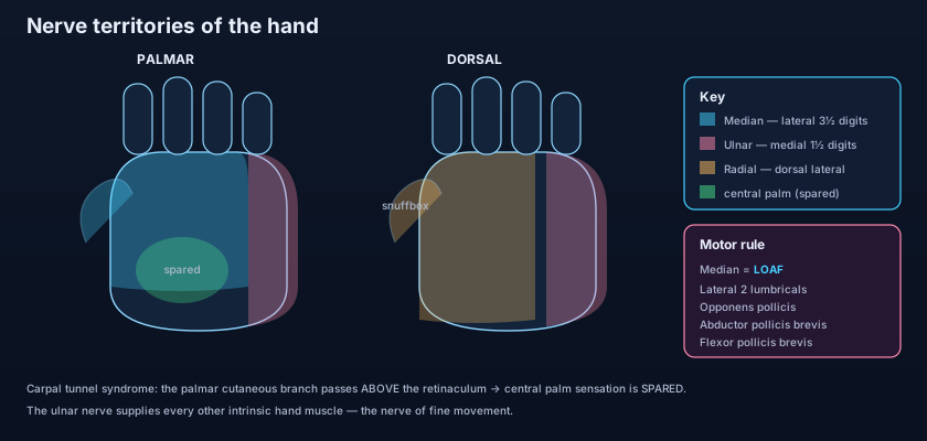
Sensation is where the median nerve hides its single most examinable detail. About five centimetres above the wrist, before the tunnel, the nerve gives off its palmar cutaneous branch, which crosses over the flexor retinaculum — above the roof, not through the tunnel — to supply the skin of the central palm and the thenar eminence. Everything else is carried by the digital branches that emerge inside the hand: the palmar skin of the thumb, index, middle and the lateral half of the ring finger, plus the nail beds and dorsal tips of those same digits. So when the tunnel narrows and squeezes the nerve, the fingers go numb but the central palm keeps its sensation, because that branch took a different road. A patient whose palm is also numb has a lesion higher up the arm, not in the tunnel. It is a small anatomical detour with a very large diagnostic payoff.
Think of the carpal tunnel as a rush-hour underpass with rigid concrete walls and a rigid roof: nine tendons and one nerve must all get through, and the tendons are tough cables while the nerve is a soft bundle of wires. Swell the lining even slightly — pregnancy, hypothyroidism, rheumatoid synovitis, hours of repetitive gripping — and the softest occupant is the one that gets crushed against the wall. The palmar cutaneous branch is the commuter who took the footbridge over the top instead: when the underpass jams, he is the only one who still gets through on time.
When the median nerve fails
Carpal tunnel syndrome is the commonest entrapment neuropathy in the body, and its story is unmistakable: tingling and burning in the thumb, index, middle and half the ring finger that wakes the patient at night and is relieved by shaking the hand out over the edge of the bed. Percussing over the tunnel reproduces the tingling (Tinel's sign); holding the wrists flexed for a minute does the same (Phalen's test). Late, the thenar eminence wastes away into a flat hollow and the thumb loses opposition — the patient can no longer bring the thumb pad to meet the little finger, so keys, buttons and jar lids become a struggle. Higher lesions look different. A supracondylar fracture of the humerus in a child, or a deep laceration at the elbow, knocks out both the long flexors and the thenar muscles: when the patient tries to make a fist, the index and middle fingers stay straight while the ulnar-supplied ring and little fingers curl — the "hand of benediction". With chronic thenar wasting and the thumb pulled back into the plane of the palm, the appearance is called the ape hand, because the thumb can no longer swing forward to oppose.
The ulnar nerve: the exposed one
No other major nerve in the body runs so close to the surface over a bone you bang every week. The ulnar nerve leaves the medial cord and runs down the medial side of the arm, pierces the medial intermuscular septum about halfway down to enter the posterior compartment, and then does the thing that defines it: it passes behind the medial epicondyle of the humerus, lying in a shallow bony groove covered by little more than skin. That is the "funny bone" — bang it and you compress the nerve directly against bone, and the shock fires straight to the little finger. It then enters the forearm between the two heads of flexor carpi ulnaris and runs distally under cover of that muscle, on top of flexor digitorum profundus, with the ulnar artery on its lateral side. At the wrist it does the opposite of the median: it passes superficial to the flexor retinaculum, through a separate small channel called Guyon's canal, between the pisiform and the hook of hamate — two landmarks of the bones of the hand. This is why carpal tunnel syndrome spares it entirely.
Its motor supply is short in the forearm and enormous in the hand. In the forearm it gives only flexor carpi ulnaris and the medial half of flexor digitorum profundus (ring and little fingers). In the hand, however, it supplies essentially every intrinsic muscle except LOAF: the hypothenar group (abductor, flexor and opponens digiti minimi), adductor pollicis, all the palmar and dorsal interossei, the medial two lumbricals, and palmaris brevis. That is why the ulnar is the nerve of finesse — the interossei and lumbricals covered in the intrinsic muscles of the hand are what spread and close the fingers and what straighten the interphalangeal joints while the knuckles bend. Its sensory field is the medial one and a half digits: the palmar branch takes the palmar skin over the hypothenar and the little and half the ring finger, and the dorsal cutaneous branch — which leaves the nerve well above the wrist — takes the matching strip on the back of the hand. That dorsal branch is another quiet diagnostic gift: numbness on the back of the medial hand means the lesion is above the wrist, not in Guyon's canal.
The claw, the paradox, and the sign
Damage the ulnar nerve and the hand deforms in a way that looks strange until you know the mechanics. The lumbricals and interossei normally flex the metacarpophalangeal (MCP) joints while extending the interphalangeal (IP) joints. Lose them in the ring and little fingers and the long extensors pull the MCP joints into hyperextension while the intact long flexors curl the IP joints — the ulnar claw hand. Now the counter-intuitive part. A lesion at the elbow, higher up, also paralyses the medial half of flexor digitorum profundus, so the very muscle that would have curled those fingertips is gone: the claw becomes less obvious. This is the ulnar paradox — the higher lesion looks better, and as the nerve regenerates from elbow to hand the claw deepens as recovery progresses. Finally, adductor pollicis. Ask the patient to grip a sheet of paper between thumb and index finger; with the adductor paralysed they cannot pinch flat, so they cheat by firing flexor pollicis longus (a median muscle), and the thumb's interphalangeal joint visibly buckles into flexion. That is Froment's sign, and it is one of the most elegant bedside tests in anatomy.
Here is the rule that ties the whole hand together: the median nerve is the nerve of grip and of the thumb, the ulnar nerve is the nerve of finesse. Cut the median at the wrist and the hand keeps its strength but loses its precision pinch and the feeling in the fingers it uses to explore the world. Cut the ulnar and the hand keeps most of its sensation and its power flexors but loses the fine choreography between the fingers — grip strength falls by up to a third and the hand becomes clumsy. This is also why a regional block at the wrist can be tailored to the territory: knowing exactly where each nerve lies is what makes local anaesthetics able to silence one half of a hand while leaving the other half fully awake.
The funny bone: you catch your elbow on a table edge and an electric shock runs to your little finger. Nothing is damaged — you simply squashed the ulnar nerve against the medial epicondyle, and the nerve, being a wire, reported the insult as sensation coming from the skin it serves. Handlebar palsy: a long-distance cyclist leans body weight through the heel of the hand onto the handlebars hour after hour, compressing the ulnar nerve in Guyon's canal; the fingers weaken and go clumsy, and because the lesion is below the dorsal cutaneous branch, the back of the hand stays normal. The dead hand on waking: fall asleep with the wrist folded under a pillow, or with an arm draped over the back of a chair, and a nerve is compressed just long enough to stop conducting. It tingles furiously as it recovers — which is exactly the same mechanism as carpal tunnel syndrome, only lasting seconds instead of months.
- Median nerve: C6–T1, formed by lateral + medial cord roots that straddle the axillary artery like an M.
- It gives NO branches in the arm; runs with the brachial artery, through the cubital fossa, between the heads of pronator teres, deep to FDS, then through the carpal tunnel.
- Motor: the flexor–pronator group EXCEPT flexor carpi ulnaris and the medial half of FDP.
- Anterior interosseous branch = pure motor (FPL, lateral half FDP, pronator quadratus) — tested by the "OK sign", causes no numbness.
- In the hand it supplies LOAF only: lateral two Lumbricals, Opponens pollicis, Abductor pollicis brevis, Flexor pollicis brevis.
- Sensory: lateral 3½ digits — but the palmar cutaneous branch crosses ABOVE the retinaculum, so the central palm is SPARED in carpal tunnel syndrome.
- Ulnar nerve: C8–T1, the terminal branch of the medial cord alone.
- Course: medial arm → behind the medial epicondyle (the "funny bone") → between the heads of FCU → SUPERFICIAL to the flexor retinaculum via Guyon's canal.
- Forearm motor: only FCU + the medial half of FDP (ring and little fingers).
- Hand motor: ALL intrinsics except LOAF — hypothenar, adductor pollicis, all interossei, medial two lumbricals, palmaris brevis.
- Sensory: the medial 1½ digits, palmar and dorsal — the dorsal cutaneous branch leaves above the wrist, so it is spared in Guyon's canal lesions.
- Signs: ulnar claw (digits 4–5), the ulnar paradox (higher lesion = less claw), and Froment's sign (thumb IP flexes to compensate for adductor pollicis).
- Expecting the whole median territory to be numb in carpal tunnel syndrome. The central palm is spared, because the palmar cutaneous branch passes over the retinaculum rather than through the tunnel.
- Assuming a higher nerve lesion always looks worse. In the ulnar paradox the reverse is true: an elbow lesion also paralyses FDP, so the fingers claw less than in a wrist lesion.
- Confusing the "hand of benediction" with the ulnar claw. The benediction hand appears on ATTEMPTED FIST with a high median lesion; the ulnar claw is the resting posture of digits 4–5 with an ulnar lesion.
A patient with long-standing carpal tunnel syndrome has numbness of the thumb, index and middle fingers but normal sensation over the central palm. Which anatomical fact explains the spared palm?
- The median nerve (C6–T1, lateral + medial cords) gives no branches in the arm, passes through the cubital fossa, between the heads of pronator teres and deep to FDS, then through the carpal tunnel.
- Median motor = the flexor–pronator group except FCU and the medial half of FDP, plus the pure-motor anterior interosseous branch and LOAF in the hand; sensory = lateral 3½ digits, with the central palm spared in carpal tunnel syndrome.
- The ulnar nerve (C8–T1, medial cord) runs behind the medial epicondyle and superficial to the retinaculum through Guyon's canal, supplying FCU + medial FDP and every intrinsic hand muscle except LOAF, with the medial 1½ digits front and back.
- Signature lesions: carpal tunnel syndrome and the hand of benediction for the median; the ulnar claw, the ulnar paradox and Froment's sign for the ulnar — median = grip and thumb, ulnar = finesse.
- Drake RL, Vogl AW, Mitchell AWM. Gray's Anatomy for Students — Upper limb: nerves of the forearm and hand.
- Moore KL, Dalley AF, Agur AMR. Clinically Oriented Anatomy — Median and ulnar nerve lesions.
- Netter FH. Atlas of Human Anatomy — Nerves of the upper limb: median and ulnar distribution.
- Last RJ. Last's Anatomy: Regional and Applied — The forearm and hand.
- Snell RS. Clinical Anatomy by Regions — Carpal tunnel syndrome, cubital tunnel and Guyon's canal.
- TeachMeAnatomy — The Median Nerve; The Ulnar Nerve.

