
The Brachial Plexus: The Great Rewiring
Five nerves leave the spinal cord at the base of your neck. Five nerves arrive in your arm. In between, in a hand's breadth of space behind your collarbone, those five strands split, merge, cross and recombine into a braid so intricate that generations of students have drawn it and re-drawn it, cursing. But the tangle is not chaos. It is a sorting machine — a place where fibres destined to bend the limb are separated from fibres destined to straighten it, and where every root contributes to more than one nerve so that no single injury can silence a whole arm. Every note a pianist plays, every jar you open, every fingertip that finds a light switch in the dark, passes through this braid first.
A motorcyclist comes off at speed and lands hard on his shoulder, head snapped violently the other way. He gets up, dusts himself down, and cannot lift his arm. There is no fracture on the X-ray and no cut on the skin — the damage is invisible, buried deep behind the clavicle where the nerve roots were stretched like guy-ropes in a gale. In a delivery room across town, a large baby's shoulder catches on the mother's pubic bone; gentle traction frees him, but for weeks afterwards one little arm lies limp at his side while the other waves. And in a waiting room, an anaesthetist slides a needle beside a patient's scalene muscles, injects a few millilitres of clear fluid, and within minutes the whole arm is warm, heavy, and completely without sensation — ready for surgery while the patient stays wide awake. Three completely different scenes, one anatomical structure: the brachial plexus.
Five levels, one mnemonic: Real Texans Drink Cold Beer
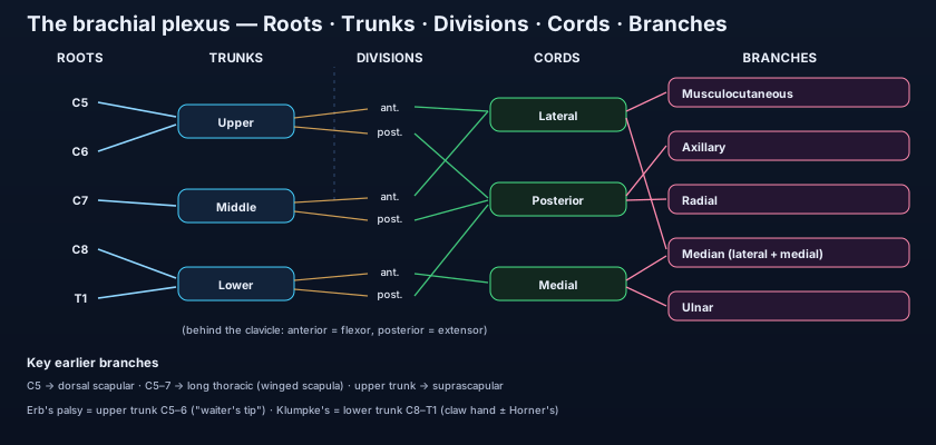
Roots → Trunks → Divisions → Cords → Branches. Learn the ladder first; the detail hangs off it. The brachial plexus is a network formed by the anterior (ventral) rami of spinal nerves C5, C6, C7, C8 and T1 — five roots. Those five combine into three trunks, each trunk splits into two divisions, the six divisions regroup into three cords, and the cords give the terminal branches. The classic mnemonic — Real Texans Drink Cold Beer — fixes the order: Roots, Trunks, Divisions, Cords, Branches. Notice one crucial word: these are the anterior rami. The posterior rami peel off earlier to supply the deep muscles and skin of the back and never enter the limb at all — which is why the plexus, for all its size, carries nothing that belongs to the trunk's own back wall. This is the general design principle of every nerve plexus described in the plan of the nervous system: mix the segments so that function, not segment, defines the final nerve.
The roots: two branches leave before the braid even begins
Two important nerves come straight off the roots, before any mixing. The dorsal scapular nerve (C5) runs backwards to supply the rhomboids and levator scapulae — the muscles that retract and elevate the scapula. The long thoracic nerve (C5, C6, C7) descends on the surface of serratus anterior to supply it; remember it as "C5, 6, 7 keeps the wing from heaven." Serratus anterior is the muscle that holds the medial border of the scapula flat against the chest wall and swings the glenoid upward when you raise your arm overhead. Damage the long thoracic nerve — a blow to the side of the chest, a mastectomy with axillary clearance, or the shoulder straps of a heavy rucksack pressing for hours — and the scapula lifts off the back like a folded wing every time the patient pushes against a wall. This is winged scapula, and it is one of the most visually striking signs in clinical anatomy. The scapula it deforms is described in the pectoral girdle.
The trunks: five become three in the posterior triangle
In the posterior triangle of the neck, above the clavicle, the roots merge into three trunks with beautiful simplicity. C5 and C6 join to form the upper trunk. C7 continues alone as the middle trunk. C8 and T1 join to form the lower trunk. Two branches arise here, both from the upper trunk: the suprascapular nerve (C5, C6), which passes through the suprascapular notch to supply supraspinatus and infraspinatus — two of the rotator cuff muscles that start abduction and externally rotate the shoulder — and the small nerve to subclavius (C5, C6). Everything else that will ever happen to the plexus happens below this level. Because the trunks lie superficially in the neck, covered only by skin, platysma and fascia, they can be felt, blocked with local anaesthetic, and — in the wrong circumstances — stretched or torn.
The divisions: the great functional sorting behind the clavicle
This is the single most elegant idea in the whole plexus — and the level with no branches at all. Behind the clavicle each of the three trunks splits into an anterior and a posterior division: three anterior, three posterior. This is not a bureaucratic subdivision — it is the moment the limb's wiring is sorted by function. In the developing embryo the limb bud has a flexor (anterior) compartment and an extensor (posterior) compartment, and the divisions separate the fibres accordingly: all anterior divisions carry fibres destined for the flexors and the skin over them; all posterior divisions carry fibres destined for the extensors. Everything downstream obeys this rule, which is why the posterior cord gives only the two great extensor nerves. No branch arises at the divisional level — nothing is supplied here, because this is a sorting station, not a destination. Learn this one idea and half the plexus stops being memorisation.
Think of the plexus as a great railway junction. Five lines (the roots) arrive from the spinal cord, each carrying a mixed load of passengers bound for different destinations. At the first station the lines are consolidated into three (the trunks). Then comes the sorting yard behind the clavicle: every train is split in two, and all the "bend the limb" carriages are shunted onto one set of rails while all the "straighten the limb" carriages go onto another. Reassembled by destination rather than by origin, they leave as three new trains (the cords), which finally fan out into the five lines that actually serve the arm. Nobody boards or alights in the sorting yard — that is exactly why the divisions have no branches.
The cords: named for where they sit around an artery
In the axilla the six divisions regroup into three cords, and their names are purely positional — they describe where each cord lies relative to the second part of the axillary artery. The three posterior divisions all unite into the posterior cord (behind the artery). The anterior divisions of the upper and middle trunks unite into the lateral cord (lateral to the artery). The anterior division of the lower trunk continues alone as the medial cord (medial to the artery). Each cord gives pre-terminal branches. The lateral cord gives the lateral pectoral nerve to pectoralis major. The posterior cord gives, in order down the subscapularis, the upper subscapular nerve, the thoracodorsal nerve (to latissimus dorsi), and the lower subscapular nerve (to subscapularis and teres major). The medial cord gives the medial pectoral nerve (to pectoralis minor and major) and the two purely sensory nerves that will supply the skin of the limb: the medial cutaneous nerve of the arm and the medial cutaneous nerve of the forearm. The artery those cords are wrapped around is the subject of the arteries of the upper limb.
The branches: the five nerves that own the arm
Five terminal nerves, and the letter M that draws them. The musculocutaneous nerve (C5–C7) leaves the lateral cord, pierces coracobrachialis, and supplies the three flexors of the front of the arm — biceps brachii, brachialis and coracobrachialis — before continuing as the lateral cutaneous nerve of the forearm. The axillary nerve (C5, C6) leaves the posterior cord, winds round the surgical neck of the humerus through the quadrangular space with the posterior circumflex humeral vessels, and supplies deltoid and teres minor plus the skin of the "regimental badge" area over the deltoid. The radial nerve (C5–T1), the largest branch, continues from the posterior cord into the radial (spiral) groove of the humerus to supply every extensor of the arm and forearm. The median nerve (C6–T1) is formed by two roots — one from the lateral cord and one from the medial cord — that unite in front of the axillary artery in the shape of a letter M; this M, drawn by the median's two roots with the musculocutaneous above and the ulnar below, is the fastest way to recognise the plexus in a cadaver or a diagram. The ulnar nerve (C8, T1, often with a C7 contribution) is the direct continuation of the medial cord. The first three are unpacked in the radial, axillary and musculocutaneous nerves; the last two in the median and ulnar nerves.
The journey: through a gap, over a rib, under a bone
Anatomy is geography, and the plexus travels through three narrow places, each one a potential trap. First it emerges from the neck through the interscalene groove — the gap between the anterior and middle scalene muscles — accompanied by the subclavian artery, while the subclavian vein passes in front of anterior scalene. Second, the trunks cross the upper surface of the first rib, wedged between bone below and the clavicle above. Third, they pass beneath the clavicle and the subclavius muscle into the axilla, where the cords wrap themselves around the axillary artery inside the axillary sheath — a fascial tube continuous with the deep cervical fascia, which is precisely what lets a single injection of local anaesthetic spread and bathe the whole bundle. The corridor from the neck into the armpit is described in the axilla, cubital fossa and carpal tunnel.
When the braid tears: Erb's, Klumpke's, and the outlet
Injuries obey the anatomy: pull the head away from the shoulder and the top of the plexus goes; pull the arm upward and the bottom goes. Erb's palsy is an injury of the upper trunk (C5–C6), caused by excessive separation of the head and shoulder — a fall onto the shoulder point, or shoulder dystocia during a difficult delivery. The paralysed muscles are those of C5–C6: deltoid and supraspinatus (abduction), infraspinatus and teres minor (lateral rotation), biceps and brachialis (elbow flexion), and supinator. The unopposed muscles pull the limb into the classic "waiter's tip" posture — the arm adducted and medially rotated, the elbow extended, the forearm pronated, the palm turned backwards as though discreetly accepting a gratuity. Klumpke's palsy is an injury of the lower trunk (C8–T1), caused by traction on an abducted arm — a fall in which the person grabs a branch or a ledge to save themselves, or a delivery in which the arm is pulled overhead. C8 and T1 supply the small intrinsic muscles of the hand, so their loss gives a claw hand: the metacarpophalangeal joints hyperextended and the interphalangeal joints flexed, because the long flexors and extensors are intact but the lumbricals and interossei that balance them are not. And because T1 also carries the sympathetic fibres ascending to the head, a Klumpke lesion at root level may add Horner's syndrome — ptosis, miosis and anhidrosis on the same side.
The plexus can also be squeezed rather than torn. Thoracic outlet syndrome is compression of the lower trunk (and often the subclavian vessels alongside it) as they cross the first rib — by a cervical rib, a fibrous band, a hypertrophied scalene muscle, or simply by shoulders dragged down for years. Patients describe tingling along the medial forearm and little finger, weakness of grip, and symptoms that worsen when they carry a heavy bag or work with the arms overhead. Fractures of the clavicle — the most commonly broken long bone in the body — threaten the plexus and the subclavian vessels lying immediately beneath, which is why the middle third fracture is examined not just for the bump but for the pulse and the sensation below it. And a penetrating wound of the axilla can divide cords, producing bizarre combinations that make no sense at root level but perfect sense once you know which cord was cut.
💡 The plexus's greatest gift to medicine is that it is a bundle you can switch off from the outside. Because every nerve of the limb funnels through one fascial sleeve, an anaesthetist can render an entire arm insensate with a single injection — no ventilator, no unconsciousness, no opioids. The approach is chosen by which levels must be blocked: interscalene (at the roots/trunks) for shoulder surgery, but it spares the C8–T1 fibres of the hand and almost always paralyses the ipsilateral diaphragm by blocking the phrenic nerve; supraclavicular (at the trunks/divisions, the "spinal anaesthetic of the arm") for anything below the shoulder, with pneumothorax as its price of admission; and axillary (at the terminal branches) for the forearm and hand, the safest of the three but one that misses the musculocutaneous nerve unless it is blocked separately. The drug doing the work is explained in how local anaesthetics block the nerve signal.
The motorcyclist from the opening scene has a traction injury of the upper trunk: he cannot abduct his shoulder, cannot bend his elbow, and the arm hangs in the waiter's-tip position — a textbook Erb's palsy, and the commonest severe plexus injury in adults, because the helmet-and-tarmac mechanism drives the head and shoulder apart exactly as the anatomy fears. The newborn from the delivery room has the same lesion by a different route: shoulder dystocia stretched C5–C6, and with physiotherapy most such infants recover well over months because the nerve is stretched rather than avulsed. The student who walks to campus with a heavy backpack, straps digging into her shoulders, develops aching, tingling and clumsiness in her hands after an hour — "rucksack palsy," a pressure neuropathy of the upper plexus and long thoracic nerve that resolves entirely if she starts using both straps, tightens them high, and carries less. The same braid, three very different insults, each one predictable from where the fibres run.
- The plexus is built from the anterior rami of C5–T1, in five levels: Roots → Trunks → Divisions → Cords → Branches (Real Texans Drink Cold Beer).
- Root branches: dorsal scapular nerve (C5) to rhomboids; long thoracic nerve (C5–C7) to serratus anterior — injury gives a winged scapula.
- Trunks in the posterior triangle: upper (C5–6), middle (C7), lower (C8–T1); the upper trunk gives the suprascapular nerve and the nerve to subclavius.
- Divisions behind the clavicle sort fibres by function: three anterior = flexors, three posterior = extensors. No branches arise here.
- Cords are named for their position around the second part of the axillary artery: lateral, posterior, medial.
- Cord branches: lateral pectoral (lateral); upper subscapular, thoracodorsal, lower subscapular (posterior); medial pectoral + the two medial cutaneous nerves (medial).
- The five terminal nerves: musculocutaneous (lateral cord), axillary + radial (posterior cord), median (lateral + medial cords), ulnar (medial cord).
- The median nerve's two roots draw the letter M in front of the axillary artery — the fastest landmark for identifying the plexus.
- Course: between anterior and middle scalene, over the first rib, under the clavicle, into the axilla inside the axillary sheath.
- Erb's palsy = upper trunk (C5–6): waiter's tip — arm adducted, medially rotated, elbow extended, forearm pronated.
- Klumpke's palsy = lower trunk (C8–T1): claw hand from loss of the intrinsics, ± Horner's syndrome from T1 sympathetic fibres.
- Thoracic outlet syndrome compresses the lower trunk over the first rib (cervical rib, fibrous band, scalene hypertrophy).
- Saying the plexus comes from spinal nerves. It comes from their anterior rami only — the posterior rami supply the back and never enter the limb.
- Expecting branches at the level of the divisions. The divisions are a sorting station only; every named branch arises from a root, a trunk, or a cord.
- Swapping Erb's and Klumpke's. Erb's is the UPPER trunk (C5–6) with the waiter's tip; Klumpke's is the LOWER trunk (C8–T1) with the claw hand and possible Horner's.
A newborn who suffered shoulder dystocia holds one arm adducted, medially rotated, with the elbow extended and the forearm pronated. Which part of the brachial plexus is injured?
- The brachial plexus is formed by the anterior rami of C5–T1 and organised in five levels — Roots, Trunks, Divisions, Cords, Branches.
- Trunks (upper C5–6, middle C7, lower C8–T1) split into three anterior (flexor) and three posterior (extensor) divisions behind the clavicle, then regroup as the lateral, posterior and medial cords around the axillary artery.
- Five terminal nerves emerge: musculocutaneous, axillary, radial, median (the M) and ulnar — plus key pre-terminal branches such as the long thoracic, suprascapular and thoracodorsal nerves.
- Its course between the scalenes, over the first rib and under the clavicle explains Erb's palsy (upper trunk, waiter's tip), Klumpke's palsy (lower trunk, claw hand ± Horner's), thoracic outlet syndrome, and the interscalene/supraclavicular/axillary anaesthetic blocks.
- Drake RL, Vogl AW, Mitchell AWM. Gray's Anatomy for Students — Upper limb: the brachial plexus.
- Moore KL, Dalley AF, Agur AMR. Clinically Oriented Anatomy — Axilla and brachial plexus, with clinical boxes on Erb's and Klumpke's palsies.
- Netter FH. Atlas of Human Anatomy — Plates of the brachial plexus: schema and axillary dissection.
- Last RJ. Last's Anatomy: Regional and Applied — Nerves of the upper limb.
- Snell RS. Clinical Anatomy by Regions — The brachial plexus and its lesions.
- TeachMeAnatomy — The Brachial Plexus: roots, trunks, divisions, cords and branches.

