
Muscles of the Forearm: The Engine Room of the Hand
Look at your hand and ask a simple question: where are the muscles that move it? Squeeze a fist and you will feel the answer — not in your palm, but halfway up your forearm, where a mass of muscle tightens and swells. The great movers of your fingers do not live in the hand at all. They sit in the forearm and reach forward on long, glistening tendons, like a puppeteer working from offstage. That single design decision is why the human hand is slim enough to slip into a coat pocket, thread a needle, and still crush a walnut.
A violinist's left hand hovers over the fingerboard. Four fingers fall and lift independently, each landing within a millimetre, dozens of times a second. Watch her forearm and you will see the truth of it: the muscle bellies rippling under the skin of her forearm are the ones doing the playing. Her fingers themselves are almost hollow — skin, bone, joint capsules and tendon, with barely any muscle. Nature faced a hard engineering problem here. To move twenty-seven bones with precision you need many powerful muscles; but pack those muscles into the hand and you get a swollen paw that can no longer reach into a crevice or wrap around a branch. So the body moved the engines upstream, into the forearm, and ran the controls down as cables. The hand became a puppet of extraordinary delicacy — and the forearm became its engine room.
Two compartments, two nerves: the master plan
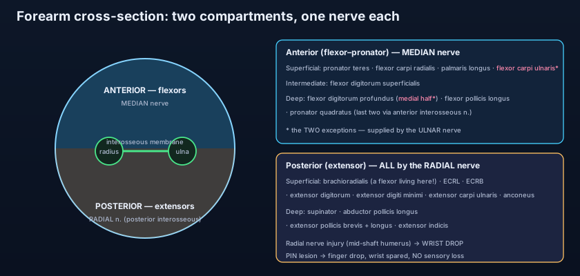
Before memorising twenty muscles, learn the two boxes they live in. The forearm is divided by the radius, the ulna and the fibrous interosseous membrane stretched between them, plus sheets of deep fascia, into two functional compartments. In front lies the anterior (flexor–pronator) compartment: the muscles that bend the wrist and fingers and roll the palm downward into pronation. Behind lies the posterior (extensor) compartment: the muscles that straighten the wrist and fingers and turn the palm upward into supination. The organising beauty is this: each compartment is essentially run by one nerve. The anterior compartment answers to the median nerve (with two famous exceptions), and the posterior compartment answers, without a single exception, to the radial nerve. Learn the bones first in the humerus, radius and ulna and the compartments almost draw themselves — every muscle here begins on a bony landmark you already know.
The anterior compartment, superficial layer: the common flexor origin
Put your thumb on the bony bump on the inner side of your elbow — the medial epicondyle of the humerus. Four muscles fan out from this single point, the common flexor origin. Running from lateral to medial: pronator teres, which crosses obliquely to the lateral surface of the radius and pronates the forearm as well as weakly flexing the elbow (median nerve). Flexor carpi radialis, whose tendon you can feel as the prominent cord at the front of your wrist on the thumb side; it flexes and abducts (radially deviates) the wrist (median nerve). Palmaris longus, a slender, largely vestigial muscle that tenses the palmar aponeurosis and is simply absent in roughly 15% of people — press your thumb and little finger together and flex the wrist to see whether you own one (median nerve). And flexor carpi ulnaris, the most medial, which flexes and adducts (ulnarly deviates) the wrist — and here is the first exception to the median rule, because it is supplied by the ulnar nerve.
Intermediate and deep: the muscles that actually grip
One layer bends the middle joints; the layer beneath it bends the tips. The intermediate layer holds a single large muscle: flexor digitorum superficialis (FDS), arising from the medial epicondyle, the ulnar coronoid process and the radius, and inserting by four tendons into the middle phalanges of the fingers. It therefore flexes the proximal interphalangeal (PIP) joints — the middle knuckles — and is supplied by the median nerve. The deep layer holds three. Flexor digitorum profundus (FDP) runs from the ulna and interosseous membrane to the distal phalanges, flexing the fingertips (the distal interphalangeal joints), and it carries the most instructive nerve supply in the whole limb: a split supply, with the medial half (to the ring and little fingers) served by the ulnar nerve — exception two — and the lateral half (index and middle) by the anterior interosseous branch of the median nerve. Flexor pollicis longus runs from the radius to the distal phalanx of the thumb and bends the thumb tip; pronator quadratus, a flat square sheet deep at the wrist, is the prime pronator and holds the radius and ulna together. Both are anterior interosseous, and therefore median. When these tendons finally reach the palm they hand over to the small muscles described in the intrinsic muscles of the hand, which supply the fine positioning the long tendons cannot.
Think of the forearm as the engine room of a ship and the hand as the rudder and propellers up on deck. You would never bolt a diesel engine onto the rudder — it would be far too heavy to steer with. Instead you put the engines low and amidships, where weight does no harm, and you run steel control cables the length of the vessel to the moving parts. That is exactly the forearm: heavy muscle bellies kept proximal, thin tendon cables running distally through guides. And like any cable-driven machine, it needs its cables held down at the corners so they do not bowstring away from the turn — which is precisely what the retinacula at the wrist are for.
The posterior compartment: one nerve, no exceptions
Every extensor muscle in the forearm is radial. Every single one. The posterior compartment is supplied entirely by the radial nerve, either directly or through its deep branch, which becomes the posterior interosseous nerve after it pierces supinator. The superficial group mostly arises from the lateral epicondyle by the common extensor origin. Brachioradialis is the odd one out and a classic examination quirk: it lies in the extensor compartment and is radially innervated, yet its action is to flex the elbow — it is a flexor living in the extensor's house. Extensor carpi radialis longus and extensor carpi radialis brevis extend and abduct the wrist; extensor digitorum sends four tendons to the extensor expansions of the fingers and is the main finger straightener; extensor digiti minimi gives the little finger its own independent lift; extensor carpi ulnaris extends and adducts the wrist; and anconeus, a small triangle behind the elbow, assists extension and stabilises the joint. The deep group is dominated by thumb work: supinator wraps the upper radius and supinates the forearm; abductor pollicis longus, extensor pollicis brevis and extensor pollicis longus pull the thumb out and back — the long extensor hooking around the dorsal tubercle of the radius, which is why it can rupture after a distal radius fracture; and extensor indicis gives the index finger a second, independent extensor for pointing.
The retinacula: harnessing the cables
At the wrist, all those long tendons must round a corner without springing away from the bone. Two thickened straps of deep fascia hold them down. On the palmar side, the flexor retinaculum bridges the carpal arch — from the scaphoid tubercle and trapezium laterally to the pisiform and hook of hamate medially — converting the concave carpus into the carpal tunnel. Through this unforgiving tube pass nine tendons (four FDS, four FDP, flexor pollicis longus) and the median nerve, which is the single most compressible structure in the upper limb; its confinement, and the anatomy of the spaces around it, are explored in the axilla, cubital fossa and carpal tunnel. On the dorsal side, the extensor retinaculum sends septa down to the radius and ulna, creating six synovial-lined compartments through which the extensor tendons run in strict order — the first containing abductor pollicis longus and extensor pollicis brevis, the sixth containing extensor carpi ulnaris. Naming the compartments is not trivia: surgeons and sonographers navigate the back of the wrist entirely by their numbers.
💡 Here is the principle that turns twenty muscles into three sentences: one nerve runs each compartment. Posterior = radial, no exceptions at all. Anterior = median, with exactly two ulnar exceptions — flexor carpi ulnaris, and the medial half of flexor digitorum profundus. That is why the ulnar nerve, when it is crushed at the elbow, weakens your grip on the ring and little fingers specifically, and why the classic "ulnar claw" is worse in a low lesion at the wrist than a high one at the elbow: cut the nerve high and you also paralyse the very FDP bellies that would otherwise curl those fingers into the claw. Trace both nerves through the limb in the median and ulnar nerves and every muscle in this article acquires an address.
Making a fist — and the trick your hand does for free
Watch what happens when you make a strong fist. The finger flexors curl the digits, but notice that your wrist does not collapse into flexion — it actually extends slightly. That is the wrist extensors firing as synergists, holding the wrist back so the long flexors are not slack. Length matters enormously to a muscle: pull your wrist fully into flexion and try to grip hard, and your grip strength collapses, because the flexor tendons have gone slack over the front of a bent wrist. This is the same physics of overlap and tension described in how muscles pull on bone to move you. The reverse of the trick has a name — the tenodesis effect. Let your wrist drop into extension and the fingers curl closed on their own; flex the wrist and the fingers open. No nerve signal to the fingers is needed; the tendons are simply too short to allow anything else. Patients with a C6 spinal cord injury who have lost finger muscles but kept wrist extension are taught to use exactly this to pick up a cup.
A tennis player with pain over the outer elbow that flares when she lifts a kettle with a straight arm has lateral epicondylitis — tennis elbow — a degenerative tendinopathy of the common extensor origin, classically at extensor carpi radialis brevis. A golfer with mirror-image pain over the inner elbow has medial epicondylitis at the common flexor origin: same story, opposite epicondyle. A new mother with sharp pain at the base of the thumb when she lifts her baby under the arms has de Quervain's tenosynovitis — inflammation of abductor pollicis longus and extensor pollicis brevis in the first extensor compartment, confirmed by Finkelstein's test (tuck the thumb into the fist and deviate the wrist ulnarly; the pain leaps). And a man who fell asleep with his arm draped over the back of a chair, compressing the radial nerve in the spiral groove of the humerus, wakes with wrist drop — the hand hanging limp because the entire posterior compartment has gone quiet at once. That, in a single sign, is the whole logic of one-nerve-per-compartment; the nerve's course is followed in the radial, axillary and musculocutaneous nerves.
- Two compartments: anterior (flexor–pronator) and posterior (extensor), separated by radius, ulna and the interosseous membrane.
- Anterior superficial (common flexor origin, medial epicondyle): pronator teres, flexor carpi radialis, palmaris longus (absent in ~15%), flexor carpi ulnaris.
- Anterior intermediate: flexor digitorum superficialis → middle phalanges → PIP flexion.
- Anterior deep: flexor digitorum profundus (DIP flexion), flexor pollicis longus, pronator quadratus.
- Anterior nerve rule: median — except flexor carpi ulnaris and the medial half of FDP (ulnar).
- Anterior interosseous nerve (median branch) = FPL + pronator quadratus + lateral half of FDP.
- Posterior superficial (common extensor origin, lateral epicondyle): brachioradialis, ECRL, ECRB, extensor digitorum, extensor digiti minimi, extensor carpi ulnaris, anconeus.
- Posterior deep: supinator, abductor pollicis longus, extensor pollicis brevis, extensor pollicis longus, extensor indicis.
- Posterior nerve rule: ALL radial — directly, or via the deep branch / posterior interosseous nerve.
- Brachioradialis is the quirk: an elbow FLEXOR sitting in the extensor compartment with radial supply.
- Flexor retinaculum → carpal tunnel (9 tendons + median nerve); extensor retinaculum → six dorsal compartments.
- Clinical anchors: tennis vs golfer's elbow (two origins), de Quervain's (APL + EPB, compartment 1), radial palsy → wrist drop.
- Assuming the whole anterior compartment is median. Two muscles break the rule: flexor carpi ulnaris entirely, and the medial half of flexor digitorum profundus — both ulnar.
- Swapping the two flexor digitorum muscles. SUPERFICIALIS inserts on the MIDDLE phalanx (PIP flexion); PROFUNDUS travels deeper and further to the DISTAL phalanx (DIP flexion) — it is the only muscle that can bend a fingertip.
- Filing brachioradialis with the extensors because of where it lives. It is radially innervated and sits in the extensor compartment, but it FLEXES the elbow.
A patient cannot flex the distal interphalangeal joints of the ring and little fingers, but flexion of the index and middle fingertips is normal. Which nerve is injured?
- The muscles that move the fingers sit in the forearm and act through long tendons — that is why the hand is slim, light and precise.
- Anterior (flexor–pronator) compartment: superficial (pronator teres, FCR, palmaris longus, FCU), intermediate (FDS), deep (FDP, FPL, pronator quadratus) — median nerve, except FCU and the medial half of FDP (ulnar).
- Posterior (extensor) compartment: superficial (brachioradialis, ECRL/ECRB, extensor digitorum, EDM, ECU, anconeus) and deep (supinator, APL, EPB, EPL, extensor indicis) — every one supplied by the radial nerve / posterior interosseous nerve.
- The retinacula harness the tendons (carpal tunnel in front, six dorsal compartments behind), and the compartment rule explains tennis and golfer's elbow, de Quervain's, wrist drop and the tenodesis effect.
- Drake RL, Vogl AW, Mitchell AWM. Gray's Anatomy for Students — Upper limb: the forearm.
- Moore KL, Dalley AF, Agur AMR. Clinically Oriented Anatomy — Forearm: anterior and posterior compartments.
- Netter FH. Atlas of Human Anatomy — Muscles of the forearm, superficial and deep layers.
- Last RJ. Last's Anatomy: Regional and Applied — The forearm and its fascial compartments.
- Snell RS. Clinical Anatomy by Regions — The forearm: flexor and extensor groups and their nerve supply.
- TeachMeAnatomy — Muscles of the Anterior and Posterior Forearm.

