
The Doorways of the Skull: Which Nerve Uses Which Hole
Look down at the floor of an emptied skull and you are looking at the busiest few square centimetres in the human body. Every nerve that connects the brain to the face, the eye, the tongue, the throat and the gut — and every artery that feeds the brain and every vein that drains it — has to pass through this floor, and the floor is solid bone. So the bone is drilled through with holes, each one a named doorway with a fixed list of what may use it. A surgeon who finds a patient with a numb cheek, a drooping eyelid and a squint is not guessing at random: those three deficits share one hole, and naming the hole names the lesion. Learn the doorways of the skull and you can read a scatter of cranial nerve palsies backwards to the single square millimetre where something has gone wrong.
A woman of fifty is referred with double vision. On examination her right eye will not turn out; when she is asked to look up, down or in, that eye lags in every direction; the upper lid droops; the pupil is fixed and a little wide. The skin over her right forehead and cheek is numb to a wisp of cotton, but her jaw is strong and her lower lip is normal. A neurologist could recite the involved nerves without a scan: the third, the fourth and the sixth — every nerve of eye movement — together with the first two divisions of the fifth, but not the third division. Then comes the question that turns a list of nerves into a diagnosis: where do those exact nerves, and only those nerves, run together? Third, fourth, ophthalmic, maxillary and sixth all pass through the wall of one structure on their way to the superior orbital fissure — the cavernous sinus. The mandibular division is spared because it never enters that wall; it leaves the skull lower down, through a different hole. Five deficits, one place. The lesion is in the cavernous sinus, and the anatomy of the foramina told her so before any machine did.
The floor of the skull is a map in three tiers
Do not memorise the holes as a list. Group them by which fossa they open into, and the list learns itself. The interior of the cranial base descends like a shallow staircase from front to back, in three steps — the ANTERIOR, MIDDLE and POSTERIOR cranial fossae — described as bony architecture in the vault and base of the skull. Almost every foramen belongs to one of these three floors, and the twelve cranial nerves are handed out fossa by fossa in the order they leave the brain. The ANTERIOR fossa, floored by the ethmoid and the orbital plates of the frontal bone, transmits only one nerve — the olfactory, CN I. The MIDDLE fossa, built around the body and greater wings of the sphenoid, is the crowded one: it transmits CN II through VI on their way to the orbit and face, together with the arteries that feed the dura. The POSTERIOR fossa, the deepest step, floored by the occipital bone and the petrous temporal, transmits CN VII through XII and, through its great central opening, the junction of brain and spinal cord with the vertebral arteries. Fix those three tiers in mind and every hole has an address before you have named it.
Anterior fossa: one plate, one nerve
The whole anterior fossa transmits a single cranial nerve, and it does so not through one hole but through a sieve. The CRIBRIFORM PLATE of the ethmoid — cribriform means "sieve-like" — is a thin shelf perforated by twenty or so tiny openings, and through each one passes a bundle of the fine, unmyelinated fibres of the OLFACTORY NERVE, CN I, running up from the olfactory epithelium in the roof of the nose to the olfactory bulb sitting on top of the plate. This arrangement has one blunt clinical consequence. A blow to the front of the head that fractures the cribriform plate shears these delicate filaments as they cross it, and the patient loses the sense of smell — anosmia — often permanently. The same fracture tears the meninges that are stuck to the plate, and cerebrospinal fluid can then trickle down into the nose: CSF rhinorrhoea, a clear drip that is worse on leaning forward and that opens a direct route for bacteria to reach the meninges. One thin sieve of bone, and its fracture explains both a lost sense and a leaking brain.
Middle fossa: the crowded floor
Front to back across the sphenoid, seven doorways in a row — and each carries a fixed cargo. The OPTIC CANAL, in the lesser wing of the sphenoid, carries the OPTIC NERVE, CN II, together with the OPHTHALMIC ARTERY — the first branch of the internal carotid and the sole arterial supply of the retina, which is why the eye is an end-organ that goes suddenly and painlessly blind when that artery occludes. Just below and lateral is the SUPERIOR ORBITAL FISSURE, the busiest doorway of all, transmitting the OCULOMOTOR (III), the TROCHLEAR (IV), the OPHTHALMIC division of the trigeminal (V1) with its LACRIMAL, FRONTAL and NASOCILIARY branches, the ABDUCENS (VI), and the SUPERIOR OPHTHALMIC VEIN draining back into the cavernous sinus — the reason its contents suffer together in the syndrome that opened this article, and the reason infections of the face can track back into the skull. All of this feeds the orbit set out in the orbit and its contents. Then come the pure trigeminal holes: the FORAMEN ROTUNDUM transmits the MAXILLARY division (V2) forward towards the cheek, and the FORAMEN OVALE transmits the MANDIBULAR division (V3) — the mnemonic "OVALe" packs its contents neatly: the Otic ganglion sits just below it, V3 passes through it, and with V3 go the Accessory meningeal artery and the Lesser petrosal nerve. Behind ovale sits the small FORAMEN SPINOSUM, and through it passes a vessel out of all proportion to its size: the MIDDLE MENINGEAL ARTERY (with the meningeal branch of V3), the artery whose rupture floods the extradural space. Finally the FORAMEN LACERUM — a ragged hole that in life is plugged with cartilage and transmits nothing straight through it; the INTERNAL CAROTID ARTERY does not pass through lacerum but runs ACROSS its upper part, having entered the skull through the carotid canal. The three trigeminal divisions and their exits are drawn together in the trigeminal nerve: V1 rotundum's neighbour at the fissure, V2 rotundum, V3 ovale.
Think of the cavernous sinus as a narrow service corridor running along each side of the pituitary gland, with the internal carotid artery threaded down its middle and a bundle of nerves pinned to its outer wall on their way to the superior orbital fissure. Pinned to that wall, from top to bottom, are the oculomotor, the trochlear, the ophthalmic and the maxillary divisions of the trigeminal — while the abducens hangs free in the lumen right beside the artery. Anything that swells inside that corridor — a clot in the sinus, an aneurysm of the carotid, a tumour pressing from the side — squeezes the whole bundle at once. That is why cavernous sinus disease produces its unmistakable signature: a frozen eye that cannot move in any direction, a drooping lid, a numb forehead and cheek, all on one side. The mandibular division escapes only because it never joins this corridor; it has already dropped out of the skull through foramen ovale, below and behind, before the others reach the wall.
Posterior fossa: the great outflow
The deepest tier hands out the last six nerves. High on the posterior surface of the petrous temporal bone opens the INTERNAL ACOUSTIC MEATUS, transmitting the FACIAL NERVE (VII), the VESTIBULOCOCHLEAR NERVE (VIII) and the small LABYRINTHINE ARTERY that feeds the inner ear; the further course of the seventh from here is followed in the facial nerve. Below and in front lies the large JUGULAR FORAMEN, a composite opening transmitting three cranial nerves — the GLOSSOPHARYNGEAL (IX), the VAGUS (X) and the ACCESSORY (XI) — together with the venous confluence in which the INFERIOR PETROSAL SINUS and the SIGMOID SINUS meet and become the INTERNAL JUGULAR VEIN, the great vein whose whole course is traced in the carotid and the jugular. Just above the front edge of the foramen magnum, the HYPOGLOSSAL CANAL pierces the occipital bone and transmits the HYPOGLOSSAL NERVE (XII) to the muscles of the tongue. And at the centre of it all is the FORAMEN MAGNUM — the largest opening in the skull — through which the MEDULLA OBLONGATA becomes the spinal cord, flanked by the two VERTEBRAL ARTERIES ascending to form the basilar artery, the ANTERIOR and POSTERIOR SPINAL ARTERIES, and the SPINAL ROOTS of the ACCESSORY NERVE (XI), which ascend through the foramen magnum from the upper cervical cord to join the cranial part and then leave again, moments later, through the jugular foramen. It is the one nerve that enters the skull only to turn round and exit by another hole.
Three nerves through one hole means three signs in one patient. The clean beauty of the jugular foramen is that a single lesion there — a tumour, a clot, a base-of-skull fracture — takes out CN IX, X and XI as a set, and the deficit is instantly recognisable at the bedside: loss of the gag reflex and taste on the back of the tongue (IX), a hoarse voice with a palate that pulls to the good side and swallowing that fails (X), and weakness turning the head and shrugging the shoulder (XI, to sternocleidomastoid and trapezius). That triad is Vernet's jugular foramen syndrome, and its power is diagnostic economy: the moment you see IX, X and XI together, you do not think of three separate nerve injuries — you think of one hole. The same logic runs through the whole cranial base. A cluster of palsies is not a coincidence to be explained nerve by nerve; it is a signpost, and the arrow points at the single foramen through which those exact nerves all pass.
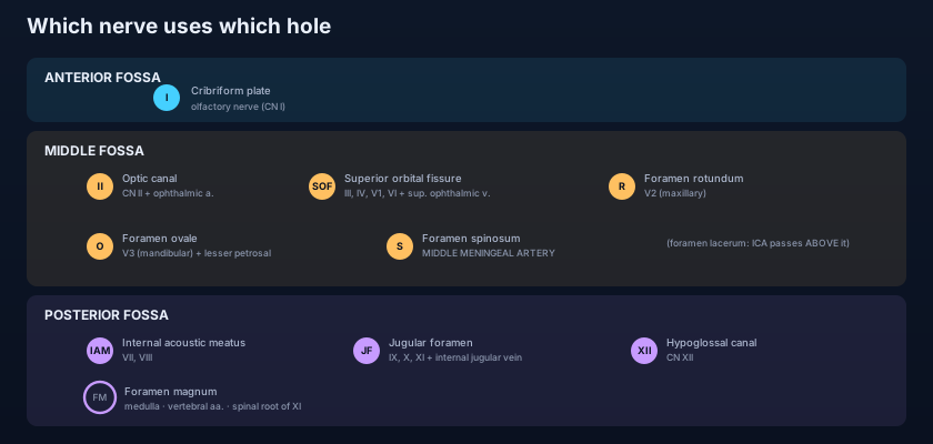
- ANTERIOR fossa: cribriform plate of the ethmoid → OLFACTORY nerve (CN I) only. A fracture here causes anosmia and CSF rhinorrhoea.
- OPTIC CANAL → optic nerve (CN II) + ophthalmic artery. SUPERIOR ORBITAL FISSURE → CN III, IV, V1 (lacrimal, frontal, nasociliary), VI + superior ophthalmic vein.
- FORAMEN ROTUNDUM → maxillary V2. FORAMEN OVALE → mandibular V3 (+ accessory meningeal artery, lesser petrosal nerve; otic ganglion just below). FORAMEN SPINOSUM → MIDDLE MENINGEAL ARTERY (+ meningeal branch of V3).
- FORAMEN LACERUM is cartilage-filled and transmits nothing straight through; the internal carotid artery runs ACROSS its upper part after entering through the carotid canal.
- INTERNAL ACOUSTIC MEATUS → CN VII, VIII + labyrinthine artery. JUGULAR FORAMEN → CN IX, X, XI + the inferior petrosal and sigmoid sinuses forming the internal jugular vein.
- HYPOGLOSSAL CANAL → CN XII. FORAMEN MAGNUM → medulla oblongata, the two vertebral arteries, the anterior and posterior spinal arteries, and the spinal roots of the accessory nerve (XI) ascending to join the cranial part.
The smaller doorways
Beyond the great foramina, a scatter of smaller passages, each with one thing to remember. The STYLOMASTOID FORAMEN, tucked between the styloid and mastoid processes on the underside of the skull, is where the FACIAL NERVE (VII) finally leaves the skull to enter the parotid gland and fan out over the face — the site whose swelling explains the facial palsy of Bell's. The CAROTID CANAL carries the INTERNAL CAROTID ARTERY (with its sympathetic plexus) up into the middle fossa, passing above foramen lacerum. Small EMISSARY veins connect the intracranial venous sinuses to the veins outside the skull through the CONDYLAR CANAL (behind the occipital condyle) and the MASTOID CANAL (through the mastoid) — clinically important because they let infection pass either way across the bone. In the hard palate, the GREATER and LESSER PALATINE FORAMINA transmit the corresponding palatine nerves and vessels to the roof of the mouth, and the INCISIVE CANAL, in the midline just behind the incisor teeth, carries the nasopalatine nerve and the terminal sphenopalatine vessels. And running forward through the sphenoid to the back of the nose is the PTERYGOID CANAL, carrying the nerve of the pterygoid canal (the combined greater and deep petrosal nerves) to the pterygopalatine ganglion. None of these carries a numbered cranial nerve trunk, but each is a named answer to a specific question — which is exactly how examiners use them.
Reading the palsies backward to the hole
The whole point of the foramina is localisation, and each of the great syndromes is a foramen speaking through its contents. CAVERNOUS SINUS SYNDROME, as in the opening scene, strikes the nerves pinned to that sinus wall on their way to the superior orbital fissure — III, IV, V1, V2 and VI — sparing V3, which has already left through foramen ovale. VERNET'S (JUGULAR FORAMEN) SYNDROME takes out IX, X and XI together where they share that hole. An ACOUSTIC NEUROMA — properly a vestibular schwannoma — grows on the eighth nerve and slowly WIDENS the internal acoustic meatus, first deafening and unbalancing the patient (VIII), then, as it enlarges, catching the seventh nerve beside it and later the fifth at the adjacent cerebellopontine angle; the audiovestibular story belongs with the ear. A tear of the MIDDLE MENINGEAL ARTERY as it enters through foramen spinosum — usually with a fracture across the thin pterion — bleeds into the extradural space and produces the classic lucid interval followed by rapid deterioration. And BASE-OF-SKULL METASTASES, seeding the bony floor, pick off cranial nerves one or two at a time in patterns named for the region they invade — the numb chin of the mental nerve, the orbital apex, the jugular foramen — so that the neurologist works backwards from which nerves have failed to which part of the floor the tumour has reached. In every case the reasoning is the same: name the deficits, find the hole they share, and you have found the lesion.
A young man is struck on the side of the head with a cricket ball, over the temple. He is briefly dazed, then gets up, feels well and refuses hospital. An hour later he complains of a worsening headache, becomes drowsy, and within the next hour his right pupil blows to a fixed dilated globe and his left arm weakens. This is the textbook course of an EXTRADURAL (epidural) HAEMATOMA. The temple overlies the PTERION, the thin H-shaped junction of four bones, and immediately deep to it runs the MIDDLE MENINGEAL ARTERY, which has entered the skull through the FORAMEN SPINOSUM. A fracture across the pterion tears the artery; arterial blood strips the tough dura off the bone under pressure, and a CT scan shows the classic biconvex (lens-shaped) collection that does not cross the suture lines to which the dura is anchored. The "lucid interval" is the treacherous hour between the concussion wearing off and the expanding clot raising intracranial pressure enough to cone the brain — the dilating pupil is the uncal third-nerve palsy of impending herniation. Contrast this with the crescentic, slower SUBDURAL haematoma of torn bridging veins, which follows dural venous anatomy rather than an arterial foramen — the venous side is laid out in the meninges and dural venous sinuses. One small hole, foramen spinosum, admits the artery whose rupture is a neurosurgical emergency.
- Group the foramina by fossa: ANTERIOR transmits only CN I; MIDDLE transmits CN II–VI plus the meningeal vessels; POSTERIOR transmits CN VII–XII plus the vertebral arteries. That grouping is the whole memory system.
- "OVALe" for foramen ovale: Otic ganglion (just below), V3, Accessory meningeal artery, Lesser petrosal nerve. Foramen ROTUNDUM = V2; both trigeminal exits sit in the middle fossa.
- CAVERNOUS SINUS SYNDROME = III, IV, V1, V2, VI (the superior-orbital-fissure-bound nerves pinned to the sinus wall); V3 is SPARED because it leaves through foramen ovale, not the fissure.
- VERNET'S (JUGULAR FORAMEN) SYNDROME = IX + X + XI together: lost gag and posterior-tongue taste, hoarseness with palatal deviation and dysphagia, weak head-turn and shoulder-shrug.
- ACOUSTIC NEUROMA widens the INTERNAL ACOUSTIC MEATUS (VIII first, then VII, then V at the cerebellopontine angle); a MIDDLE MENINGEAL tear through FORAMEN SPINOSUM at the pterion gives an extradural haematoma with a lucid interval.
- The FORAMEN MAGNUM is the only opening transmitting the medulla, the paired vertebral arteries, the anterior/posterior spinal arteries AND the spinal roots of XI (which then re-exit through the jugular foramen).
- Saying the internal carotid artery passes THROUGH the foramen lacerum. It does not: foramen lacerum is plugged with cartilage in life and transmits nothing straight through. The internal carotid enters via the carotid canal and runs ACROSS the upper part of lacerum — a distinction examiners love.
- Putting the middle meningeal artery through foramen ovale. Ovale transmits V3 (with the accessory meningeal artery and lesser petrosal nerve); it is the smaller foramen SPINOSUM, just behind it, that transmits the MIDDLE MENINGEAL ARTERY — the vessel torn in an extradural haematoma.
- Forgetting that V3 is spared in cavernous sinus syndrome. Only V1 and V2 run in the cavernous sinus wall towards the superior orbital fissure and rotundum; V3 has already dropped out of the skull through foramen ovale, so a cavernous sinus lesion leaves the jaw and lower lip normal.
A patient develops paralysis of all eye movements on the left with a drooping lid, a numb left forehead and cheek, but a normal left jaw with a strong bite and normal sensation over the chin. Where is the lesion, and which foramen has its contents spared?
- Every cranial nerve and every vessel crossing the skull base uses a named foramen, so localising a lesion is often simply asking "which hole do these deficits share?". Group the foramina by fossa and the twelve nerves fall out in order.
- ANTERIOR fossa: cribriform plate → CN I only. MIDDLE fossa: optic canal (II + ophthalmic artery), superior orbital fissure (III, IV, V1, VI + superior ophthalmic vein), rotundum (V2), ovale (V3 + accessory meningeal a. + lesser petrosal n.), spinosum (middle meningeal artery); foramen lacerum is cartilage-filled and the carotid runs above it.
- POSTERIOR fossa: internal acoustic meatus (VII, VIII + labyrinthine a.), jugular foramen (IX, X, XI + the sinuses forming the internal jugular vein), hypoglossal canal (XII), and the foramen magnum (medulla, vertebral arteries, spinal + posterior spinal arteries, spinal roots of XI). Smaller passages: stylomastoid (VII exiting), carotid canal, condylar/mastoid emissary veins, palatine and incisive canals, pterygoid canal.
- The syndromes are foramina speaking: cavernous sinus (III, IV, V1, V2, VI — V3 spared), Vernet's jugular foramen (IX, X, XI), acoustic neuroma widening the internal acoustic meatus (VIII→VII→V), a middle meningeal tear through spinosum giving an extradural haematoma with a lucid interval, and base-of-skull metastases picking off nerves region by region.
- Drake RL, Vogl AW, Mitchell AWM. Gray's Anatomy for Students — The Head and Neck: the cranial fossae and the foramina of the skull base with their contents.
- Moore KL, Dalley AF, Agur AMR. Clinically Oriented Anatomy — The skull: interior of the cranial base; foramina and the structures transmitted.
- Netter FH. Atlas of Human Anatomy — Base of the skull: interior view; foramina and cranial nerve exits.
- Last RJ. Last's Anatomy: Regional and Applied — The skull and the cranial fossae; the foramina and their transmitted nerves and vessels.
- Snell RS. Clinical Anatomy by Regions — The skull: the interior of the base and the clinical significance of the foramina.
- TeachMeAnatomy — The Base of the Skull; Foramina of the Cranial Fossae.

