
The Ankle and the Arches: The Engineering of a Step
You take somewhere near eight thousand steps a day, and every single one of them asks the same impossible thing of your foot: be soft, then be hard. Soft on landing, so the foot can spread and mould itself to a kerb, a stone, a stair edge without breaking. Hard at push-off, a fraction of a second later, so that the calf can drive a rigid lever into the ground and throw your whole body weight forward. No engineer has ever built a structure that changes its stiffness twenty times a minute, for eighty years, without a single moving part being replaced. Your foot does it with twenty-six bones, three arches, a sheet of fibrous tissue on the sole, and one beautifully simple trick involving your toes.
A basketball player comes down from a rebound and lands, for a quarter of a second, on the edge of someone else's shoe. His foot is pointed downwards in mid-air, and it rolls inwards. There is a sound like tearing cloth, and he sits down on the court holding the outside of his ankle. Two thousand miles away, a woman gets out of bed and puts her heel to the floor for the first time that morning, and a hot line of pain runs from her heel into the arch — by her third or fourth step it has almost gone, and by evening it is back. A third person, sixty years old, notices that the inner border of her right foot has quietly flattened over two years, and that she can no longer rise onto the toes of that side. Three completely different complaints, and all three are the same story: the story of an asymmetric joint sitting on top of a suspension bridge.
The mortise: a carpenter's joint made of bone
The ankle proper is one of the purest hinges in the body — and it is built like joinery. The talocrural (ankle) joint is a synovial hinge between the trochlea of the talus below and a deep bracket above. That bracket — the mortise — has three walls: the inferior articular surface of the tibia (the plafond, or ceiling) on top, the medial malleolus of the tibia on the inside, and the lateral malleolus of the fibula on the outside. Both bones are described in the tibia and fibula, and the crucial detail is that the lateral malleolus reaches about two centimetres further down than the medial one, so the outer wall of the mortise is deeper. Tibia and fibula are lashed together just above the joint by the distal tibiofibular syndesmosis — the interosseous ligament plus the anterior and posterior tibiofibular ligaments — a fibrous joint whose only job is to stop the two walls of the mortise springing apart when the talus is driven up between them. Because the walls grip the talus on both sides, the only movements available here are dorsiflexion (toes up) and plantarflexion (toes down). Not a degree of side-to-side. It is a uniaxial hinge, and it is uniaxial by architecture, not by ligament.
Why the ankle is safest when the toes point up
Look down on the trochlea of the talus from above and you find it is not a rectangle: it is a wedge, distinctly wider in front than behind. Now put that wedge into a mortise of fixed width and the consequence writes itself. In dorsiflexion, the wide anterior part of the talus is drawn up into the mortise, the malleoli are pressed apart against the springy syndesmosis, and the joint is packed tight — this is the close-packed, maximally stable position. In plantarflexion, the narrow posterior part sits in the mortise instead, there is slack on both sides, and the talus can rock and rotate a little within its bracket. That is the loose-packed, vulnerable position. Which is why almost nobody sprains an ankle walking up a slope with the toes pulled up, and why almost everybody sprains it coming down from a jump, going down stairs, or stepping off a kerb — all moments when the foot is plantarflexed and the mortise has let go.
The great asymmetry: one strong sheet, three weak bands
If you learn one thing about the ankle, learn that its two sides are not equals. On the medial side there is a single, thick, triangular sheet: the deltoid ligament. It fans from the medial malleolus down to a continuous line of attachment on the navicular, the talus and the calcaneus, and it is described in four parts — tibionavicular, tibiocalcaneal, and the anterior and posterior tibiotalar bands. It is so strong that when a violent eversion force is applied, the ligament usually does not tear at all: it pulls the medial malleolus off the tibia with it. A ligament that avulses bone rather than fail is a rare thing in the body, and it tells you everything about the medial side. The lateral side is the opposite in every way: three separate, slimmer bands running from the lateral malleolus. The anterior talofibular ligament (ATFL) runs forward to the neck of the talus and is the weakest and by far the most commonly torn ligament in the entire human body. The calcaneofibular ligament (CFL) runs downwards and backwards to the calcaneus, and tears second, in more severe injuries. The posterior talofibular ligament (PTFL) is the strongest of the three, runs horizontally backwards, and is rarely injured except in frank dislocation.
Think of the mortise as a wooden clamp holding a wedge-shaped block. On one side the clamp is bolted with a single wide steel plate; on the other, with three thin straps of leather, and the shortest of them is the one nearest the front. Push the block straight up and the plate and the straps share the load happily. But let the block sit low, where it is narrow, and twist it inwards, and the whole force falls on the front leather strap alone. It snaps. That strap is the anterior talofibular ligament, and that is the story of nearly every rolled ankle on every football pitch in the world.
Inversion and eversion happen below the ankle, not at it
Students often say "the ankle turns the sole inwards", and it is one of the most useful errors to correct, because the whole clinical picture depends on it. Inversion (sole turned to face medially) and eversion (sole turned laterally) occur at two joints below the talocrural. The subtalar (talocalcaneal) joint, between the talus and the calcaneus, works about an oblique axis and provides most of it. Just in front of it lies the transverse tarsal (midtarsal) joint — really two joints in a single S-shaped line across the foot: the talonavicular medially and the calcaneocuboid laterally, described further in the bones of the foot. Together these give the foot the ability to keep the sole flat on a sloping surface while the leg stays vertical. Walk across a rocky beach and the ankle hinge stays busy with forward progress, while the subtalar and transverse tarsal joints do all the invisible adapting underneath. Add the small gliding movements of the intertarsal, tarsometatarsal and intermetatarsal joints, plus flexion and extension at the metatarsophalangeal and interphalangeal joints, and you have a platform that can conform to almost anything — a comparison that reads beautifully against the far more mobile, far less weight-bearing arrangement of the wrist and hand joints.
Three arches and the things that hold them up
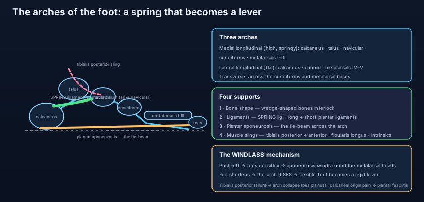
The foot does not rest on the ground like a plank. It rests on it like a bridge. Weight arriving down the tibia lands on the talus and is then distributed forwards and backwards along vaults. The medial longitudinal arch is the high, springy one: calcaneus, talus, navicular, the three cuneiforms and the first three metatarsals. Its keystone is the head of the talus, and it is the arch that absorbs shock and gives the foot its recoil. The lateral longitudinal arch is much flatter and more rigid: calcaneus, cuboid, and the fourth and fifth metatarsals — built for transmitting load, not for spring, which is why the outer border of a wet footprint touches the ground and the inner border does not. Across the foot runs the transverse arch, highest at the level of the cuneiforms and cuboid, its wedge-shaped bones locking against each other like the voussoirs of a stone vault. Three things hold all of this up. First, bone shape — the wedges themselves. Second, the plantar ligaments: the spring (plantar calcaneonavicular) ligament, a thick fibrocartilaginous hammock slung from the sustentaculum tali of the calcaneus to the navicular, directly under the head of the talus and the true guardian of the medial arch; the short plantar (plantar calcaneocuboid) ligament; the long plantar ligament, the longest in the foot; and the plantar aponeurosis, a broad tie-beam of dense fascia from the medial tubercle of the calcaneus forward into the toes. Third, muscle: tibialis posterior, whose tendon fans out onto the navicular and the cuneiforms and actively lifts the medial arch, tibialis anterior in front, fibularis longus crossing the sole obliquely from lateral to medial to sling the transverse arch, and the layered intrinsic muscles of the foot beneath.
The windlass: the most elegant idea in the foot
Here is how a foot changes its stiffness without changing its parts. The plantar aponeurosis is anchored behind on the calcaneus and in front on the plantar plates and bases of the proximal phalanges, so it passes underneath the metatarsal heads on its way to the toes. When you reach the end of a step and the heel lifts, the body rolls forward over the toes and the metatarsophalangeal joints are forced into dorsiflexion. The aponeurosis is then wound around the metatarsal heads like a rope around a drum — a windlass. Winding shortens it; shortening pulls the calcaneus and the metatarsal heads towards each other; and drawing the two ends of an arch together can only raise it. In that instant the arch rises, the tarsal bones lock, the whole foot stiffens, and the calf muscles of the leg get a rigid lever to push against. Let the toes come flat again and the windlass unwinds, the arch lowers, and the foot becomes a soft, adaptable platform once more. This is why toe-off feels springy, why sprinters' spikes are stiff under the forefoot, and why a stiff big toe (hallux rigidus) ruins the mechanics of the entire limb.
The single sentence that unlocks the ankle: the mortise is tightest in dorsiflexion and loosest in plantarflexion, and the lateral ligament is three thin bands while the medial is one thick sheet. Put those together and you have predicted the commonest musculoskeletal injury on earth — the inversion sprain in plantarflexion that tears the anterior talofibular ligament — and you have also predicted its mirror image, because a violent eversion force cannot tear the deltoid, so it snaps the medial malleolus off instead and then, if the force continues, spirals up to break the fibula too. Injury patterns are not memorised facts; they are ligament strength read backwards. It is the same lesson the knee teaches with its ligaments, only here the bone gives way before the ligament does.
The rolled ankle: the basketball player lands plantarflexed and inverted, tears the ATFL, and swells over the front of the lateral malleolus. He is examined against the Ottawa ankle rules — an X-ray is only needed if there is bone tenderness over the posterior edge or tip of either malleolus, over the navicular or the base of the fifth metatarsal, or if he cannot bear weight for four steps — and most such patients need no film at all. The broken ankle: a foot fixed on the ground while the body rotates over it produces a Pott's-type fracture, classified by Weber according to how the fibular fracture sits relative to the syndesmosis (below it, at it, or above it) — the higher the fibular break, the more likely the syndesmosis is torn and the mortise unstable. The first step in the morning: the woman with heel pain has plantar fasciitis — degeneration and inflammation at the origin of the plantar aponeurosis on the medial calcaneal tubercle. Overnight the foot rests plantarflexed and the fascia shortens; the first steps stretch it violently, which is exactly why the pain is worst at the start and eases with walking, and why rest, stretching, heel support and short courses of anti-inflammatory drugs are the usual approach. The collapsing arch: the sixty-year-old with a flattening medial border has tibialis posterior tendon dysfunction — lose the active sling and the spring ligament slowly stretches, the head of the talus drops medially, the heel drifts into valgus, and she can no longer perform a single-leg heel raise, because the windlass has no arch left to raise.
- The talocrural (ankle) joint is a uniaxial hinge: the talar trochlea in a mortise of tibial plafond + medial malleolus + lateral malleolus, with only dorsiflexion and plantarflexion.
- The lateral malleolus descends about 2 cm lower than the medial, and the distal tibiofibular syndesmosis binds the two walls of the mortise together.
- The talus is WIDER ANTERIORLY: dorsiflexion is the close-packed, stable position; plantarflexion is loose-packed and vulnerable.
- Medial (deltoid) ligament = one strong triangular sheet in four parts (tibionavicular, tibiocalcaneal, anterior and posterior tibiotalar) — it avulses the medial malleolus rather than tear.
- Lateral ligament = three weaker bands: ATFL (weakest, most commonly torn), CFL, PTFL (strongest) — hence the inversion sprain in plantarflexion.
- Inversion and eversion do NOT occur at the ankle: they happen at the subtalar and transverse tarsal (talonavicular + calcaneocuboid) joints.
- Medial longitudinal arch: calcaneus, talus, navicular, three cuneiforms, metatarsals 1–3 — high and springy, keystone = head of the talus.
- Lateral longitudinal arch: calcaneus, cuboid, metatarsals 4–5 — flatter and made for load transmission; the transverse arch runs across the cuneiforms and cuboid.
- Passive supports: wedge-shaped bones, the spring (plantar calcaneonavicular) ligament under the talar head, the short and long plantar ligaments, and the plantar aponeurosis.
- Active supports: tibialis posterior (the key medial arch sling), tibialis anterior, fibularis longus crossing the sole for the transverse arch, and the intrinsic muscles.
- Windlass mechanism: dorsiflexing the toes winds the plantar aponeurosis around the metatarsal heads, shortens it, raises the arch and makes the foot a RIGID LEVER for push-off.
- Clinical anchors: ATFL sprain and the Ottawa rules, Pott's/Weber fractures, plantar fasciitis (first-step pain), pes planus from tibialis posterior dysfunction, Achilles rupture.
- Saying the ankle inverts and everts. The talocrural joint only dorsiflexes and plantarflexes; inversion and eversion belong to the subtalar and transverse tarsal joints.
- Assuming the ankle is most stable when relaxed and pointed. The opposite is true: the wedge-shaped talus makes DORSIFLEXION the close-packed position, and plantarflexion the loose, sprain-prone one.
- Treating the arch as a purely ligamentous structure. Bone shape, ligaments AND muscle slings all hold it — which is why tibialis posterior failure alone can collapse a lifelong normal arch.
A footballer lands from a jump with the foot plantarflexed and the sole turning inwards, and is tender in front of the lateral malleolus. Which single anatomical feature best explains why this position is the one that produces ankle sprains?
- The ankle is a hinge: the talus held in a mortise of tibial plafond, medial malleolus and the lower-reaching lateral malleolus, bound by the distal tibiofibular syndesmosis — dorsiflexion and plantarflexion only.
- Stability is asymmetric: one strong deltoid sheet medially, three weak bands laterally (ATFL, CFL, PTFL), and a wedge-shaped talus that makes plantarflexion the loose position — hence the inversion sprain of the ATFL.
- Inversion and eversion belong to the subtalar and transverse tarsal joints, which let the foot mould itself to uneven ground while the leg stays vertical.
- Three arches (medial and lateral longitudinal, transverse) are held by bone shape, the spring and plantar ligaments and the plantar aponeurosis, and the slings of tibialis posterior, tibialis anterior and fibularis longus — and the windlass mechanism turns the flexible foot into a rigid lever the moment the toes dorsiflex.
- Drake RL, Vogl AW, Mitchell AWM. Gray's Anatomy for Students — Lower limb: the ankle and joints of the foot.
- Moore KL, Dalley AF, Agur AMR. Clinically Oriented Anatomy — Ankle and foot joints; arches of the foot.
- Netter FH. Atlas of Human Anatomy — Ligaments and tendons of the ankle and foot.
- Last RJ. Last's Anatomy: Regional and Applied — The ankle joint and the tarsus.
- Snell RS. Clinical Anatomy by Regions — Ankle sprains, malleolar fractures and the plantar aponeurosis.
- TeachMeAnatomy — The Ankle Joint; The Arches of the Foot.

