
Muscles of the Leg: Three Compartments and the Strongest Tendon in the Body
Stand up and rise onto your toes. In that single unremarkable second, a pair of muscles at the back of your leg has just lifted your entire body weight — every kilogram of you — through a tendon no thicker than your thumb. You will do it perhaps ten thousand times before you sleep tonight: every step you take is a small, private feat of strength. The leg, the segment between knee and ankle, is where the body converts the loose swing of the thigh into precise, gripping contact with the ground. It does this with only twelve muscles, packed into three sealed compartments, each compartment with its own nerve, its own job and its own way of failing.
A man in his forties plays his weekly game of badminton. He lunges for a drop shot, pushes off hard — and feels a violent blow to the back of his calf, as if someone standing behind him had kicked him with real malice. He spins round. There is nobody there. He tries to walk and the foot flops; he cannot push off, cannot rise onto that toe at all. What he heard, and what the players at the next court also heard, was a snap. His calcaneal tendon — the Achilles, the thickest and strongest tendon in the human body — has torn clean across, about five centimetres above its insertion, in the narrow zone where its blood supply is poorest. A tendon built to carry twelve times body weight failed on a badminton court. That is the paradox of the leg: it is engineered for enormous force, and every one of its failures is a failure of something enormous.
Three compartments, three nerves, one rule
Learn the walls first and the muscles almost sort themselves. The skeleton of the leg is two bones — the weight-bearing tibia and the slender, non-weight-bearing fibula described in the tibia and fibula — strapped together by a tough interosseous membrane. Around them a sleeve of deep fascia (the crural fascia) sends two intermuscular septa inwards to the fibula. Those partitions, plus the membrane and the bones themselves, divide the leg into three watertight compartments: anterior, lateral and posterior. The rule that makes the whole region learnable is this: each compartment has ONE nerve and ONE dominant action. Anterior = deep fibular nerve = lift the foot. Lateral = superficial fibular nerve = turn the sole outwards. Posterior = tibial nerve = push the ground away. This is exactly the compartmental logic you already met in the muscles of the forearm, only here the compartments are stiffer, the pressures higher, and the consequences of swelling far more dangerous.
The anterior compartment: the muscles that lift the foot
Four muscles occupy the narrow gutter between the tibia and fibula in front, all supplied by the deep fibular nerve and fed by the anterior tibial artery. Tibialis anterior is the giant of the group: it arises from the lateral surface of the tibia and the interosseous membrane, runs down as the most medial tendon at the ankle, and inserts onto the medial cuneiform and the base of the first metatarsal. Because it crosses the ankle in front and the subtalar joint medially, it does two things at once — dorsiflexion and inversion. Extensor hallucis longus arises from the middle of the medial fibula and the membrane and inserts on the base of the distal phalanx of the great toe; extensor digitorum longus arises from the lateral tibial condyle and the anterior fibula and fans into four tendons that reach the middle and distal phalanges of the lateral four toes through their extensor expansions. Fibularis tertius is really a split-off part of extensor digitorum longus, running to the base of the fifth metatarsal, and it is absent in a good number of people. All four tendons are pinned down at the ankle by the superior and inferior extensor retinacula — thickenings of fascia that stop the tendons bowstringing away from the joint when you pull your toes up.
Its most important job is not lifting at all — it is letting down gently. Watch a person walk and you will see tibialis anterior working twice in every stride. First it holds the toes clear of the ground during swing, so the foot does not catch. Then, the instant the heel strikes, it takes the whole weight of the foot on an eccentric contraction and lowers the sole to the floor over about a tenth of a second — braking, not lifting. Paralyse it and that control vanishes: the foot slaps audibly onto the ground with every step. That slap is one of the earliest clinical signs of a deep fibular nerve lesion, and it is often heard by a relative long before the patient complains.
The lateral compartment: the two evertors
The smallest compartment holds only two muscles, both arising from the lateral surface of the fibula, both supplied by the superficial fibular nerve, and both eversion specialists — they turn the sole outwards and, just as importantly, resist the sole being turned inwards. This compartment is unique in one respect: it has no artery of its own. It is supplied by perforating branches from the fibular (peroneal) artery of the posterior compartment, which is why it is the compartment most vulnerable to ischaemia when pressure rises. Fibularis longus arises from the head and upper two-thirds of the fibula, and does something no other muscle in the leg does: its tendon passes behind the lateral malleolus, hooks around the cuboid, and then crosses the entire sole obliquely to insert on the medial cuneiform and the base of the first metatarsal — the very same landmarks as tibialis anterior, approached from the opposite side. That strap under the foot is the main dynamic support of the transverse arch. Fibularis brevis, deeper and shorter, arises from the lower two-thirds of the fibula and inserts on the tuberosity of the fifth metatarsal.
That fifth metatarsal insertion earns its own paragraph, because it explains one of the commonest injuries in any emergency department. Roll your ankle inwards — step off a kerb badly, land awkwardly from a jump — and the everting muscles fire violently to stop the inversion. Fibularis brevis pulls so hard on the tuberosity of the fifth metatarsal that it can rip the bone fragment off with it: an avulsion fracture. This is why every patient with a "sprained ankle" should have the base of the fifth metatarsal pressed as part of the examination, and why that bony point is written into the Ottawa ankle rules. The joints being wrenched in that moment are described in the ankle, foot joints and arches.
The posterior compartment: the engine of walking
A deep transverse fascia splits this compartment into a superficial half you can see and a deep half you cannot. Everything behind that fascia is supplied by the tibial nerve and the posterior tibial artery. The superficial group is the calf — the visible, sculpted mass that gives the leg its shape. Gastrocnemius is the showman: two heads arising from the popliteal surfaces of the medial and lateral femoral condyles, which means it crosses the knee as well as the ankle. It is therefore both a plantarflexor and a weak knee flexor, and it can only generate full power when the knee is straight — bend the knee and you slacken it, which is the entire basis of the bedside test. Beneath it lies soleus, a broad flat sheet arising from the soleal line of the tibia, the posterior tibia and the head and upper shaft of the fibula. Soleus crosses only the ankle, so it works equally well with the knee bent or straight; it is the postural workhorse that keeps you from toppling forwards while you stand still. Plantaris, the small vestigial one, has a tiny belly and a very long thin tendon that runs between the two — famous mostly for being mistaken for a nerve by students and for being harvested as a tendon graft. All three converge on the calcaneal tendon.
The calcaneal tendon is the strongest and thickest tendon in the human body, roughly fifteen centimetres long, inserting into the middle of the posterior surface of the calcaneus. It routinely transmits forces of six to twelve times body weight during running and jumping. Yet it has one structural flaw: a relatively avascular zone two to six centimetres above its insertion, where the blood supply from above and below thins out and meets. That is where degenerative tendinopathy settles, and that is where it ruptures. Beneath the superficial group, and separated from it by the transverse fascia, lies the deep group, whose members are far less glamorous and far more important than their size suggests.
Four deep muscles: one unlocks the knee, three hold up the foot. Popliteus is the odd one out — it never reaches the foot at all. It arises from the lateral femoral condyle inside the knee capsule and runs downwards and medially to the posterior tibia above the soleal line. When you stand with the knee fully extended, the joint is screwed home and locked; popliteus laterally rotates the femur on the fixed tibia to unlock it before flexion can begin, which is why it is called the key of the knee joint. Flexor hallucis longus arises from the posterior fibula and interosseous membrane, its tendon grooving the back of the talus and running under the sustentaculum tali to reach the base of the distal phalanx of the great toe — it is the true push-off muscle, delivering the final thrust of every step. Flexor digitorum longus arises from the posterior tibia, crosses superficial to the tibialis posterior tendon at the ankle, then crosses deep to flexor hallucis longus in the sole, and inserts into the distal phalanges of the lateral four toes. And deepest of all, sandwiched between the two flexors on the interosseous membrane and both bones, lies tibialis posterior: it inverts and plantarflexes, and its tendon fans out into the navicular tuberosity, all three cuneiforms, the cuboid and the middle three metatarsal bases — the great sling under the medial longitudinal arch, and the single most important dynamic support the arch has. Lose it and the arch collapses into an adult acquired flatfoot; the bones it holds up are laid out in the bones of the foot.
Think of the leg as a mast with three sets of rigging. The anterior lines pull the bow of the foot up; the lateral lines pull it outwards; the posterior lines, thickest and strongest by far, haul it down and back. Now think of the foot itself as a suspension bridge: the medial longitudinal arch is the span, the plantar ligaments are the static cables that hold it whatever happens, and tibialis posterior is the single active cable with a winch on it, tightening the span every time you load it. Static cables never tire but never adjust; the winch adjusts constantly — and, like any working machine, it is the part that wears out.
The tarsal tunnel: everything passing behind the ankle
Behind the medial malleolus, held down by the flexor retinaculum, everything that travels from the deep posterior compartment into the sole passes through a single fibro-osseous corridor: the tarsal tunnel, the ankle's answer to the carpal tunnel. The order from front to back is fixed, and generations have memorised it as Tom, Dick And Very Nervous Harry: Tibialis posterior tendon, flexor Digitorum longus tendon, posterior tibial Artery, tibial Vein, tibial Nerve, flexor Hallucis longus tendon. That order matters. The artery is where you feel the posterior tibial pulse, one of the two pedal pulses that tell you whether the arteries of the lower limb are still delivering. The nerve, compressed in that tunnel by swelling, a ganglion or a badly healed fracture, produces tarsal tunnel syndrome: burning pain and tingling in the sole, worse on standing and at night, with a positive Tinel's sign over the tunnel.
Soleus is often called the peripheral heart, and the name is not a metaphor. It is riddled with wide, thin-walled venous sinuses; every time it contracts it squeezes them flat and drives blood upwards through one-way valves, and every time it relaxes they refill from below. Walking therefore pumps venous blood from foot to groin against gravity — the calf-muscle pump. Stop the pump and the system stalls: this is precisely why hours of immobility on a long flight, in a hospital bed or in a plaster cast let blood pool and clot in the deep veins of the calf, the beginning of the deep vein thrombosis described in the veins and lymphatics of the lower limb. The advice to "get up and walk about" is not comfort advice. It is a prescription to restart a pump.
When compartments fail
The very fascia that gives these muscles their leverage is what makes them dangerous. Fascia does not stretch. Bleed or swell inside a closed compartment — most often after a tibial shaft fracture, a crush injury or a reperfused ischaemic limb — and the pressure climbs until it exceeds capillary perfusion pressure. Muscle and nerve then die inside an intact, pulsatile limb, because the pulse in a large artery persists long after the capillaries have been shut down. This is acute compartment syndrome, and its cardinal sign is pain out of all proportion to the injury, made agonising by passive stretching of the muscles in that compartment. Waiting for pulselessness is waiting for the diagnosis to be too late; the treatment is urgent fasciotomy. The chronic cousin is far commoner and far more benign: chronic exertional compartment syndrome and medial tibial stress syndrome — "shin splints" — where the pain of the anterior or deep posterior compartment builds predictably over a set distance of running and settles with rest.
The other classic failure is neurological, and it has an elegant anatomical explanation. The common fibular nerve, one of the two terminal branches of the sciatic nerve, winds round the neck of the fibula in a place where it is covered by nothing but skin, and only there does it divide into its deep and superficial branches. So a single injury at that one point — a tight plaster, a fibular neck fracture, a poorly padded operating table, prolonged squatting or crossing the legs — paralyses BOTH the anterior and the lateral compartments at once. The foot can neither be lifted nor everted: foot drop, with the high-stepping gait of a patient who must lift the whole limb to clear the toes. It is the exact lower-limb twin of wrist drop, and it is dissected fully in the sciatic, tibial and fibular nerves.
Return to the man with the torn Achilles. He is laid face down with his feet hanging over the end of the couch, and the examiner squeezes his calf. On the normal side, squeezing the calf deforms soleus and gastrocnemius, tugs the intact tendon and the foot plantarflexes visibly. On the injured side, nothing moves — the chain between muscle and heel is broken. That is Simmonds' (Thompson's) test, and it is more reliable than asking the patient to push down, because flexor hallucis longus and tibialis posterior can still weakly plantarflex and fool you. Two everyday manoeuvres complete the bedside examination of this whole region: ask the patient to walk on tiptoe, which tests the posterior compartment and the tibial nerve, and then to walk on their heels, which tests the anterior compartment and the deep fibular nerve. A patient who cannot do the first has a tibial problem; one who cannot do the second has foot drop. Two requests, ten seconds, and three compartments examined.
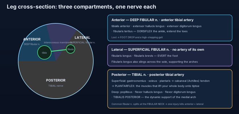
- Anterior compartment — deep fibular nerve, anterior tibial artery: tibialis anterior (dorsiflexes AND inverts; eccentrically lowers the foot after heel strike), extensor hallucis longus, extensor digitorum longus, fibularis tertius; all pass under the superior and inferior extensor retinacula.
- Lateral compartment — superficial fibular nerve, NO artery of its own: fibularis longus (crosses the sole to the medial cuneiform and 1st metatarsal, supporting the transverse arch) and fibularis brevis (to the tuberosity of the 5th metatarsal).
- Posterior superficial — gastrocnemius (two heads from the femoral condyles, so it also flexes the knee and weakens with the knee bent), soleus (soleal line and fibula, crosses the ankle only) and plantaris, all into the calcaneal tendon.
- Posterior deep — popliteus (unlocks the extended knee), flexor hallucis longus (the push-off muscle), flexor digitorum longus, and tibialis posterior, the deepest: inverts, plantarflexes and is the key dynamic support of the medial longitudinal arch.
- The whole posterior compartment is tibial nerve + posterior tibial artery, split into superficial and deep halves by the deep transverse fascia.
- Tarsal tunnel order behind the medial malleolus — Tom, Dick And Very Nervous Harry: Tibialis posterior, flexor Digitorum longus, posterior tibial Artery, tibial Vein, tibial Nerve, flexor Hallucis longus.
- The calcaneal (Achilles) tendon is the strongest and thickest tendon in the body; it ruptures in the relatively avascular zone 2–6 cm above its insertion.
- Simmonds'/Thompson's test: squeezing the calf of a prone patient fails to plantarflex the foot when the tendon is ruptured.
- Soleus is the "peripheral heart": the calf-muscle pump drives venous return, so immobility — flights, casts, hospital beds — predisposes to DVT.
- Acute compartment syndrome (classically after a tibial shaft fracture): pain out of proportion, worse on passive stretch, in a limb that still has a pulse — treat by fasciotomy, do not wait for pulselessness.
- The common fibular nerve divides at the fibular neck, so one lesion there paralyses the anterior AND lateral compartments: foot drop with loss of both dorsiflexion and eversion.
- Bedside screen: walking on tiptoe tests the posterior compartment and tibial nerve; walking on the heels tests the anterior compartment and deep fibular nerve.
- Assuming tibialis anterior and tibialis posterior are opposites in every way. Both INVERT the foot — they differ in the sagittal plane, where tibialis anterior dorsiflexes and tibialis posterior plantarflexes. Eversion belongs to the fibular muscles alone.
- Testing calf power with the knee bent and concluding the calf is weak. A flexed knee slackens gastrocnemius, because it arises from the femur; soleus, crossing only the ankle, is the muscle you are actually testing in that position.
- Waiting for a lost pulse before diagnosing compartment syndrome. Pressure high enough to kill muscle and nerve is still far below the pressure needed to occlude a major artery, so the limb stays warm and pulsatile while it is dying.
A 30-year-old is treated for a fractured neck of the fibula. On review she cannot dorsiflex or evert the right foot and has numbness over the dorsum, but plantarflexion, inversion and sensation of the sole are all normal. Which single nerve lesion explains all of these findings?
- The leg has three fascial compartments, each with one nerve: anterior (deep fibular — dorsiflexors), lateral (superficial fibular — evertors, and no artery of its own), posterior (tibial — plantarflexors).
- Anterior: tibialis anterior (dorsiflexion + inversion, and eccentric lowering after heel strike), EHL, EDL, fibularis tertius. Lateral: fibularis longus (to the medial cuneiform across the sole) and brevis (to the 5th metatarsal tuberosity, avulsed in inversion injury).
- Posterior superficial (gastrocnemius, soleus, plantaris) forms the calcaneal tendon — the strongest in the body; posterior deep (popliteus, FHL, FDL, tibialis posterior) unlocks the knee, pushes off and holds up the medial longitudinal arch. Behind the medial malleolus: Tom, Dick And Very Nervous Harry.
- Clinical signature: Achilles rupture with a positive Simmonds/Thompson test, DVT when the calf pump stops, compartment syndrome after a tibial fracture, shin splints, and foot drop from a common fibular lesion at the fibular neck — screened in seconds by walking on tiptoe then on the heels.
- Drake RL, Vogl AW, Mitchell AWM. Gray's Anatomy for Students — Lower limb: compartments of the leg.
- Moore KL, Dalley AF, Agur AMR. Clinically Oriented Anatomy — The leg: anterior, lateral and posterior compartments; compartment syndrome.
- Netter FH. Atlas of Human Anatomy — Muscles of the leg: superficial and deep dissections; cross-sections.
- Last RJ. Last's Anatomy: Regional and Applied — The leg and the tarsal tunnel.
- Snell RS. Clinical Anatomy by Regions — Achilles tendon rupture, foot drop and the common fibular nerve.
- TeachMeAnatomy — Muscles of the Anterior, Lateral and Posterior Leg.

