
The Hip Bone and the Bony Pelvis: The Foundation We Stand On
Everything above your waist — your head, your arms, your ribcage, the weight of every meal you have ever eaten — has to reach the ground somehow, and it does so through a single ring of bone no wider than your shoulders. The shoulder girdle solved the same problem by refusing to solve it: it floats on muscle, barely touching the skeleton, and buys itself extraordinary freedom. The pelvic girdle made the opposite bargain. It fused. It locked itself to the spine, sealed its own joints, and gave up almost all movement in exchange for the right to carry you. That trade is the whole story of standing upright, and you can read it in every ridge and hollow of the bone.
Sit down on a hard wooden chair and stay there for an hour. Long before the hour is up, you will start to shift, to lean, to slide one buttock forward and then the other. What is complaining is not the chair — it is two blunt bony knobs, one under each buttock, taking the entire weight of your torso through a patch of skin no bigger than a coin. Those are the ischial tuberosities, and evolution built them to be sat on. Now stand up. In that single movement the load leaves those two knobs, travels back through the pelvic ring, turns a right angle at two deep cup-shaped sockets, and pours down into your thigh bones. The same piece of bone is a chair when you sit and an archway when you stand. No other bone in the body changes job so completely, so often, without moving at all.
Three bones that agreed to become one
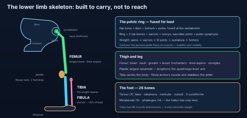
The hip bone (os coxae) is not born as one bone — it is assembled. In a child, three separate bones — the ilium, the ischium and the pubis — meet at a Y-shaped cartilage in the floor of the hip socket. Somewhere between the middle and the end of the teenage years that triradiate cartilage ossifies, and the three fuse into the single irregular bone we call the os coxae. The point where they meet is the acetabulum, the deep cup that receives the head of the femur, and it is no accident that the fusion happens precisely there: the meeting place had to be the strongest part, because it is where the entire load turns the corner. Everything else about the hip bone is shaped around that fact. It is a textbook example of the principle set out in the classification and structure of bones — trabeculae align themselves along the lines of force, and the outer cortex thickens exactly where the pull of muscle and the push of body weight demand it.
The ilium: the great fan
The ilium is the broad upper blade, and it is the part you can find on yourself right now: rest your hands on your hips and the ridge under your thumbs is the iliac crest. That crest runs from the anterior superior iliac spine (ASIS) in front — the bony point that a trouser waistband catches on, and the anchor of the inguinal ligament and sartorius — back to the posterior superior iliac spine (PSIS), marked on the skin by the two dimples above the buttocks. Below each of these sits a smaller inferior spine: the anterior inferior iliac spine (AIIS) giving origin to the straight head of rectus femoris, and the posterior inferior iliac spine (PIIS) just above the greater sciatic notch. On its inner surface the ilium is scooped into the smooth iliac fossa, home of the iliacus muscle; behind that fossa lies the ear-shaped auricular surface, the only part of the hip bone that articulates with the sacrum, and behind that again the roughened iliac tuberosity for the immensely strong interosseous sacroiliac ligaments. On the outer surface, three curved gluteal lines — posterior, anterior and inferior — divide the field between the gluteal muscles that clothe it. And at the back the bone is cut away into the deep greater sciatic notch, the gateway out of the pelvis.
The ischium and the pubis: the seat and the strut
The ischium is the posteroinferior part. Its body forms roughly two-fifths of the acetabulum; behind that body a sharp ischial spine projects medially, separating the greater sciatic notch above from the lesser sciatic notch below. Then comes the ischial tuberosity — thick, blunt, weight-bearing, the origin of the hamstrings and the bone you sit on — and from it the ischial ramus runs forward to join the inferior pubic ramus. The pubis is the anterior part: a flattened body carrying the symphyseal surface where the two hip bones meet in the midline, topped by the pubic crest and, at its lateral end, the pubic tubercle — the landmark that tells a hernia surgeon whether a groin lump is inguinal (above and medial) or femoral (below and lateral). From the body a superior pubic ramus runs back to the acetabulum and an inferior pubic ramus runs down to meet the ischium. Together the ischial and pubic rami enclose the obturator foramen, the large gap that makes the hip bone lighter without weakening it, closed almost completely by the obturator membrane and leaving only a small obturator canal at its upper edge for the obturator nerve and vessels — the neurovascular pair traced in the femoral and obturator nerves.
The acetabulum: a cup that grips
Compare it, for one moment, with the shoulder — and the whole design philosophy becomes visible. The acetabulum is a deep hemispherical socket on the lateral surface of the hip bone, facing laterally, downwards and slightly forwards. Its articular part is the horseshoe-shaped lunate surface, coated in thick hyaline cartilage; the non-articular centre is the acetabular fossa, filled with fat and the ligament of the head of the femur. Inferiorly the rim is deficient — the acetabular notch — and that gap is bridged by the transverse acetabular ligament, converting the notch into a foramen through which vessels reach the joint. The result is a socket that encloses more than half of the femoral head and physically grips it. The glenoid cavity of the scapula is a shallow saucer that holds barely a third of the humeral head and dislocates readily; the acetabulum almost never dislocates without violence great enough to break something. Depth bought stability, and stability cost range of motion — the same bargain the pelvis made with the spine, written small.
Think of the bony pelvis as a stone bridge, and of the sacrum as its keystone. Push down on a keystone and it does not fall through — it wedges itself tighter, and drives its load outwards into the two arches on either side. The sacrum is wedge-shaped in exactly this way, wider above than below and wider in front than behind, so the weight of the whole trunk drives it down into the gap between the two hip bones and jams it there. The sacroiliac ligaments are the mortar, and they are among the strongest ligaments in the body. This is why the pelvis is a ring rather than two struts: a ring cannot be broken in one place only. Crack a stone bridge at a single point and it stays standing; break it in two and it collapses. That is precisely the rule for pelvic fractures — if you see one break in the ring on an X-ray, look immediately for the second.
The ring: two hip bones, a sacrum, a coccyx
The bony pelvis is completed behind by the sacrum and coccyx. Each hip bone meets the sacrum at a sacroiliac joint — anatomically a synovial joint, but with irregular interlocking surfaces and ligaments so strong that in the adult it moves only a few degrees. In front, the two pubic bones meet at the pubic symphysis, a secondary cartilaginous joint in which a fibrocartilaginous interpubic disc is gripped between two layers of hyaline cartilage and reinforced above and below by the superior pubic and arcuate pubic ligaments. Together these three joints turn four bones into one continuous ring — and an oblique plane through that ring, running from the sacral promontory along the arcuate line and the pecten pubis to the upper border of the symphysis, is the pelvic brim or inlet. Above it lies the greater (false) pelvis, walled by the iliac fossae and really belonging to the abdomen; below it lies the lesser (true) pelvis, the funnel that contains the bladder, rectum and reproductive organs, and whose lower opening is the pelvic outlet, bounded by the pubic arch, the ischial tuberosities, the sacrotuberous ligaments and the tip of the coccyx.
Two ligaments that build two doorways
The notches on the back of the hip bone are not holes until ligament makes them so. Two great ligaments run from the sacrum outwards. The sacrospinous ligament passes from the lateral sacrum and coccyx to the ischial spine; the sacrotuberous ligament, longer and stronger, passes from the same region to the ischial tuberosity. Between them they cross the greater and lesser sciatic notches and convert each into a foramen. The greater sciatic foramen, above the sacrospinous ligament, is the main exit from the pelvis into the gluteal region: through it pass piriformis, the superior gluteal nerve and vessels above the muscle, and below it the inferior gluteal nerve and vessels, the posterior femoral cutaneous nerve, the nerves to quadratus femoris and obturator internus, the pudendal nerve and internal pudendal vessels, and — largest of all — the sciatic nerve, the thickest nerve in the human body, whose whole subsequent journey is followed in the sciatic, tibial and common fibular nerves. The lesser sciatic foramen, below the sacrospinous ligament, is the way back in: the tendon of obturator internus leaves through it, and the pudendal nerve and internal pudendal vessels, having curved round the ischial spine, re-enter the perineum through it. Those two ligaments also resist the upward tilt of the sacral tip when body weight pushes the promontory down — the very thing that stops the keystone from rotating out of its arch.
The male and female pelvis: a bone shaped by childbirth
The female pelvis is the only part of the human skeleton whose shape is dictated by an event that happens to only half the species. Because a full-term head must pass through the true pelvis, the female pelvis is broader and shallower: the inlet is oval or transversely elliptical rather than the male heart shape, the cavity is wider and more cylindrical rather than funnel-shaped, the sacrum is shorter, wider and less curved, the greater sciatic notch is wider (approaching 90° rather than 70°), the ischial spines are less inwardly projecting, the ischial tuberosities are everted rather than turned in, and — the single most reliable difference — the subpubic angle beneath the pubic arch is wide and rounded, roughly 80–90°, where the male angle is a narrow 50–60° V. Obstetricians measure these dimensions because they decide whether a head can descend, and forensic anthropologists read them because they survive when almost nothing else does. It is a striking thought: the widest, most generous shape in the human skeleton exists so that another skeleton can get out.
How weight actually gets to the ground
Trace the load. Body weight descends the vertebral column to the fifth lumbar vertebra and passes into the base of the sacrum. From the sacrum it does not go straight down — it turns outwards through the two sacroiliac joints into the ilia. From each ilium it runs along a thickened bar of bone, the sacropubic arch, to the acetabulum, and there it turns downwards again into the head and neck of the femur, described in the femur and patella. When you stand, that is the end of the story: hip to knee to ankle to floor. When you sit, the path is different — the load leaves the sacroiliac joints and travels down the ilio-ischial bar to the ischial tuberosities instead, bypassing the hip joints entirely. Two struts oppose these lines and stop the ring from being pulled apart: the horizontal bar of the pubic bodies and superior rami in front, and the ischiopubic rami below. The consequence for the hip joint is that its socket is permanently under compression, which is exactly the loading that cartilage tolerates best.
The iliac crest is one of the most useful landmarks in clinical medicine, and it earns its keep three times over. A line drawn between the highest points of the two crests — the intercristal or Tuffier's line — crosses the spine at about the level of the fourth lumbar vertebra, which is how an anaesthetist finds a safe interspace for a lumbar puncture or spinal block, well below the end of the spinal cord. The ASIS is the fixed point from which true leg length is measured, and the reference for the inguinal ligament. And the posterior iliac crest, just deep to the PSIS dimple, is thick cancellous bone packed with red marrow — which makes it the standard site for a bone-marrow aspirate and trephine biopsy, the test that looks directly at the cell factory discussed in haematopoiesis and its drug targets. One ridge of bone: a spinal roadmap, a measuring point, and a window into the blood.
The sprinter in the blocks: a 17-year-old explodes out of a starting position and feels a sudden tearing at the front of the hip. In an adult this would be a hamstring or rectus femoris strain; in an adolescent whose apophyses have not yet fused, the tendon is stronger than the growth plate, and the bone gives way first — an avulsion fracture, most often of the AIIS by rectus femoris, the ASIS by sartorius, or the ischial tuberosity by the hamstrings. The X-ray shows a flake of bone pulled clean off the pelvis. The motorcyclist: a high-energy impact crushes the pelvic ring in two places. The danger is not the bone; it is the enormous presacral venous plexus and the branches of the internal iliac artery running along the inner wall. A pelvis can hide several litres of blood, and death here is from haemorrhage, not fracture — which is why the first treatment at the roadside is a binder that squeezes the ring back into shape and reduces its volume. The grandmother in the kitchen: an 82-year-old with osteoporosis falls sideways onto her hip and cannot stand. The pelvic ring is intact; what has failed is the femoral neck just beyond it, and the injury is a race against the fragile blood supply of the femoral head.
- The hip bone (os coxae) = ilium + ischium + pubis, fused at the triradiate cartilage in the floor of the acetabulum during adolescence.
- Ilium: iliac crest, ASIS, AIIS, PSIS, PIIS, iliac fossa, three gluteal lines, auricular surface for the sacrum, and the greater sciatic notch.
- Ischium: body, ischial spine (separating greater from lesser sciatic notch), ischial tuberosity (the sitting bone, hamstring origin) and ramus.
- Pubis: body with symphyseal surface, pubic crest and tubercle, superior and inferior rami.
- The obturator foramen is closed by the obturator membrane except for the obturator canal, which transmits the obturator nerve and vessels.
- The acetabulum has an articular lunate surface, a non-articular fossa, and a notch bridged by the transverse acetabular ligament.
- The pelvis is a closed ring: two hip bones + sacrum + coccyx, joined at two sacroiliac joints and the pubic symphysis.
- The pelvic brim (inlet) divides the greater (false) pelvis above from the lesser (true) pelvis below; the lower opening is the pelvic outlet.
- The sacrospinous and sacrotuberous ligaments convert the two sciatic notches into the greater and lesser sciatic foramina.
- Greater sciatic foramen = the main exit (sciatic nerve, gluteal nerves and vessels, pudendal bundle, piriformis); lesser = the way back in (obturator internus tendon, pudendal bundle re-entering the perineum).
- Female pelvis: oval inlet, wide subpubic angle (80–90°), wider greater sciatic notch, everted ischial tuberosities, shorter and less curved sacrum.
- Load path: spine → sacrum → sacroiliac joints → ilia → acetabula → femora when standing; sacrum → ilio-ischial bars → ischial tuberosities when sitting.
- Calling the whole hip bone the "ilium". The ilium is only the upper blade; the hip bone is ilium + ischium + pubis, and the acetabulum belongs to all three.
- Assuming the greater sciatic foramen is a hole in the bone. It is a notch in the bone that only becomes a foramen once the sacrospinous ligament roofs it off — the same is true of the lesser foramen and the sacrotuberous ligament.
- Confusing the false pelvis with the true pelvis. The greater (false) pelvis lies ABOVE the brim and holds abdominal viscera; the lesser (true) pelvis lies below it and is the one whose dimensions matter in childbirth.
A pelvic radiograph shows a fracture through the left superior pubic ramus. The reporting registrar is asked what to look for next, and why. Which statement is the best reason?
- The hip bone is three bones — ilium, ischium and pubis — fused at the acetabulum, whose lunate surface, fossa, notch and transverse acetabular ligament together grip more than half of the femoral head.
- Its landmarks are a clinical map: iliac crest (≈ L4) and ASIS for measurement and injection, ischial tuberosity for sitting and the hamstrings, pubic tubercle for hernias, obturator foramen and canal for the obturator nerve.
- Two hip bones, the sacrum and the coccyx form a closed ring joined at the sacroiliac joints and pubic symphysis; the brim divides greater (false) from lesser (true) pelvis, and the sacrospinous and sacrotuberous ligaments create the greater and lesser sciatic foramina.
- Weight runs spine → sacrum → sacroiliac joints → acetabula → femora when standing and to the ischial tuberosities when sitting; the female pelvis is remodelled for childbirth (oval inlet, wide subpubic angle, everted tuberosities), and a ring that breaks rarely breaks only once.
- Drake RL, Vogl AW, Mitchell AWM. Gray's Anatomy for Students — Pelvis and perineum: the pelvic girdle.
- Moore KL, Dalley AF, Agur AMR. Clinically Oriented Anatomy — The bony pelvis, sexual dimorphism and pelvic fractures.
- Netter FH. Atlas of Human Anatomy — Bony framework of the pelvis; male and female pelvis compared.
- Last RJ. Last's Anatomy: Regional and Applied — The hip bone and sacroiliac joint.
- Snell RS. Clinical Anatomy by Regions — Pelvic walls, sciatic foramina and clinical notes on pelvic trauma.
- TeachMeAnatomy — The Hip Bone; The Bony Pelvis; The Sacroiliac Joint.

