
The Pectoral Girdle: Clavicle and Scapula
Reach up and touch the ceiling. Everything your arm just did — the swing, the lift, the last few degrees of stretch — was carried by a limb that is attached to the rest of your skeleton at exactly one small joint, no wider than a fingertip, at the base of your neck. The hip is locked into a deep bony socket; the shoulder is not locked into anything. Your arm hangs from your trunk on a single strut of bone and a sling of muscle, and that daring piece of engineering is precisely why you can throw, climb, paint a ceiling, and scratch the middle of your own back. The price of that freedom is fragility — and the story of the pectoral girdle is the story of the trade.
A cyclist clips a kerb at speed and puts a hand out to save herself. The impact travels up the palm, through the wrist, along the forearm, up the arm — and stops at a slender curved bone above her collar, which snaps. In the emergency department the diagnosis takes about two seconds: the shoulder has dropped, there is a step you can see through the skin, and she cannot lift the arm. It is the commonest fracture in the human body, and it happens because that bone is the only bony bridge between her arm and her chest. Break it, and the limb loses its strut and sags under its own weight. Everything else holding the arm on — and there is a great deal of it — is muscle.
One bony joint, and a limb that floats
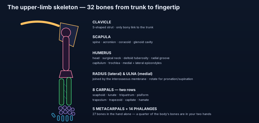
The pectoral girdle is two bones on each side: the clavicle in front, the scapula behind. Together the clavicle (collar bone) and the scapula (shoulder blade) form the pectoral girdle, the bony ring that connects the free upper limb to the axial skeleton. But look carefully at where that ring actually touches the trunk and you find a single point: the sternoclavicular joint, where the medial end of the clavicle meets the manubrium of the sternum. That is it. The scapula touches no bone of the trunk at all — it rides on a bed of muscle over the back of the ribcage. The pelvic girdle, by contrast, is fused into the sacrum on both sides and locked at the pubic symphysis in front: a rigid ring built for weight-bearing. The pectoral girdle sacrifices that stability deliberately. Because the scapula can slide, tilt and rotate freely on the chest wall, the socket it carries can be aimed almost anywhere in space — so the shoulder becomes the most mobile joint in the body, at the cost of being the most commonly dislocated. Structure follows purpose, exactly as it does everywhere in the living scaffold of the skeleton.
Think of a construction crane mounted on a flatbed truck. The clavicle is the outrigger — a rigid strut that pushes the whole assembly out and away from the cab so the boom has room to swing without hitting the bodywork. The scapula is the turntable: it is not bolted to the chassis, it rides on it, free to swivel and tilt so the boom can be aimed at anything. Remove the outrigger and the turntable collapses inward against the cab, the arc of the boom shrinks, and every lift becomes weaker and clumsier. That is exactly what a person with a badly malunited clavicle experiences — a shoulder that has quietly moved forward and downward, and an arm that no longer reaches as far or as strongly.
The clavicle: the strut that holds your shoulder out
Run a finger along your collar bone from the notch at the base of your throat out to the point of your shoulder and you can feel its entire length — the clavicle is subcutaneous from end to end, palpable in every living person. It is shaped like a flattened letter S: convex forward in its medial two-thirds, concave forward in its lateral third. The rounded, bulky sternal (medial) end articulates with the manubrium at the sternoclavicular joint; the flattened acromial (lateral) end articulates with the acromion of the scapula at the acromioclavicular joint. Its upper surface is smooth (the skin slides over it), while the under surface is roughened by the ligaments and muscles that grip it. On that inferior surface, near the lateral end, sit the two marks of the coracoclavicular ligament: the conoid tubercle, a small cone-shaped prominence for the conoid part, and running laterally from it the trapezoid line, a low ridge for the trapezoid part. These two bands are the real suspensory ligaments of the limb — they, far more than the small acromioclavicular joint capsule, transmit the weight of the arm up to the clavicle. Medially, a shallow subclavian groove on the inferior surface marks where subclavius muscle lies, and near the sternal end an impression receives the strong costoclavicular ligament tethering the bone to the first rib.
💡 The clavicle keeps two records at once, and both are strange. It is the first bone in the entire body to begin ossifying — around the fifth intrauterine week, before the vertebrae, before the skull — and it is the only long bone that ossifies in membrane rather than from a cartilage model (though its two ends later form secondary cartilaginous centres). It is also the last bone to finish: the medial epiphysis fuses at about 25 years, which makes it a genuinely useful bone for estimating age in forensic radiology. And for the exam, remember the counterpart fact — it is the commonest fractured bone in the human body, and one of the very few bones a doctor can fully evaluate by sight and touch alone.
Why the clavicle breaks where it breaks
The junction of the middle and lateral thirds is the bone's structural weak point. The usual mechanism is not a direct blow but a fall onto the outstretched hand or onto the point of the shoulder: force runs up the limb and is delivered to the clavicle along its length. The bone fails at the junction of the middle and lateral thirds, where the forward convexity reverses into the backward-facing curve, where the cross-section changes from prismatic to flattened, and where no muscle covers it. What follows is one of the most reliably reproduced deformities in all of orthopaedics. The medial fragment is pulled upward by sternocleidomastoid, which hauls on the sternal end. The lateral fragment is pulled downward by the sheer dead weight of the limb (and drawn medially by pectoralis major and latissimus dorsi), because the coracoclavicular ligament that would normally hold it up is attached distal to the break. The result is a visible step: the medial end riding high, the shoulder drooping down, forward and inward, and a patient who instinctively supports the elbow with the other hand to take the weight off. Most heal well in a simple sling. What makes the fracture worth respecting is what lies behind it.
Immediately posterior to the medial two-thirds of the clavicle, separated from it only by the thin sheet of subclavius and a fascial plane, run the great vessels and nerves of the limb: the subclavian vein in front, the subclavian artery behind it, and behind that the trunks of the brachial plexus as they cross the first rib toward the axilla. The apex (cupola) of the lung sits just deeper still. This is why a displaced clavicular fracture is rare but never trivial — a sharp fragment can, in principle, lacerate the subclavian vessels, injure the plexus, or puncture the pleura and produce a pneumothorax. It is also why the subclavian vein is such a favoured route for a central venous line: the operator can use the palpable clavicle as a landmark, knowing exactly what lies behind it. Distal to this crossing, the same vessel continues as the axillary artery, the trunk line described in the arteries of the upper limb.
The scapula: a flat triangle skating on the ribcage
A flat bone with three borders, three angles, three processes, and four fossae — every one of them a muscle attachment. The scapula is a thin triangular plate of bone lying on the posterolateral chest wall, spanning roughly the second to the seventh ribs. Its three borders are the superior border (short, thin, interrupted near its lateral end by the suprascapular notch), the medial or vertebral border (the long edge you can trace parallel to the spine), and the lateral or axillary border (thick and strong, running up toward the socket). Its three angles are the superior angle, the inferior angle (the lowest point, easily felt), and the lateral angle, which is expanded to carry the glenoid cavity. Across the back of the bone runs the spine of the scapula, a shelf of bone that divides the posterior surface into a smaller supraspinous fossa above and a much larger infraspinous fossa below. The spine sweeps laterally and flattens into the acromion, the bony roof of the shoulder that you feel as the outermost point of your shoulder and that articulates with the clavicle. Projecting forward from the upper border, like a bent finger, is the coracoid process. The whole anterior (costal) surface is a shallow concavity, the subscapular fossa, that faces the ribs.
Now the socket, and the two tubercles that flank it. The glenoid cavity is a shallow, pear-shaped, laterally facing socket at the lateral angle — strikingly small relative to the humeral head it receives, which is the bony reason the shoulder trades stability for range and depends so heavily on the glenoid labrum, the capsule and the rotator cuff. Just above its rim is the supraglenoid tubercle, the origin of the long head of biceps brachii, whose tendon then dives into the joint; just below the rim is the infraglenoid tubercle, the origin of the long head of triceps brachii. Note how elegantly this works: both of these are two-joint muscles that cross the shoulder as well as the elbow, and by taking origin from the scapula rather than the humerus they help hold the head of the humerus in its shallow socket while they move the elbow. The rest of the bone's surfaces are equally busy — supraspinatus fills the supraspinous fossa, infraspinatus the infraspinous, subscapularis the subscapular fossa, and teres major and minor arise near the lateral border. The muscles themselves, their nerves and their actions are the subject of the shoulder and scapular muscles.
The scapulothoracic "joint": the joint that isn't one
Between the subscapular surface of the scapula and the posterior chest wall lies a plane of loose areolar tissue and two bursae, across which the bone glides. Anatomists call this the scapulothoracic joint, but it is a functional articulation, not a true synovial one: there is no joint capsule, no articular cartilage, no synovial membrane — a bone simply sliding on muscle sliding on muscle. Yet it is indispensable. Raising your arm overhead is not one movement but two, performed together: about 120° of true abduction happens at the glenohumeral joint, and the last 60° comes from the scapula rotating upward on the chest wall, swinging the glenoid to face the sky. This is the scapulohumeral rhythm, classically 2:1 — two degrees at the shoulder for every one at the scapula. It is driven by a force couple: the upper fibres of trapezius pull the acromion up while serratus anterior and the lower fibres of trapezius pull the inferior angle forward and around. Paralyse one of those muscles and full elevation becomes impossible, however healthy the ball-and-socket joint itself may be — a vivid demonstration of the principle that motion is produced by muscles pulling on levers rather than by joints alone.
Bones you can feel: the girdle at the bedside
Almost the whole girdle is palpable — which makes it a map of surface landmarks. Start at the jugular notch, trace the clavicle laterally to the small step of the acromioclavicular joint, then onto the flat acromion itself. Drop about 2–3 cm below the lateral third of the clavicle, press firmly and slightly laterally in the deltopectoral triangle, and you will feel the coracoid process — deep, tender, and the only part of the scapula palpable from the front. Behind, follow the spine of the scapula medially and it leads you to the root of the spine, which lies opposite the T3 spinous process; the superior angle lies at about T2, and the inferior angle at about T7 — the classic landmark for counting ribs, positioning a stethoscope, and marking the level for a chest drain or a thoracotomy. In a thin person you can watch the whole bone glide as the arm moves. And clinically the girdle is a place where anaesthetists work as well as surgeons: a supraclavicular or infraclavicular brachial plexus block is placed by reference to the clavicle and the first rib, using the agents described in local anaesthetics and how they block the nerve signal.
A rugby player lands hard on the point of his shoulder. The clavicle is intact but there is a tender step at its lateral end and the bone seems to sink when pressed — a shoulder separation: an acromioclavicular joint injury, and if the coracoclavicular ligaments have torn too, the whole limb is now hanging from nothing but muscle and the clavicle rides visibly high. Next, a young woman who cannot lift her arm past shoulder height since a lymph-node biopsy in the axilla; when she pushes against a wall, her right scapula lifts off the chest like a bird's wing — a winged scapula from injury to the long thoracic nerve (C5, C6, C7) supplying serratus anterior, the muscle that normally pins the medial border to the ribs and rotates the scapula upward. Without it, the scapulothoracic mechanism fails and the last 60° of elevation is lost. Third, a newborn delivered with a difficult shoulder: a palpable lump over the clavicle a fortnight later is the callus of a birth fracture, the commonest fracture of delivery, and it needs nothing but reassurance. Three injuries, one girdle, and each one comprehensible the moment you know what attaches where.
- The pectoral girdle = clavicle + scapula. Its ONLY bony link to the axial skeleton is the sternoclavicular joint; everything else is muscle — hence extreme mobility and relative instability.
- The clavicle is S-shaped, subcutaneous along its whole length, and acts as a strut holding the shoulder out from the trunk.
- Clavicular landmarks: sternal (medial) and acromial (lateral) ends; conoid tubercle and trapezoid line inferolaterally; subclavian groove medially.
- It is the first bone to ossify, the only long bone to ossify in membrane, and the commonest fractured bone in the body.
- Fracture at the middle/lateral third junction: medial fragment pulled UP by sternocleidomastoid, lateral pulled DOWN by the weight of the limb.
- Subclavian vessels and the brachial plexus pass BEHIND the clavicle — rarely injured, but the reason the fracture is respected.
- Scapula borders: superior, medial (vertebral), lateral (axillary). Angles: superior, inferior, lateral (carries the glenoid).
- Processes: the spine, continuing laterally as the acromion (articulates with the clavicle), and the forward-hooking coracoid process.
- Fossae: supraspinous and infraspinous behind (divided by the spine), subscapular in front; the suprascapular notch interrupts the superior border.
- Supraglenoid tubercle = long head of biceps; infraglenoid tubercle = long head of triceps. The glenoid itself is small and shallow.
- The scapulothoracic "joint" is functional, not synovial — yet scapular upward rotation supplies the final ~60° of arm elevation (2:1 scapulohumeral rhythm).
- Surface levels: superior angle ≈ T2, root of the spine ≈ T3, inferior angle ≈ T7 — used for counting ribs and chest procedures.
- Calling the acromioclavicular joint the shoulder joint. The shoulder (glenohumeral) joint is between the humeral head and the glenoid cavity; the AC joint is between the acromion and the clavicle, and a "separated shoulder" is an AC injury, not a dislocation of the shoulder itself.
- Confusing the acromion with the coracoid process. The acromion is the flat lateral roof continuous with the spine and felt from above; the coracoid is a hooked finger of bone projecting forward, felt only by pressing deep below the clavicle.
- Assuming the shoulder abducts to 180° at the glenohumeral joint alone. Only about 120° comes from the ball-and-socket; the rest requires upward rotation of the scapula, which is why a winged scapula limits overhead reach.
In the classic fracture at the junction of the middle and lateral thirds of the clavicle, which muscle elevates the medial fragment?
- The pectoral girdle (clavicle + scapula) attaches the whole upper limb to the trunk through one small joint — the sternoclavicular — with muscle doing everything else; that is the anatomical source of the shoulder's unmatched mobility.
- The clavicle is an S-shaped subcutaneous strut with sternal and acromial ends, a conoid tubercle, a trapezoid line and a subclavian groove; it is the first bone to ossify, the only long bone to ossify in membrane, and the most commonly fractured bone in the body.
- Classic clavicular fracture deformity: medial fragment pulled up by sternocleidomastoid, lateral fragment pulled down by the limb's weight — with the subclavian vessels, brachial plexus and lung apex lying immediately behind the bone.
- The scapula carries the shallow glenoid on its lateral angle and glides on the ribs at the non-synovial scapulothoracic "joint"; its upward rotation supplies the last ~60° of overhead reach, which is why serratus anterior palsy (winged scapula) cripples elevation.
- Drake RL, Vogl AW, Mitchell AWM. Gray's Anatomy for Students — Upper Limb: bones of the shoulder girdle.
- Moore KL, Dalley AF, Agur AMR. Clinically Oriented Anatomy — Pectoral girdle: clavicle and scapula, and clinical blue boxes on clavicular fracture.
- Netter FH. Atlas of Human Anatomy — Plates: clavicle, scapula and the shoulder region.
- Sinnatamby CS. Last's Anatomy: Regional and Applied — The upper limb: osteology of the pectoral girdle.
- Snell RS. Clinical Anatomy by Regions — The upper limb: surface anatomy and bony landmarks.
- TeachMeAnatomy — The Clavicle; The Scapula.

