
The Lumbosacral Plexus: The Wiring Diagram of the Whole Lower Limb
Every step you have ever taken was authorised by a bundle of nerves you cannot see, buried in the back of your own abdomen and pelvis. Before a single muscle of the thigh contracts, the signal has already passed through a switchboard that gathers the roots of the lower back, shuffles them, splits them into front and back, and sends them out as named cables — the femoral, the obturator, the sciatic. This is the lumbosacral plexus, and it is the reason a disc that slips in your lower back can make your great toe weak, why a tight belt can burn the outside of your thigh, and why an anaesthetist can silence an entire leg for surgery while you stay awake and talking. Learn the switchboard, and the whole limb — every muscle, every patch of skin, every reflex — falls into a pattern you can recite.
A man of forty bends to lift a box the wrong way and feels something give in his lower back. Within a day a line of fire runs from his buttock, down the back of his thigh, past the outside of his calf and into the top of his foot — and when the doctor asks him to pull his great toe up against resistance, it gives way. A pregnant woman in her third trimester describes a strange burning patch on the outside of one thigh, the size of a hand, numb to touch but painful to clothing. In an operating theatre a few streets away, a needle slides between two lumbar vertebrae and a woman's legs go warm, then heavy, then still — while she stays fully awake for her caesarean. Three unrelated stories, and each one is the lumbosacral plexus speaking. The lower limb is not wired at random any more than the hand is: it is wired by a plan laid down in the embryo, and once you know the plan, the pattern of weakness and numbness tells you the level of the lesion before any scan does.
The lower-limb twin of the brachial plexus
Same idea, simpler execution — and built in two places instead of one. Every limb faces the same engineering problem: several spinal nerves must be mixed so that a single named nerve can carry fibres from more than one segment, and so that one muscle is never left dependent on one root alone. In the arm this is solved by the brachial plexus, with its famous five-stage ladder of roots, trunks, divisions, cords and branches. The lower limb solves the same problem with the lumbosacral plexus — but three differences matter. First, it is simpler: there are no trunks and no cords, only roots that divide into anterior and posterior portions and reassemble straight into named nerves. Second, it is split into two parts — a lumbar plexus (L1–L4) and a sacral plexus (L4–S4) — linked by a bridge called the lumbosacral trunk. Third, and most memorably, the two halves are built in two entirely different rooms: the lumbar plexus is formed inside the substance of psoas major, on the posterior abdominal wall; the sacral plexus is formed on the front of piriformis, inside the pelvis. If you can picture those two muscles, you can picture the whole plexus. It is a beautiful application of the general layout described in the plan of the nervous system: segments in, named cables out.
The lumbar plexus: built inside psoas major
The anterior rami of L1–L4 (usually with a contribution from T12) enter the substance of psoas major and there form the lumbar plexus. Its branches then emerge from the muscle's borders like roots pushing out of soil, and where each one emerges is a fact worth owning. From above downwards: the iliohypogastric and ilioinguinal nerves (L1) emerge from the lateral border and run around the abdominal wall, supplying the lower abdominal muscles and the skin over the pubis and groin — they are the nerves at risk in an appendicectomy or hernia-repair incision. The genitofemoral nerve (L1–L2) is unique: it is the only branch to pierce the anterior surface of psoas, then splits into a genital branch (supplying cremaster — the efferent limb of the cremasteric reflex, whose afferent limb is the femoral branch of the same nerve) and a femoral branch to the skin over the femoral triangle. The lateral femoral cutaneous nerve of the thigh (L2–L3) emerges from the lateral border, crosses iliacus and passes under the lateral end of the inguinal ligament — the exact spot where it gets trapped. Then come the two giants.
Femoral and obturator: same roots, opposite divisions, opposite borders. The femoral nerve (L2–L4) is formed from the POSTERIOR divisions and emerges from the LATERAL border of psoas, in the groove between psoas and iliacus. It passes under the inguinal ligament — outside the femoral sheath — to enter the femoral triangle, where it supplies the anterior compartment: the great knee extensors and hip flexors. The obturator nerve (L2–L4) is formed from the ANTERIOR divisions and emerges from the MEDIAL border of psoas, runs along the lateral pelvic wall and escapes through the obturator canal to reach the medial compartment: the adductors. Two nerves, identical root value, opposite everything else — and that single contrast is the cleanest way to remember the whole division rule. Their courses, territories and lesions are followed in detail in the femoral and obturator nerves. A short accessory obturator nerve (L3–L4) is present in about one person in ten, crossing over the superior pubic ramus instead of through the canal.
Think of psoas major as a thick cable duct running down the back wall of the abdomen, and the lumbar plexus as the junction box buried inside it. Cables enter from the spine, are spliced together in the dark, and then punch out through the duct's walls at different points: two out of the top of the side wall to serve the abdominal wall, one straight out of the front, one out of the side to run along the floor to the outer thigh — and the two heaviest cables leave through opposite walls, the femoral out of the lateral side and the obturator out of the medial side. Now you understand why a psoas abscess or a bleed into the muscle behaves like a fault in the duct itself: it does not knock out one appliance, it takes down a whole set of circuits at once, and the femoral nerve — running inside the duct for the longest stretch — is usually the first to fail.
The sacral plexus: built on piriformis
Part of L4 does not stay with the lumbar plexus. It descends over the ala of the sacrum, joins the whole of L5, and together they form the lumbosacral trunk — the bridge that carries the lumbar contribution down into the pelvis. There the trunk meets the anterior rami of S1–S4, and on the anterior surface of piriformis the sacral plexus takes shape as a broad triangle whose apex points at the greater sciatic foramen. Almost everything it produces leaves the pelvis through that foramen, and the single most useful landmark in the whole region is piriformis itself: the superior gluteal nerve leaves ABOVE the muscle; everything else leaves BELOW it. The superior gluteal nerve (L4–S1) supplies gluteus medius, gluteus minimus and tensor fasciae latae — the abductors that hold the pelvis level when you stand on one leg, which is why losing it produces a Trendelenburg sign. The inferior gluteal nerve (L5–S2) leaves below piriformis and supplies gluteus maximus alone, the great hip extensor of stairs and standing up. Both are followed further in the muscles of the gluteal region.
Then the largest nerve in the human body. The sciatic nerve (L4–S3) is the sum of almost everything the sacral plexus has left. As thick as a thumb where it emerges below piriformis, it is really two nerves travelling inside one sheath — a tibial part (anterior divisions) and a common fibular part (posterior divisions) — which is why it eventually splits, and why the two halves behave so differently after injury; that story continues in the sciatic, tibial and fibular nerves. Alongside it run the smaller members of the family: the posterior cutaneous nerve of the thigh (S1–S3), which takes the skin of the back of the thigh and the lower buttock; the nerve to quadratus femoris (L4–S1), which passes deep to the sciatic nerve and also supplies inferior gemellus; the nerve to obturator internus (L5–S2), which supplies superior gemellus and then loops around the ischial spine to reach obturator internus from the perineal side; and the pudendal nerve (S2–S4), the nerve of the perineum, of continence and of sexual function, which leaves through the greater sciatic foramen, hooks around the ischial spine and re-enters through the lesser sciatic foramen into the pudendal canal. Its root value is worth chanting: S2, 3, 4 keeps the pelvis off the floor.
The rule that makes the whole thing memorable
Here is the organising principle, and it is worth more than any list. Each root splits into an ANTERIOR division and a POSTERIOR division, and those two destinations are fixed for the whole limb. ANTERIOR divisions go to flexors and ADDUCTORS: the obturator nerve to the adductor compartment, the tibial nerve to the hamstrings and to the calf flexors. POSTERIOR divisions go to extensors and ABDUCTORS: the femoral nerve to the knee extensors, the superior and inferior gluteal nerves to the abductors and extensor of the hip, the common fibular nerve to the dorsiflexors and evertors. Notice that this looks upside-down compared to the arm — in the upper limb the anterior compartments flex and the posterior extend, neatly. In the lower limb the embryonic limb bud rotates medially by about ninety degrees during development, dragging the extensors round to the front and the flexors round to the back, and twisting the dermatomes into their spiral. The division rule survives the rotation intact; the surface anatomy does not. That is the single fact that explains why the knee bends backwards and the elbow forwards.
The plexus exists precisely so that no muscle depends on one spinal segment. Cut a single anterior ramus and you will find surprisingly little weakness, because every major nerve of the limb carries fibres from three or four roots — the femoral from L2, L3 and L4, the sciatic from L4 all the way to S3. That redundancy is why a disc prolapse rarely paralyses a leg, and why root lesions and nerve lesions look completely different: a root lesion gives a stripe of dermatomal numbness with mild multi-muscle weakness, whereas a nerve lesion gives dense weakness of one named group with a sharply mapped patch of numbness. Knowing exactly where each nerve lies is also what lets an anaesthetist place a femoral or a sciatic block, using local anaesthetics to switch off one leg for hours while the patient chats through the operation.
Dermatomes and myotomes: the map on the skin
The dermatomes spiral down the limb in a strip that begins in the groin and ends in the perineum, and eight of them are worth naming outright. L1 takes the groin; L2 the front of the thigh; L3 the knee; L4 the medial side of the leg and the medial malleolus — and, crucially, the knee jerk. L5 takes the dorsum of the foot and the great toe; S1 the lateral border of the foot and the sole — and the ankle jerk. S2 takes the back of the thigh, and S3–S5 the perineum in concentric rings around the anus. The myotomes are just as tidy: L2–L3 hip flexion, L3–L4 knee extension, L4–L5 dorsiflexion, L5 great-toe extension, S1–S2 plantarflexion. Chain them together and you have a walk-down examination: ask the patient to lift the thigh, straighten the knee, pull the foot up, pull the great toe up, then push down as if on an accelerator, and you have tested L2 to S2 in five movements. Add the knee jerk (L3–L4) and the ankle jerk (S1) and you have levelled the lesion without touching a scanner.
A lumbar intervertebral disc prolapses posterolaterally — the weakest corner of the annulus — and compresses the traversing root, which is the root numbered BELOW the disc. So an L4/L5 disc hits the L5 root, and an L5/S1 disc hits S1. This is the single commonest mistake in the whole subject, and it is easy once you see why: the L4 root has already exited above the L4/L5 disc and is out of the way, while L5 is still travelling down the canal to leave one level lower. Now the clinical picture writes itself. An L5 lesion gives numbness of the dorsum of the foot and the great toe with weak great-toe extension and weak dorsiflexion, and both ankle reflexes stay normal. An S1 lesion gives numbness of the lateral foot and sole, weak plantarflexion — the patient cannot rise onto tiptoe on that leg — and an absent ankle jerk. Raise the straight leg of either patient and the stretched root fires: pain shooting down the back of the thigh at thirty or forty degrees is the classic straight-leg raise, and it is nothing more than pulling on an already-tethered nerve.
When the wiring is squeezed
Meralgia paraesthetica is the plexus's most everyday complaint: the lateral femoral cutaneous nerve is trapped where it passes under the lateral end of the inguinal ligament, and the patient reports a burning, tingling, numb patch on the outer thigh, sharply bounded, with no weakness at all — because the nerve is purely sensory. Pregnancy, obesity, a tight belt, a heavy tool belt, or tight jeans are the usual culprits, and losing weight or loosening the waistband is often the whole cure. At the other extreme lies cauda equina syndrome, a genuine emergency: a large central disc prolapse or a tumour compresses the whole bundle of roots below the cord, and the red flags are saddle anaesthesia (numbness over the perineum and inner thighs, the S3–S5 territory), new bladder or bowel disturbance, and bilateral leg symptoms. That combination needs imaging and decompression within hours, not days. In between sit the plexus injuries that come from its neighbours: a psoas abscess tracking down from spinal tuberculosis, or a retroperitoneal haematoma in an anticoagulated patient, each compressing the femoral nerve inside the muscle and producing a weak, unstable knee with numbness down the medial leg. And because the roots are so accessible in the midline, they are also the target of spinal and epidural anaesthesia — deliberately blocking the same wiring that disease blocks by accident.
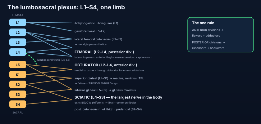
- The lumbar plexus (L1–L4, ± T12) is formed WITHIN psoas major; the sacral plexus (L4–S4) is formed ON piriformis in the pelvis; the lumbosacral trunk (L4–L5) bridges the two.
- Lumbar branches: iliohypogastric and ilioinguinal (L1), genitofemoral (L1–L2 — the only one to pierce the FRONT of psoas; cremasteric reflex), lateral femoral cutaneous (L2–L3).
- Femoral nerve (L2–L4) = POSTERIOR divisions, emerges LATERAL to psoas. Obturator nerve (L2–L4) = ANTERIOR divisions, emerges MEDIAL to psoas.
- Sacral branches: superior gluteal (L4–S1, ABOVE piriformis), inferior gluteal (L5–S2), sciatic (L4–S3, the largest nerve in the body), posterior cutaneous nerve of the thigh (S1–S3), nerves to quadratus femoris and obturator internus, and pudendal (S2–S4).
- Piriformis is the landmark: the superior gluteal nerve leaves ABOVE it, everything else BELOW it — including the sciatic nerve.
- THE RULE: anterior divisions → flexors and ADDUCTORS (obturator, tibial); posterior divisions → extensors and ABDUCTORS (femoral, superior/inferior gluteal, common fibular).
- Dermatomes: L1 groin, L2 anterior thigh, L3 knee, L4 medial leg and medial malleolus, L5 dorsum of foot and great toe, S1 lateral foot and sole, S2 back of thigh, S3–S5 perineum.
- Reflexes: knee jerk = L3–L4 (femoral nerve); ankle jerk = S1 (tibial nerve). Both are quick level-checks at the bedside.
- Myotomes: L2–L3 hip flexion, L3–L4 knee extension, L4–L5 dorsiflexion, L5 great-toe extension, S1–S2 plantarflexion.
- A posterolateral disc prolapse compresses the root numbered BELOW it: L4/L5 disc → L5 root; L5/S1 disc → S1 root.
- Meralgia paraesthetica = entrapment of the lateral femoral cutaneous nerve under the inguinal ligament: burning numb outer-thigh patch, NO weakness (purely sensory).
- Cauda equina syndrome is the red flag: saddle anaesthesia, new bladder/bowel disturbance and bilateral leg symptoms — image and decompress within hours.
- Saying a prolapsed disc compresses the root of the same number. It compresses the TRAVERSING root, which is the one numbered below — an L4/L5 disc produces an L5 syndrome, not an L4 one.
- Swapping the femoral and obturator divisions. Both are L2–L4, but the femoral is POSTERIOR division and lateral to psoas, while the obturator is ANTERIOR division and medial to psoas.
- Assuming the sacral plexus behaves like the brachial plexus, with trunks and cords. It has neither — only roots, anterior and posterior divisions, and named branches, formed in two separate sites rather than one.
A 45-year-old man has weakness of great-toe extension and dorsiflexion, with numbness over the dorsum of the foot. Both ankle jerks are normal. Which disc and which root are most likely involved?
- The lumbosacral plexus is the lower-limb twin of the brachial plexus, but simpler (no trunks or cords) and built in two places: the lumbar plexus (L1–L4) inside psoas major, the sacral plexus (L4–S4) on piriformis, joined by the lumbosacral trunk (L4–L5).
- Lumbar output: iliohypogastric and ilioinguinal, genitofemoral (pierces the front of psoas), lateral femoral cutaneous, and the two giants — femoral (posterior divisions, lateral border) and obturator (anterior divisions, medial border).
- Sacral output: superior gluteal above piriformis; below it the inferior gluteal, the sciatic (L4–S3, the largest nerve in the body), the posterior cutaneous nerve of the thigh and the pudendal (S2–S4).
- One rule and one trap: anterior divisions serve flexors and adductors, posterior divisions serve extensors and abductors — and a prolapsed disc hits the root numbered BELOW it (L4/L5 → L5).
- Drake RL, Vogl AW, Mitchell AWM. Gray's Anatomy for Students — Lower limb: the lumbar and sacral plexuses.
- Moore KL, Dalley AF, Agur AMR. Clinically Oriented Anatomy — Posterior abdominal wall and pelvis: nerves of the lower limb.
- Netter FH. Atlas of Human Anatomy — Lumbar plexus, sacral and coccygeal plexuses.
- Last RJ. Last's Anatomy: Regional and Applied — The posterior abdominal wall; the pelvis.
- Snell RS. Clinical Anatomy by Regions — Dermatomes, myotomes and intervertebral disc herniation.
- TeachMeAnatomy — The Lumbar Plexus; The Sacral Plexus.

