
The Uterus: An Organ That Multiplies Its Own Size Twenty-Fold
It is the size of a small pear, weighs about seventy grams, and its cavity is not a chamber but a slit — the two walls touch. Nine months later it is an organ that fills the abdomen to the ribs, weighs a kilogram, and holds a fetus, a placenta and a litre of fluid. Then, over six weeks, it goes back. No other organ in the human body does this, and does it repeatedly, and does it without scarring. And the anatomy that permits it — the interlacing muscle bundles, the ligaments that are not ligaments, the artery that crosses above a ureter two centimetres from the cervix — is also the anatomy that explains a torrential postpartum haemorrhage, a ruptured ectopic pregnancy, and the single most feared accident in gynaecological surgery.
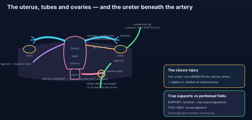
The operation has gone well. A hysterectomy for heavy bleeding in a fifty-year-old woman, an hour in, no surprises. The surgeon is now at the base of the broad ligament, working towards the cervix, and the field is bloodier than she would like. She reaches for the clamp — and stops. She puts the sucker down, asks for the pelvic sidewall to be exposed, and traces with a fingertip along the wall of the pelvis until she feels the soft, worm-like cord that gives a slow peristaltic wriggle under her finger. There it is. Two centimetres lateral to the cervix, running forwards and medially, and directly above it, crossing it at right angles, the uterine artery she was about to clamp. She lifts the artery clear, ligates it where she can see the ureter beneath, and the moment passes. Nothing dramatic happened, and that is exactly the point: the most consequential centimetre in the female pelvis is one where nothing dramatic is supposed to happen — and where a clamp placed blind, on a wet field, in a hurry, can cost a kidney.
Four parts, from the top down
Name the parts in the order a surgeon meets them, and every clinical fact hangs on one of them. The FUNDUS is the rounded dome lying ABOVE the level at which the uterine tubes enter — that definition matters, because the fundus is defined by the tubes, not by shape. The angle at each side where the tube meets the uterus is the CORNU (horn), and three structures converge on it: the uterine tube, the ligament of the ovary behind, and the round ligament in front. Below the fundus is the BODY (corpus), the thick muscular bulk, tapering downwards. Where it narrows, about a centimetre above the cervix, is the ISTHMUS — an unremarkable band in the non-pregnant uterus and one of the most important pieces of tissue in obstetrics, because during pregnancy the isthmus is taken up and stretched to form the LOWER UTERINE SEGMENT: thin, relatively avascular, covered by loose peritoneum that can be reflected off it, and therefore the site through which almost every caesarean section is performed. Finally the CERVIX, a firm cylinder of fibrous tissue with only a little smooth muscle, and the only part of the uterus you can see and touch in a living woman. It has a SUPRAVAGINAL portion above the vaginal attachment and a VAGINAL portion projecting into the vault, surrounded by the fornices. Its canal opens above into the uterine cavity at the INTERNAL OS and below into the vagina at the EXTERNAL OS — round in a woman who has never delivered, a transverse slit in one who has. The cavity itself is a slit: triangular in coronal section, with the tubal openings at the upper corners and the internal os at the apex below, and its two walls in apposition until something distends them.
Why it leans forward
The uterus is not a vertical organ. It sits in two bends, and the two have different names because they are measured between different pairs of structures. ANTEVERSION is the angle between the axis of the cervix and the axis of the VAGINA — about 90°, so the whole uterus is tipped forwards on the vagina. ANTEFLEXION is the angle between the axis of the BODY and the axis of the CERVIX — about 120°, so the body is additionally bent forwards on the cervix. Put the two together and the normal uterus lies almost horizontally, resting its anterior surface on the superior surface of the bladder, which is why a full bladder pushes it upright and why a transabdominal pelvic ultrasound is done with the bladder deliberately filled. This posture is not accidental. It means that any rise in intra-abdominal pressure — a cough, a lift, a sneeze — presses the uterine body DOWN onto the bladder and forwards, rather than driving the cervix straight down the axis of the vagina like a piston. Anteversion, in other words, is part of the anti-prolapse mechanism, and the structure that maintains it is the round ligament. In roughly one woman in five the uterus is RETROVERTED and often retroflexed instead — tipped backwards into the pouch of Douglas. It is a normal anatomical variant in the great majority: it does not cause infertility, and the older habit of blaming it for backache and dyspareunia has not survived scrutiny. It matters practically rather than pathologically — it changes the direction in which a sound, a coil or a curette must be passed, and a retroverted uterus that is FIXED (rather than freely mobile) is a different matter altogether, because fixity suggests adhesions or endometriosis tethering it in the pouch of Douglas.
Three layers, and the one that saves lives
From outside in: the PERIMETRIUM is simply peritoneum, adherent over most of the uterus but loose and easily stripped over the lower segment anteriorly — the surgeon's plane at caesarean. The MYOMETRIUM is the bulk of the organ: thick smooth muscle arranged in interlacing, criss-crossing bundles rather than neat circular and longitudinal coats, and it is this arrangement that does the work. During pregnancy its cells undergo enormous hypertrophy (and some hyperplasia), and in labour it contracts with a retraction that shortens the fibres permanently with each contraction rather than letting them return to length. After the placenta separates, the spiral arteries that fed it are open, raw vessels carrying something like five hundred millilitres of blood per minute. Nothing sutures them. What closes them is the myometrium itself: the interlacing bundles contract around the vessels running between them and strangle them shut — the LIVING LIGATURE, and the reason a woman does not exsanguinate in the ten minutes after delivery. Innermost is the ENDOMETRIUM, a glandular mucosa in two strata: a superficial FUNCTIONAL layer, supplied by the coiled spiral arteries, which proliferates, secretes and is shed each menstruation; and a deep BASAL layer, supplied by short straight arteries that do not respond to the hormonal withdrawal, which survives every period and regenerates the functional layer from below. That two-layer design is why menstruation is a controlled demolition and not an injury — and why destroying or scarring the basal layer, by over-vigorous curettage or infection, produces the permanent amenorrhoea of intrauterine adhesions.
Think of two ways to stop bleeding from a hosepipe running through a wall. The first is to find the pipe and tie it — that is what a surgeon does, and it needs the vessel to be visible, accessible and few in number. The second is to build the wall out of something that clenches: pack the pipe inside a mass of interwoven elastic cords and then pull all the cords tight at once, and every pipe running between them is crushed shut wherever it happens to lie, without anyone having to find it. The myometrium is the second design. That is the only workable answer when the bleeding surface is the whole placental bed and the vessels number in the hundreds. It also tells you instantly what to do when it fails. An atonic uterus — soft, boggy, high — is a wall that has stopped clenching, and the treatment is not a clamp but anything that makes the muscle contract again: rubbing the fundus through the abdominal wall, emptying the bladder, oxytocin, ergometrine, misoprostol, and if all else fails compressing the uterus between two hands or a suture. You are not repairing a pipe. You are restarting a grip.
The ligaments that are not ligaments
Half the confusion in this topic comes from calling a fold of peritoneum a ligament. The BROAD LIGAMENT is a double fold of peritoneum draped over the uterus and tubes and running out to the lateral pelvic wall — a sheet, not a strut. It is a fold of peritoneum like any other, and it supports the uterus about as effectively as a bedsheet supports a sleeping body. Its value is as a container and a map. Named subdivisions: the MESOSALPINX, the part between the tube above and the ovarian ligament below; the MESOVARIUM, the short posterior fold that attaches the ovary (note that the ovary is NOT inside the broad ligament — it hangs behind it); and the MESOMETRIUM, the large remainder below. Running within it are the uterine tube along its upper free edge, the round ligament passing forwards, the ligament of the ovary passing backwards, the uterine artery and venous plexus, lymphatics and nerves, and — critically — the URETER in the base, close to the cervix. At the lateral end, a fold running up over the ovarian vessels as they cross the pelvic brim is the SUSPENSORY LIGAMENT OF THE OVARY (infundibulopelvic ligament), described with the ovaries. The ROUND LIGAMENT is a genuine fibromuscular cord, but not a support in the vertical sense. It runs from the uterine cornu, forwards and laterally within the broad ligament, over the pelvic brim, through the DEEP INGUINAL RING, along the inguinal canal, and out of the superficial ring to fan into the LABIUM MAJUS. It is the female remnant of the GUBERNACULUM — the same structure that in the male drags the testis to the scrotum — and its job is to hold the fundus forwards, maintaining anteversion. Because it is a cord that must stretch as the uterus rises out of the pelvis, it is the source of ROUND LIGAMENT PAIN in the second trimester: a sharp, brief, one-sided groin pain on standing up, coughing or rolling over in bed, following exactly the line of the inguinal canal.
The TRUE supports are quite different structures, and they act at the level of the CERVIX and upper vagina — low down, where the leverage is. They are condensations of the pelvic fascia (the endopelvic fascia) carrying smooth muscle, and they are strong. The CARDINAL LIGAMENTS — also called the transverse cervical ligaments, or Mackenrodt's — fan out from the cervix and upper vagina to the lateral pelvic wall on each side, and they are the principal suspension of the uterus; the ureter and the uterine artery run in the upper part of this fascial mass. The UTEROSACRAL LIGAMENTS run backwards and upwards from the cervix and upper vagina, on either side of the rectum, to the front of the sacrum; they pull the cervix backwards, which is precisely what keeps the body of the uterus tipped forwards, and they raise the visible rectouterine folds that bound the pouch of Douglas. They also carry the sensory fibres from the cervix, which is why they are the target of uterosacral nerve ablation for dysmenorrhoea and why endometriotic deposits on them cause deep dyspareunia. The PUBOCERVICAL ligaments run forwards from the cervix to the back of the pubis, supporting the bladder neck along the way. And beneath all of them lies the true floor: the levator ani of the pelvic floor, whose fibres form a muscular shelf that the pelvic organs rest on. The clinical formula is worth stating plainly: the ligaments SUSPEND, the pelvic floor SUPPORTS, and prolapse follows failure of either — which is why hysterectomy alone does not cure a prolapse, and why the cardinal and uterosacral ligaments are deliberately re-attached to the vaginal vault when the uterus is removed.
The tube: four parts and one fatal preference
Each uterine tube is about 10 cm long, runs in the upper free border of the broad ligament, and is described in four parts from the ovary inwards. The INFUNDIBULUM is the funnel-shaped lateral end, opening into the peritoneal cavity through the abdominal ostium and fringed by finger-like FIMBRIAE that sweep over the ovarian surface at ovulation; one of them, the OVARIAN FIMBRIA, is longer than the rest and attached to the ovary, guiding the released oocyte to the opening. The AMPULLA is the next and by far the largest part — thin-walled, wide, tortuous, occupying about two thirds of the length — and it is the NORMAL SITE OF FERTILISATION. The ISTHMUS is the short, narrow, thick-walled segment nearest the uterus, and the INTRAMURAL (interstitial) part is the final centimetre or so tunnelling through the wall of the uterus itself to open at the cornu — the narrowest point in the whole passage. The wall is muscular and lined with a mucosa thrown into elaborate longitudinal folds and carpeted with ciliated and secretory cells: the cilia beat towards the uterus, the muscle performs slow peristalsis, and together they move the ovum medially over several days while the sperm travel the other way. Notice the anatomical oddity this creates. The tube is the only place where the peritoneal cavity opens to the outside world, so infection can climb from the vagina, through the uterus and tube, to the peritoneum — the route of pelvic inflammatory disease — and, in the other direction, the shed oocyte is briefly free in the peritoneal cavity before the fimbriae catch it.
A woman of twenty-eight presents with six weeks of amenorrhoea, a positive pregnancy test, and one-sided lower abdominal pain that has become severe over an hour. She is pale and tachycardic, her abdomen is tender, and she says her right shoulder tip aches. The anatomy explains every line of it. Fertilisation normally occurs in the AMPULLA, and if the conceptus is delayed there — by scarring from previous pelvic inflammatory disease, by damaged cilia after chlamydial salpingitis, by previous tubal surgery — it implants where it sits. The ampulla is therefore the commonest site of ectopic pregnancy. Its wall is thin and it cannot undergo the decidual and muscular adaptation that the uterus does, so at around six to eight weeks the trophoblast erodes through it and the tube ruptures. What follows is determined by the fact that the tube lies within the peritoneal cavity: the bleeding is INTRAPERITONEAL, not concealed, and it is arterial. Blood runs to the most dependent part, which in a woman lying or sitting is the pouch of Douglas — hence the exquisite pain on moving the cervix and the bulging, tender posterior fornix. As blood accumulates it reaches the undersurface of the diaphragm, whose parietal peritoneum is supplied by the PHRENIC nerve from C3–C5, and the pain is referred to the shoulder tip. Note the exception that matters most: an ectopic in the INTERSTITIAL (cornual) part sits inside the muscular uterine wall, so it can grow larger and rupture later — typically at ten to sixteen weeks — and when it does, it tears into the uterine and ovarian anastomosis and bleeds catastrophically.
- Parts: FUNDUS (above the tubal entries), BODY, ISTHMUS (becomes the lower uterine segment in pregnancy — the caesarean incision site), and CERVIX with supravaginal and vaginal portions, internal os, cervical canal and external os. The cavity is a triangular SLIT, not a chamber.
- Normal position = ANTEVERSION (cervix on vagina, about 90°) plus ANTEFLEXION (body on cervix, about 120°), so the uterus lies forward over the bladder. RETROVERSION occurs in about 20% and is a normal variant unless the uterus is fixed.
- Three layers: perimetrium (peritoneum, loose over the lower segment), MYOMETRIUM (interlacing smooth muscle — the LIVING LIGATURE that strangles the spiral arteries after delivery), and ENDOMETRIUM (functional layer shed each cycle, basal layer regenerating it).
- The BROAD LIGAMENT is a peritoneal fold, NOT a support: it contains the tube, round ligament, ligament of the ovary, uterine vessels, and the URETER in its base; subdivisions are mesosalpinx, mesovarium and mesometrium.
- The ROUND LIGAMENT (gubernaculum remnant) runs from the cornu through the deep inguinal ring and inguinal canal to the labium majus, maintains anteversion, and stretches in pregnancy to give round ligament pain. The TRUE supports are the CARDINAL (Mackenrodt's), UTEROSACRAL and pubocervical ligaments, plus the pelvic floor.
- Tube in four parts: infundibulum with fimbriae (one longer ovarian fimbria), AMPULLA (widest and longest — normal fertilisation site and commonest ectopic site), isthmus, and intramural/interstitial part (narrowest; a cornual ectopic ruptures late and bleeds catastrophically).
Blood, and the crossing that costs kidneys
Two arteries approach the uterus from opposite directions and meet in the middle. The UTERINE ARTERY is a branch of the anterior division of the internal iliac artery, described with the internal iliac artery. It runs medially on the levator ani, in the base of the broad ligament, to reach the side of the cervix; there it gives a descending vaginal branch and then turns UPWARDS, running a markedly tortuous course along the lateral border of the uterus — the tortuosity being slack that will be taken up as the organ enlarges in pregnancy. It gives arcuate arteries that encircle the uterus in the myometrium, from which radial arteries dive inwards and end as the straight arteries of the basal endometrium and the SPIRAL arteries of the functional layer. Near the cornu it ends by anastomosing with the OVARIAN artery, which comes from the opposite direction entirely: a direct branch of the abdominal aorta at about L2, reflecting the ovary's origin high on the posterior abdominal wall, descending over the pelvic brim in the suspensory ligament. The tubal branches of both meet in the mesosalpinx, so the tube is supplied from both ends. And here is the relation that dominates pelvic surgery: as the uterine artery crosses medially at the base of the broad ligament, the URETER passes BENEATH it, running forwards and medially about 2 CM LATERAL TO THE CERVIX on its way to the bladder. The mnemonic is WATER UNDER THE BRIDGE — water (the ureter) under the bridge (the artery). This crossing is the classic site of ureteric injury during hysterectomy, and it is also why a caesarean hysterectomy or a bleeding broad ligament haematoma is so dangerous: the tissue is oedematous, the field is bloody, and the ureter is displaced. The corresponding rule in surgery is absolute — identify the ureter before ligating the uterine artery, tracing it as described with the kidneys and ureters. Venous drainage follows a rich uterine venous plexus in the broad ligament, draining to the internal iliac veins; the ovarian veins take the other route, the right to the inferior vena cava and the left to the left renal vein.
Lymphatic drainage of the uterus is the one topic students try to simplify and cannot, because the uterus does not drain to one place — it drains according to which piece of it is asking, and each route is a fossil of its development or of a ligament passing through it. The FUNDUS and upper body follow the ovarian vessels back up to their origin and drain to the PARA-AORTIC nodes at L1–L2, exactly as the ovary and testis do, because these structures began high on the posterior abdominal wall. Most of the BODY drains to the EXTERNAL ILIAC nodes. A small but famous trickle from the CORNU follows the ROUND LIGAMENT through the inguinal canal to the SUPERFICIAL INGUINAL nodes — which is why a woman may present with an enlarged groin node from a uterine tumour. The CERVIX drains in three directions: laterally in the cardinal ligaments to the INTERNAL ILIAC and obturator nodes, forwards to the EXTERNAL ILIAC nodes, and backwards along the uterosacral ligaments to the SACRAL nodes. Read that list once more as a surgeon: it is the reason a radical hysterectomy for cervical carcinoma must take the parametrium and a pelvic node dissection, while an endometrial carcinoma of the fundus demands attention to the para-aortic chain that a pelvic dissection alone would miss entirely.
Two kinds of pain from one organ
The uterus is supplied by the UTEROVAGINAL PLEXUS, an extension of the INFERIOR HYPOGASTRIC (pelvic) plexus described in the autonomic nerves of the pelvis. Its sympathetic fibres come from T10–L1 through the hypogastric nerves and superior hypogastric plexus; its parasympathetic fibres come from the PELVIC SPLANCHNIC nerves, S2–S4. Motor control of the myometrium is largely hormonal rather than neural — a denervated uterus still labours and still delivers, which is why a woman with a complete spinal cord transection can have an entirely normal vaginal delivery. What the nerves carry that matters clinically is PAIN, and the visceral afferents divide at a line that runs roughly through the internal os. Pain from the BODY of the uterus — the pain of a contraction, of dysmenorrhoea, of a degenerating fibroid — travels with the SYMPATHETIC fibres back to spinal segments T10–L1. It is therefore referred to the dermatomes of those segments: the lower abdomen from umbilicus to pubis, the sacral and low back region, and sometimes the upper thigh. Pain from the CERVIX and upper vagina — the pain of cervical dilatation, and the stimulus of passing an instrument through the os — travels the other way, with the PARASYMPATHETIC pelvic splanchnics to S2–S4. This split is not academic. It is the reason a PUDENDAL NERVE BLOCK, which anaesthetises the perineum for an instrumental delivery or an episiotomy, does absolutely nothing for the pain of contractions: the pudendal nerve is S2–S4 somatic, and contraction pain has already gone home to T10–L1. It is also why epidural analgesia in labour must reach as high as T10 to abolish first-stage pain, and why paracervical block relieves cervical dilatation pain but not uterine cramping.
Caesarean section was once performed through a vertical incision in the upper body of the uterus — the classical caesarean — and it carried a fearsome rate of infection, adhesions and later rupture. The modern operation cuts transversely through the LOWER UTERINE SEGMENT instead, and every advantage is anatomical. The lower segment is derived from the isthmus, so its muscle is thin and it is comparatively avascular, lying away from the ascending uterine arteries at the sides. It is covered by loosely attached peritoneum that can be incised and pushed down, taking the bladder with it, so the uterine wound is later covered by peritoneum and bladder rather than lying free against bowel — which is why adhesions are fewer. It is the passive segment in labour: it stretches rather than contracts, so a scar there is under far less tension in a subsequent pregnancy, and the risk of rupture in a later labour is a fraction of that after a classical incision. And it is behind the bladder, which is exactly why the bladder must be reflected and why bladder injury is the commonest visceral complication of a repeat caesarean, when adhesions have pulled it up onto the scar. As detailed in the anatomy of childbirth, the whole operation is an argument about which piece of uterine wall you choose to cut, and the answer is the piece that will not have to contract afterwards.
- The UTERINE ARTERY (from the internal iliac) runs in the base of the broad ligament to the side of the cervix, then ascends tortuously to anastomose with the OVARIAN artery (from the aorta at L2, descending in the suspensory ligament).
- WATER UNDER THE BRIDGE: the ureter passes BENEATH the uterine artery about 2 cm lateral to the cervix — the classic site of ureteric injury at hysterectomy. Identify the ureter before clamping.
- Lymphatics differ by region: fundus and upper body → PARA-AORTIC (along the ovarian vessels); most of the body → external iliac; cornu → superficial inguinal (along the round ligament); cervix → internal iliac, external iliac and sacral nodes.
- Nerve supply = uterovaginal plexus from the inferior hypogastric plexus. Pain from the BODY travels with SYMPATHETICS to T10–L1 (referred to lower abdomen and back); pain from the CERVIX with PARASYMPATHETICS to S2–S4.
- A pudendal block (S2–S4 somatic) anaesthetises the perineum but NOT uterine contraction pain; an epidural must reach T10 to abolish first-stage labour pain.
- Fibroids (leiomyomas) are classified by location — submucosal (bleeding and infertility), intramural, and subserosal (pressure and pedunculated masses) — while endometriosis is functioning endometrium outside the uterus, commonly on the ovaries and uterosacral ligaments.
- Calling the broad ligament a support of the uterus. It is a peritoneal fold — a sheet that carries the tube, round ligament, uterine vessels and the ureter, and divides into mesosalpinx, mesovarium and mesometrium. The genuine suspension is the cardinal and uterosacral ligaments at cervical level, over the muscular pelvic floor.
- Confusing anteversion with anteflexion. Anteversion is the angle of the CERVIX on the VAGINA (about 90°); anteflexion is the angle of the BODY on the CERVIX (about 120°). They are measured between different pairs of structures, and a uterus can be anteverted but retroflexed.
- Reversing the ureter–artery relation. The URETER is the water and passes UNDERNEATH; the uterine ARTERY is the bridge and passes above, about 2 cm lateral to the cervix. Getting this the wrong way round in an exam is a wrong answer; getting it wrong in theatre is a divided ureter.
During a total abdominal hysterectomy the surgeon is about to clamp and ligate the uterine artery at the level of the cervix. Which structure lies immediately at risk, and what is its precise relation to the artery at that point?
- The uterus has a fundus (above the tubal entries), body, isthmus (which becomes the lower uterine segment and the caesarean incision site) and cervix (supravaginal and vaginal portions, internal os, canal, external os), with a slit-like triangular cavity; it normally lies ANTEVERTED on the vagina (about 90°) and ANTEFLEXED on the cervix (about 120°), resting on the bladder, with retroversion a normal variant in about 20%.
- Three layers: perimetrium, MYOMETRIUM — interlacing bundles that clamp the spiral arteries shut after delivery, the living ligature behind uterine massage and oxytocin for postpartum haemorrhage — and ENDOMETRIUM with its shed functional layer and regenerating basal layer.
- The broad ligament is a PERITONEAL FOLD carrying the tube, round ligament, ovarian ligament, uterine vessels and the ureter in its base; the round ligament (gubernaculum remnant) runs to the labium majus and maintains anteversion; the TRUE supports are the cardinal, uterosacral and pubocervical ligaments over the pelvic floor. The tube runs infundibulum with fimbriae → AMPULLA (fertilisation and commonest ectopic) → isthmus → intramural part.
- Uterine artery (internal iliac) anastomoses with the ovarian artery (aorta, L2), and the URETER passes BENEATH it 2 cm lateral to the cervix — water under the bridge, the classic hysterectomy injury. Lymph: fundus → para-aortic, body → external iliac, cornu → superficial inguinal, cervix → internal iliac/external iliac/sacral. Pain: body with sympathetics to T10–L1, cervix with parasympathetics to S2–S4 — so a pudendal block does not touch contraction pain.
- Drake RL, Vogl AW, Mitchell AWM. Gray's Anatomy for Students — Pelvis and Perineum: the uterus, uterine tubes and their supports.
- Moore KL, Dalley AF, Agur AMR. Clinically Oriented Anatomy — Female internal genital organs; the ureter and uterine artery relation; lymphatic drainage of the uterus.
- Netter FH. Atlas of Human Anatomy — Uterus, uterine tubes and ovaries; ligaments of the female pelvis; pelvic vasculature.
- Last RJ. Last's Anatomy: Regional and Applied — The uterus, broad ligament and endopelvic fascia.
- Snell RS. Clinical Anatomy by Regions — Clinical notes on hysterectomy, ectopic pregnancy and caesarean section.
- TeachMeAnatomy — The Uterus; The Uterine Tubes; Ligaments of the Female Reproductive Tract.

