
The Shoulder: Mobility Bought With Instability
No other joint in your body moves like this one. You can sweep your arm through a full circle, scratch the middle of your own back, hurl a ball at 150 km/h, and lift a child clean over your head — all through a single joint that has almost no bony socket to speak of. The head of the humerus does not sit inside the shoulder blade so much as it leans against it, like a golf ball resting on a tee. Every degree of that extraordinary freedom was bought by giving up bony containment, and the bill arrives as the single most commonly dislocated major joint in the human body. Understand that trade-off and the whole shoulder — its ligaments, its cuff, its injuries — falls into place.
A swimmer touches the wall, a violinist holds her bow steady through a long phrase, a painter reaches above his head for the eighth hour running. Watch a toddler being swung by the arms and laughing, then watch a rugby player go down with his arm flung out sideways and get up cradling it, the shoulder squared off and the arm held slightly away from his body. In one afternoon the same joint has behaved as a precision instrument, an endurance machine, and a catastrophe. The shoulder is where the body chose freedom over safety — and unlike the hip, which locks its ball deep into a bony cup, the shoulder leaves the head of the humerus almost out in the open, held by soft tissue alone. Everything you will learn here is a consequence of that one design decision.
The glenohumeral joint: a golf ball on a tee
A synovial ball-and-socket joint in which the socket barely deserves the name. The glenohumeral joint is formed between the large, roughly hemispherical head of the humerus and the shallow, pear-shaped glenoid cavity (glenoid fossa) on the lateral angle of the scapula — the bones described in the clavicle and scapula and the humerus. The mismatch is the whole story: the humeral head presents an articular surface roughly three to four times the area of the glenoid, so only about a third of it is in contact at any moment. Both surfaces are covered in hyaline cartilage, thicker centrally on the glenoid and thicker peripherally on the head. As a multiaxial ball-and-socket it permits flexion and extension, abduction and adduction, medial and lateral rotation, and — combining all of these — circumduction: the full cone of movement that no other joint in the body can complete. The general principle is the familiar one from how joints trade stability for range, pushed to its extreme.
The labrum, the capsule and the ligaments that barely hold
Around the rim of the glenoid runs the glenoid labrum, a fibrocartilaginous collar that deepens the socket by roughly 50% and widens the contact area, acting like a rubber gasket that grips the head. Superiorly the labrum blends with the tendon of the long head of biceps brachii — which is why a forceful traction injury can peel the labrum off from front to back, the SLAP lesion (Superior Labrum Anterior to Posterior). The fibrous capsule attaches medially to the margin of the glenoid (just beyond the labrum) and laterally to the anatomical neck of the humerus, dipping down onto the surgical neck medially. Crucially it is lax — deliberately redundant inferiorly, folding into an axillary recess that unfurls to let you take the arm overhead. Reinforcing it anteriorly are the three glenohumeral ligaments — superior, middle and inferior — thickenings of the capsule rather than separate cords; the inferior glenohumeral ligament forms a hammock-like sling that becomes the key restraint against anterior dislocation when the arm is abducted and laterally rotated. The coracohumeral ligament runs from the coracoid process to the greater tubercle and helps suspend the head against gravity, while the transverse humeral ligament bridges the greater and lesser tubercles to hold the long head of biceps inside the intertubercular (bicipital) groove.
Picture a heavy ball balanced on a shallow saucer, with four straps buckled across it — one over the top, one at the front, two at the back — but nothing at all underneath. As long as the straps are tight and pull in unison, the ball is pinned to the saucer and can spin freely in any direction. Let one strap fray, or push the ball hard downwards and forwards where there is no strap at all, and it rolls straight off the rim. Those four straps are the rotator cuff, and the missing one underneath is exactly why a dislocated shoulder almost always escapes downwards and forwards.
The rotator cuff: the real socket is made of muscle
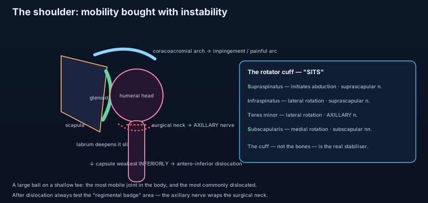
Four muscles — SITS — whose tendons fuse with the capsule and hold the head onto the glenoid. The rotator cuff is the true stabiliser of the shoulder, and its members are remembered as SITS. Supraspinatus arises from the supraspinous fossa of the scapula, passes under the acromion and inserts on the superior facet of the greater tubercle; it initiates abduction (the first ~15°) and is supplied by the suprascapular nerve (C5–C6). Infraspinatus arises from the infraspinous fossa and inserts on the middle facet of the greater tubercle; it is a lateral rotator, also supplied by the suprascapular nerve (C5–C6). Teres minor runs from the lateral border of the scapula to the inferior facet of the greater tubercle, is likewise a lateral rotator, and is the odd one out in its innervation — the axillary nerve (C5–C6). Subscapularis, the only anterior member, arises from the subscapular fossa on the costal surface of the scapula and inserts on the lesser tubercle; it is the powerful medial rotator, supplied by the upper and lower subscapular nerves (C5–C6). All four are branches of the brachial plexus, and their tendons blend into the capsule itself so that every contraction pulls the humeral head into the glenoid — dynamic compression rather than passive bracing. Note the crucial gap: the cuff covers the joint superiorly, anteriorly and posteriorly, but there is no cuff tendon inferiorly. That unguarded inferior quadrant, combined with the lax axillary recess, is why dislocation is characteristically antero-inferior. Their bulk and their partnership with deltoid and the scapular movers is taken further in the muscles that move the shoulder.
Bursae, the coracoacromial arch, and the painful arc
Above the joint, the coracoid process, the acromion and the coracoacromial ligament stretched between them form the coracoacromial arch — a rigid osseoligamentous roof that prevents upward dislocation of the humeral head but leaves only a narrow subacromial space beneath it. Gliding within that space is the subacromial bursa, which usually communicates with the subdeltoid bursa to form a single large sac lying between the supraspinatus tendon below and the acromion and deltoid above. Its job is frictionless gliding as the arm rises. Narrow that space — by a bony spur, by a thickened or inflamed cuff tendon, or by a scapula that fails to rotate properly — and the supraspinatus tendon and bursa are pinched against the arch on every elevation: subacromial impingement. Clinically it produces the painful arc, pain concentrated between roughly 60° and 120° of abduction, easing above and below because that is the range in which the tendon passes directly under the acromion. A second bursa, the subscapular bursa, lies between the subscapularis tendon and the scapular neck and normally communicates with the joint cavity itself.
The shoulder is not one joint but four
Raising your arm overhead is a committee decision. The shoulder complex is a linked chain of four articulations. The glenohumeral joint provides most of the rotation. The acromioclavicular (AC) joint is a small plane synovial joint between the lateral end of the clavicle and the acromion, with a fibrocartilaginous articular disc; it is stabilised weakly by its own acromioclavicular ligament and powerfully by the coracoclavicular ligament, which has two named parts — the conoid (medial, cone-shaped) and the trapezoid (lateral, flat). These are the true suspensory ligaments of the upper limb: they transmit the weight of the arm to the clavicle. Tear them in a fall onto the point of the shoulder and the scapula drops away from the clavicle, producing the visible step deformity of a shoulder separation (an AC joint injury — quite distinct from a dislocation of the glenohumeral joint). The sternoclavicular (SC) joint is a saddle-type synovial joint with a complete articular disc, and it is the only bony articulation between the entire upper limb and the axial skeleton — everything else is muscle. Finally the scapulothoracic 'joint' is not a true joint at all but a gliding surface where the subscapularis and serratus anterior slide over the thoracic wall, allowing the scapula to protract, retract, elevate, depress and — most importantly — rotate.
💡 Scapulohumeral rhythm is the reason you can reach the top shelf. Of the roughly 180° of full arm elevation, only about 120° happens at the glenohumeral joint — the remaining 60° comes from the scapula rotating upward on the chest wall, in a smooth ratio of about 2:1 (two degrees of humeral movement for every one degree of scapular rotation). Trapezius and serratus anterior drive that scapular rotation. This is why a patient with a paralysed serratus anterior (long thoracic nerve injury, with the classic winged scapula) cannot raise the arm fully above the head no matter how healthy the shoulder joint itself is: the socket has stopped turning to follow the ball.
The arm is abducted and forcibly externally rotated as the player is tackled — the exact position in which the inferior glenohumeral ligament is stretched taut and the head is levered forwards over the anterior rim. The head slips antero-inferiorly beneath the coracoid, tearing the anterior labrum off the glenoid (a Bankart lesion) and often denting the posterolateral humeral head against the rim (a Hill–Sachs lesion). The patient supports the limb in slight abduction, unable to adduct it; the normally rounded deltoid contour flattens into a squared-off shoulder with a sulcus below the acromion. Now examine the axillary nerve, which winds around the surgical neck of the humerus through the quadrangular space and is the nerve most at risk here: test sensation over the 'regimental badge' area on the upper lateral arm, and test deltoid power once reduced — the details of its course are laid out in the axillary and radial nerves. Always document that examination before and after reduction. Analgesia for the reduction and for the aching days afterwards usually leans on the anti-inflammatory drugs described in how NSAIDs work.
When the cuff fails, and when the capsule freezes
Rotator cuff tears most often involve supraspinatus, and they are common in the elderly for a reason worth remembering: the tendon has a relatively hypovascular zone near its insertion, it is repeatedly compressed under the coracoacromial arch, and decades of overhead use degenerate it. So while a young cuff usually tears only with major trauma, an older cuff can fail with a trivial pull or even spontaneously. Clinically the patient cannot initiate abduction — if the examiner passively lifts the arm past the first 15°, deltoid can take over and complete the movement, the classic sign of an isolated supraspinatus tear. Adhesive capsulitis ('frozen shoulder') is a different animal: the capsule itself becomes inflamed, thickened and contracted, obliterating the axillary recess, and both active and passive movement are lost — especially external rotation. It runs a long course of freezing, frozen and thawing phases over months to years, and is associated with diabetes, thyroid disease and prolonged immobilisation. The distinction is clinical and simple: a cuff tear limits active movement while passive range is preserved; a frozen shoulder limits both.
- Glenohumeral = multiaxial synovial ball-and-socket; a large humeral head on a shallow glenoid, only ~a third in contact at any time.
- The glenoid labrum deepens the socket by ~50%; the fibrous capsule is lax and redundant inferiorly to permit abduction.
- Ligaments: superior, middle and inferior glenohumeral (capsular thickenings), coracohumeral (suspends the head), transverse humeral (holds the long head of biceps in its groove).
- The coracoacromial arch (coracoid + acromion + coracoacromial ligament) roofs the joint and prevents upward dislocation.
- The subacromial/subdeltoid bursa glides under that arch; narrowing it produces impingement and a painful arc at ~60–120° of abduction.
- Rotator cuff = SITS: Supraspinatus, Infraspinatus, Teres minor, Subscapularis — tendons fused with the capsule, compressing the head into the glenoid.
- Actions/nerves: supraspinatus initiates abduction (suprascapular n.); infraspinatus + teres minor laterally rotate (suprascapular n. / axillary n.); subscapularis medially rotates (upper + lower subscapular nn.).
- No cuff tendon inferiorly → dislocation is typically antero-inferior, with the axillary nerve at risk (test the regimental badge area).
- The complex = glenohumeral + acromioclavicular (conoid + trapezoid coracoclavicular ligaments; separation) + sternoclavicular (the only bony link to the axial skeleton) + scapulothoracic gliding surface.
- Scapulohumeral rhythm ≈ 2:1 — ~120° glenohumeral + ~60° scapular rotation gives ~180° of elevation.
- Cuff tear limits active movement with preserved passive range; frozen shoulder (adhesive capsulitis) limits both, especially external rotation.
- Confusing a shoulder dislocation with a shoulder separation. A dislocation is the humeral head leaving the glenoid; a separation is an acromioclavicular injury with tearing of the coracoclavicular ligaments — different joint, different sign, different treatment.
- Assuming teres minor is supplied by the suprascapular nerve like its neighbours. Three of the four cuff muscles follow the suprascapular/subscapular pattern, but teres minor is supplied by the axillary nerve.
- Saying the humerus alone lifts the arm to 180°. The glenohumeral joint contributes only about 120°; the rest is scapular upward rotation — miss that and you cannot explain why a winged scapula blocks overhead reach.
A young man dislocates his shoulder while his abducted arm is forced into lateral rotation. Which nerve must be specifically tested, and where?
- The glenohumeral joint is a ball-and-socket with a large humeral head on a shallow glenoid, deepened only ~50% by the fibrocartilaginous labrum and enclosed by a deliberately lax capsule with an inferior axillary recess.
- Passive restraints are the superior, middle and inferior glenohumeral ligaments, the coracohumeral ligament and the transverse humeral ligament — but the true stabiliser is the rotator cuff (SITS), whose tendons blend with the capsule and pull the head into the socket.
- The cuff is absent inferiorly, so dislocation is characteristically antero-inferior with Bankart/Hill–Sachs lesions and axillary nerve injury; the coracoacromial arch above produces impingement and the 60–120° painful arc.
- The shoulder complex also includes the acromioclavicular joint (conoid + trapezoid ligaments; separation), the sternoclavicular joint (the only bony link to the axial skeleton) and the scapulothoracic gliding surface, working together in a ~2:1 scapulohumeral rhythm to reach overhead.
- Drake RL, Vogl AW, Mitchell AWM. Gray's Anatomy for Students — Upper Limb: the glenohumeral joint and shoulder complex.
- Moore KL, Dalley AF, Agur AMR. Clinically Oriented Anatomy — Pectoral girdle and glenohumeral joint; rotator cuff.
- Netter FH. Atlas of Human Anatomy — Shoulder: joints, ligaments and rotator cuff plates.
- Sinnatamby CS. Last's Anatomy: Regional and Applied — The shoulder region.
- Snell RS. Clinical Anatomy by Regions — The upper limb: shoulder joint and its clinical anatomy.
- TeachMeAnatomy — The Shoulder Joint and the Rotator Cuff Muscles.

