
Arteries of the Upper Limb: One Vessel, Many Names
Press two fingers into your wrist, just lateral to the tendon at the front, and you will feel it: a soft, insistent tap, sixty or seventy times a minute. That pulse has travelled a remarkable road. It began as a jet leaving the heart, curved through the arch of the aorta, slipped over the first rib, threaded the armpit, ran down the inside of your arm, split in the crease of your elbow, and arrived under your fingertip. And here is the strange, beautiful part: along that whole road it is essentially one continuous tube. It is not five arteries in series — it is one artery that changes its name every time it passes a landmark, like a river renamed at each town it flows through.
A nurse wraps a cuff around a man's arm and inflates it until the vessel beneath is squeezed shut. She places her stethoscope in the crease of his elbow, just medial to the hard cord of the biceps tendon, and lets the pressure fall. At a precise moment, sound returns — a knocking, then a whooshing, then silence again. Two numbers are written down, and from them a whole cardiovascular story is read. In the next room, an anaesthetist lifts a patient's wrist, feels for the tap of one artery, and slides in a fine cannula that will report the blood pressure beat by beat for the next six hours. Down the corridor, a surgeon is harvesting a length of that very same vessel from a forearm, to graft it onto a diseased coronary artery in the chest. Three procedures, three rooms — and all three are working on branches of a single arterial stream, one that has been quietly running the length of the limb since before any of them were born.
One stream, four names, three landmarks
Learn the three renaming points and you have learned the whole map. The limb's blood begins at the subclavian artery. On the right it arises from the brachiocephalic trunk; on the left it springs directly from the arch of the aorta — an asymmetry left over from how the embryonic arch vessels remodel, and the reason the left subclavian is a little longer. The subclavian arches over the lateral border of the FIRST RIB, and at that exact edge its name changes: it becomes the axillary artery. The axillary artery crosses the armpit and, at the lower border of TERES MAJOR, becomes the brachial artery. The brachial artery runs down the front of the arm and, in the CUBITAL FOSSA at the level of the neck of the radius, divides into its two terminal branches — the radial and ulnar arteries — which run the forearm and end by forming the arches of the palm. First rib, teres major, cubital fossa: three landmarks, and every name in the limb falls into place. The general logic of arteries, capillaries and veins that this stream obeys is laid out in the body's cardiovascular plan.
Think of a single motorway running the length of a country. It does not change its tarmac at the county lines, but its number changes: the M1 becomes the A1 at one bridge, the A1 becomes the coast road at a river, and the coast road forks at a roundabout into two lanes that rejoin later in the capital. Nobody rebuilt the road at those points — the signs simply changed. The subclavian–axillary–brachial stream is exactly that: one continuous tube of muscular artery whose name flips at the first rib, at teres major, and at the elbow. And like a good road network, it is not a dead end: the two forks — radial and ulnar — meet again in the palm, so if one lane is blocked, traffic still reaches the fingers.
The subclavian artery: the limb's doorway
Before it ever reaches the arm, the subclavian gives four branches that matter enormously — three of them to structures far from the limb. The vertebral artery climbs through the transverse foramina of the cervical vertebrae to enter the skull and supply the hindbrain. The internal thoracic artery descends behind the costal cartilages to supply the anterior chest wall (and is the workhorse graft of coronary bypass surgery). The thyrocervical trunk is a short stump that immediately splits — classically into the inferior thyroid, suprascapular and transverse cervical arteries — feeding the thyroid, the neck and the scapular region. And the costocervical trunk supplies the upper posterior intercostal spaces and the deep neck. The artery itself runs behind the anterior scalene muscle, in the groove on the first rib, with the trunks of the brachial plexus pressed against it above and behind — which is exactly why a cervical rib or a tight scalene can compress artery and nerves together in thoracic outlet syndrome.
The axillary artery: three parts, six branches
Pectoralis minor lies across the artery like a strap, and anatomists count from it. The axillary artery is divided into three parts by the overlying pectoralis minor, and the branch count is beautifully simple: one, two, three. The FIRST part (proximal to the muscle) gives one branch — the superior (supreme) thoracic artery, to the upper chest wall. The SECOND part (behind the muscle) gives two — the thoracoacromial artery, which bursts through the clavipectoral fascia and fans into four terminal twigs (acromial, clavicular, deltoid, pectoral), and the lateral thoracic artery, which runs along the lateral chest wall and supplies the breast. The THIRD part (distal to the muscle) gives three — the subscapular artery, the largest branch of them all, which divides into the circumflex scapular (passing through the triangular space to the back of the scapula) and the thoracodorsal artery to latissimus dorsi; and the two circumflex humeral arteries, anterior and posterior, which encircle the surgical neck of the humerus and anastomose with one another. The posterior circumflex humeral is the one to remember: it passes through the quadrangular space together with the axillary nerve, so a fracture of the surgical neck or an anterior shoulder dislocation can injure vessel and nerve in one stroke — the mechanism unpacked in the radial, axillary and musculocutaneous nerves.
The brachial artery: the vessel medicine touches most
Below teres major the stream becomes the brachial artery. It begins medial in the arm and gradually spirals to lie in front of the elbow, running on the medial side of the biceps, in the groove between biceps and triceps, with the median nerve crossing it from lateral to medial partway down. Its branches are three worth naming. The profunda brachii (deep artery of the arm) is the largest: it leaves early and travels with the RADIAL nerve in the radial (spiral) groove on the back of the humerus — so a mid-shaft humeral fracture threatens artery and radial nerve together. Then come the superior ulnar collateral (which accompanies the ulnar nerve behind the medial epicondyle) and the inferior ulnar collateral, both of which descend to join the rich anastomotic net around the elbow. Superficial and easily compressed, the brachial artery is where blood pressure is measured: the cuff occludes it, and the stethoscope listens over it in the cubital fossa, medial to the biceps tendon — the spot mapped in the axilla, cubital fossa and carpal tunnel.
A seven-year-old falls onto an outstretched hand and breaks the humerus just above the elbow — a supracondylar fracture, the commonest elbow fracture of childhood. The sharp proximal fragment is driven forwards, straight towards the brachial artery lying in front of it. If the vessel is torn, kinked or simply squeezed by the swelling inside a tight fascial compartment, the forearm's blood supply fails. The warning signs are the classic ones: pain out of proportion, pain on passive extension of the fingers, pallor, a cold hand, absent radial pulse. If it is not relieved in hours, the forearm flexor muscles die and heal as fibrous, shortened scar, pulling the wrist and fingers into a permanent claw — Volkmann's ischaemic contracture. This is why every child with a supracondylar fracture has the radial pulse checked, and why a white, cold, pulseless hand after an elbow injury is a surgical emergency rather than a thing to observe overnight.
Radial and ulnar: the forearm's two lanes
The fork happens in the cubital fossa, and from there the limb has two independent supplies. The RADIAL artery is the smaller but straighter of the two. It runs down the lateral forearm under the belly of brachioradialis, then emerges to lie superficially at the wrist, immediately LATERAL to the tendon of flexor carpi radialis and directly over the distal radius — the classic pulse point, palpable because a hard bone lies behind it. It then swings dorsally, crossing the floor of the anatomical snuffbox between the tendons of the thumb, before diving between the heads of the first dorsal interosseous into the palm to form the deep palmar arch. Its accessibility explains its clinical career: arterial line insertion, arterial blood gas sampling, the radial approach for coronary angiography, and harvesting as a coronary bypass graft. The ULNAR artery is the larger. It passes deep to the flexor muscles, then runs down the medial forearm alongside the ulnar nerve, entering the hand superficial to the flexor retinaculum through Guyon's canal to form the superficial palmar arch. Early on it gives the common interosseous artery, a short trunk that immediately divides into the anterior interosseous (running on the front of the interosseous membrane) and the posterior interosseous (passing above the membrane to supply the extensor compartment) — a two-vessel supply for the forearm's deep muscles, described alongside them in the muscles of the forearm. Both arteries also give recurrent branches that climb back up to join the anastomosis around the elbow.
The palmar arches: why the hand has a spare
In the palm, the two lanes rejoin — twice. The SUPERFICIAL palmar arch lies just under the palmar aponeurosis and is formed mainly by the ULNAR artery, completed laterally by the superficial palmar branch of the radial. From its convexity spring three or four common palmar digital arteries, which run distally and split at the web spaces into the proper palmar digital arteries — the pair of vessels that flank each finger along its sides, one on each border. The DEEP palmar arch lies deeper, across the bases of the metacarpals, and is formed mainly by the RADIAL artery, completed medially by the deep palmar branch of the ulnar; it gives palmar metacarpal arteries that join the common digitals. Two arches, both fed from both sides, is a remarkable piece of engineering for an organ that grips hot pans, is compressed against handlebars, and is asked to work while flexed and squeezed. It also has a direct clinical consequence: before puncturing or harvesting the radial artery, clinicians perform ALLEN'S TEST — compress both radial and ulnar arteries until the palm blanches, release the ulnar alone, and watch. If colour floods back within seconds, the ulnar supply and the arches are adequate and the radial can safely be sacrificed. If the hand stays pale, do not touch the radial artery.
The body builds detours before it needs them. Around the shoulder, the suprascapular and transverse cervical arteries (from the subclavian, via the thyrocervical trunk) anastomose freely with the circumflex scapular and thoracodorsal arteries (from the axillary) — the SCAPULAR ANASTOMOSIS. This means the third part of the axillary artery can be ligated between the origins of the subscapular and the thyrocervical branches and the limb will still be perfused: blood simply flows subclavian → suprascapular/transverse cervical → circumflex scapular → back into the axillary beyond the block. The same idea repeats at the elbow (collateral and recurrent branches weaving a net) and in the palm (the two arches). Study those anastomoses and you learn something deep about vascular design: a limb that must survive being squeezed, twisted and slept on cannot be supplied by a single unbranched pipe. When a clot does form in such a network, the drugs that fight it are mapped in the haemostasis drug map.
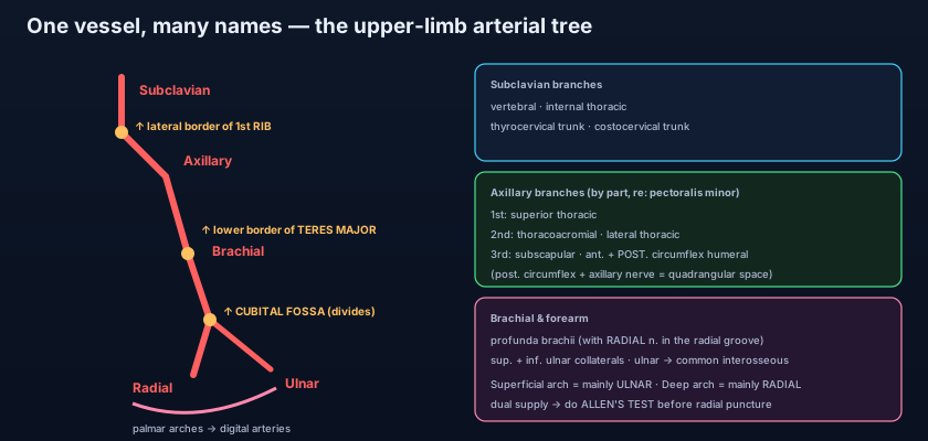
- One continuous stream renamed three times: subclavian → axillary (at the lateral border of the FIRST RIB) → brachial (at the lower border of TERES MAJOR) → radial + ulnar (dividing in the CUBITAL FOSSA).
- Right subclavian arises from the brachiocephalic trunk; left subclavian arises directly from the arch of the aorta.
- Subclavian branches: vertebral, internal thoracic, thyrocervical trunk, costocervical trunk.
- Axillary artery — one, two, three by part: 1st = superior thoracic; 2nd = thoracoacromial + lateral thoracic; 3rd = subscapular (largest) + anterior and posterior circumflex humeral.
- The posterior circumflex humeral artery travels with the axillary nerve through the quadrangular space — both at risk in surgical-neck fracture and anterior shoulder dislocation.
- Brachial branches: profunda brachii (with the RADIAL nerve in the radial groove), superior and inferior ulnar collaterals.
- Blood pressure is measured over the brachial artery; its pulse is felt in the cubital fossa MEDIAL to the biceps tendon.
- Supracondylar humeral fracture endangers the brachial artery → forearm ischaemia → Volkmann's ischaemic contracture.
- The radial pulse is felt at the wrist LATERAL to flexor carpi radialis; the radial artery then crosses the anatomical snuffbox to the deep palm.
- The ulnar artery gives the common interosseous artery, which divides into anterior and posterior interosseous arteries.
- Superficial palmar arch = mainly ULNAR (→ common then proper palmar digital arteries); deep palmar arch = mainly RADIAL.
- Allen's test checks the ulnar/arch supply before radial puncture, cannulation or graft harvest.
- Thinking the subclavian, axillary and brachial arteries are three separate vessels. They are one continuous artery whose name changes at the first rib and at teres major — nothing branches or restarts at those points.
- Swapping the two palmar arches. The SUPERFICIAL arch is mainly ULNAR; the DEEP arch is mainly RADIAL — remember that the superficial arch, like the ulnar nerve, enters the hand superficial to the retinaculum.
- Feeling for the brachial pulse in the wrong place. It is MEDIAL to the biceps tendon in the cubital fossa; the radial pulse, by contrast, is LATERAL to the flexor carpi radialis tendon at the wrist.
The axillary artery becomes the brachial artery at which anatomical landmark?
- The upper limb is supplied by one continuous artery renamed at landmarks: subclavian → axillary (lateral border of the first rib) → brachial (lower border of teres major) → radial and ulnar (division in the cubital fossa).
- The axillary artery has three parts by pectoralis minor giving one, two and three branches; the subscapular is the largest and the posterior circumflex humeral runs with the axillary nerve in the quadrangular space.
- The brachial artery gives the profunda brachii (with the radial nerve) and the ulnar collaterals; it is the site of blood-pressure measurement and is threatened by supracondylar fracture (Volkmann's contracture).
- Radial and ulnar arteries rejoin as the deep (mainly radial) and superficial (mainly ulnar) palmar arches — a double supply confirmed clinically by Allen's test and mirrored by the scapular anastomosis at the shoulder.
- Drake RL, Vogl AW, Mitchell AWM. Gray's Anatomy for Students — Upper Limb: arteries of the upper limb.
- Moore KL, Dalley AF, Agur AMR. Clinically Oriented Anatomy — Upper Limb: axillary, brachial, radial and ulnar arteries.
- Netter FH. Atlas of Human Anatomy — Plates: arteries of the shoulder, arm, forearm and hand.
- Sinnatamby CS. Last's Anatomy: Regional and Applied — The upper limb: vascular supply.
- Snell RS. Clinical Anatomy by Regions — The upper limb: blood vessels and clinical notes.
- TeachMeAnatomy — The Arterial Supply to the Upper Limb; The Palmar Arches.

