
Arteries of the Lower Limb: One Vessel That Changes Its Name Five Times
Students memorise the arteries of the leg as a list of names and then forget them by the following week. There is a far kinder truth hiding underneath: there is essentially only ONE artery. It leaves the abdomen as a single trunk and travels the whole length of the limb to the toes, and every time it slips under a ligament, through a hole in a muscle, or past the edge of a bone, anatomists give it a new name — as though a river were renamed at every bridge. Learn the landmarks and the names fall out of them for free. Learn the names alone and you will be lost at the first ankle you have to feel a pulse in.
An elderly man tells his doctor a story he thinks is unremarkable. For the last year, walking to the corner shop has begun to hurt — a deep, gripping cramp in the calf that arrives after about two hundred metres. He stops, leans on a wall, and within a minute or two it melts away completely. Then he can walk again, another two hundred metres, and it comes back at exactly the same distance. He has not fallen, he has not injured anything, and at rest he feels perfectly well. The doctor does not order a scan. She kneels down, puts two fingers behind his inner ankle bone and finds nothing; slides them onto the top of his foot and finds nothing there either. The diagnosis was made with a fingertip. Somewhere above the knee, a river has silted up — and the calf, which needs ten times its resting blood supply the moment it starts to work, is the first place in the body to complain.
Where the river begins: the abdomen hands the limb over
Every artery in the leg is a descendant of the abdominal aorta, and the family tree branches only twice before the limb begins. At roughly the level of the fourth lumbar vertebra the abdominal aorta ends by dividing into the right and left common iliac arteries. Each common iliac then splits, in front of the sacroiliac joint, into an internal and an external iliac artery. The internal iliac turns down into the pelvis and is the artery of the pelvic viscera, the perineum and — importantly for us — the buttock and the back of the hip: it supplies the superior gluteal artery (which leaves above piriformis with the nerve of the same name), the inferior gluteal artery (below piriformis), and the obturator artery, which passes through the obturator canal to reach the medial thigh. The external iliac artery ignores the pelvis entirely and heads for the limb. On its way it gives just two named branches — the inferior epigastric artery, which climbs the back of the anterior abdominal wall and marks the medial boundary of the deep inguinal ring, and the deep circumflex iliac artery, which runs laterally along the iliac crest. Then it reaches a ligament, passes under it, and stops being the external iliac altogether. Nothing about the vessel changes. Only the name does. This is the whole grammar of the cardiovascular plan in one limb: the tube is continuous, the labels are geographical.
Name change one: the femoral artery
The external iliac artery passes deep to the inguinal ligament and becomes the femoral artery. The exact spot has a name you must know, because it is where you press: the mid-inguinal point, midway between the anterior superior iliac spine (ASIS) and the pubic symphysis. There the artery enters the femoral triangle, wrapped with the femoral vein in a funnel of transversalis fascia called the femoral sheath. Inside the triangle the order from lateral to medial is fixed and famous — NAVEL: Nerve, Artery, Vein, Empty space (the femoral canal), Lymphatics. Two details in that mnemonic are worth more than the mnemonic itself. First, the femoral nerve lies OUTSIDE the femoral sheath, not within it — the sheath contains only the artery, the vein and the canal. Second, the artery is LATERAL to the vein, which is exactly what makes both structures findable: feel the pulse, and the vein is one finger's breadth medial to your fingertip. Every femoral line, every cardiac catheter, every emergency venous access in a shocked patient is placed on that single relationship. The triangle itself, its floor and its contents, is mapped in the femoral triangle, popliteal fossa and gait.
The femoral artery's small branches supply skin; its one enormous branch supplies the whole thigh. Near its origin the femoral artery gives four modest superficial branches that fan out over the groin and lower abdominal wall: the superficial epigastric, the superficial circumflex iliac, and the superficial and deep external pudendal arteries. Then, about four centimetres below the inguinal ligament, it gives the branch that matters: the profunda femoris — the deep artery of the thigh — which is the principal blood supply of the entire thigh. The profunda dives backwards between the adductors and gives, in turn, the lateral circumflex femoral artery (which encircles the femur laterally, supplying the quadriceps and the greater trochanter), the medial circumflex femoral artery (which passes backwards between iliopsoas and pectineus to reach the back of the hip), and four perforating arteries that pierce adductor magnus one after another to supply the posterior compartment and the hamstrings. After giving the profunda, what remains of the femoral artery is often called the superficial femoral artery by clinicians, and it now has almost no job except to keep travelling.
The retrograde supply — and why a hip fracture kills bone
Of all the branches in the limb, one deserves to be learned on its own: the medial circumflex femoral artery. It curls round the back of the femoral neck and gives off a ring of retinacular arteries that run UP the neck, from below the head towards it — that is, retrograde, against the direction you would expect. These little vessels, hugging the bone beneath the reflected fibres of the joint capsule, are essentially the only meaningful blood supply of the head of the femur in an adult. The artery of the ligament of the head (a branch of the obturator) contributes almost nothing after childhood. Now put the anatomy and the ward together. An eighty-year-old woman with osteoporosis slips on a wet floor and fractures the neck of her femur INSIDE the capsule — an intracapsular fracture. The break does not merely separate bone; it tears the retinacular vessels running across it. The head of the femur is left with a shaft of dead-ended supply, and the bone dies: avascular necrosis. That single anatomical fact is why the surgeon's decision changes with the level of the fracture. An intracapsular fracture in an elderly patient is usually replaced (hemiarthroplasty or total hip replacement), because fixing a fragment whose blood supply has been destroyed is fixing a piece of bone that will not live. A fracture below the capsule — intertrochanteric — leaves the retinacular vessels intact and is simply fixed with a screw and plate. The bone itself, its neck angle and its trabecular architecture are covered in the femur and patella, and the capsule that carries these vessels in the hip joint.
Picture the limb's blood supply as a single motorway running from a city to the coast. It does not change: same tarmac, same traffic. But it is signposted differently in each county it crosses — the M-something becomes the A-something at the county line, and the A-something becomes a coast road when it passes the last hill. The counties here are landmarks: the inguinal ligament, the adductor hiatus, the lower border of popliteus, the ankle. The retinacular arteries of the femoral neck are something else entirely — they are a single unlit farm track that is the only way into one isolated village. Widen the motorway all you like; cut that farm track and the village starves. The head of the femur is that village.
Name change two: the adductor hiatus and the popliteal artery
Below the femoral triangle the artery slips into a tunnel: the adductor canal, also called the subsartorial canal or Hunter's canal, a triangular corridor on the medial side of the thigh roofed by sartorius and bounded by vastus medialis and the adductors. It carries the femoral artery, the femoral vein and the saphenous nerve — a protected passage that keeps the vessel deep and safe through the most muscular part of the limb. At the bottom of the canal the artery passes through the adductor hiatus, a gap in the tendon of adductor magnus, and emerges into the back of the knee. Third name: the popliteal artery. Its defining feature is depth. The popliteal artery is the DEEPEST structure in the popliteal fossa, lying directly on the popliteal surface of the femur and the capsule of the knee, with the popliteal vein superficial to it and the tibial nerve most superficial of all. Everything about it clinically follows from that: a popliteal aneurysm is felt as a deep, expansile, pulsatile mass behind the knee rather than an obvious lump; and because the artery is tethered above at the hiatus and below at the fibrous arch of soleus, it cannot move out of harm's way — a posterior knee dislocation or a supracondylar fracture of the femur can shear it, which is why every knee dislocation is treated as a vascular emergency until the foot pulses are confirmed. Along the way the popliteal artery gives the superior, middle and inferior genicular arteries, which weave together over the front of the knee as the genicular anastomosis, a standing collateral network that keeps the leg perfused when the knee is fully flexed and the main vessel is kinked.
Name changes three and four: the leg divides in two
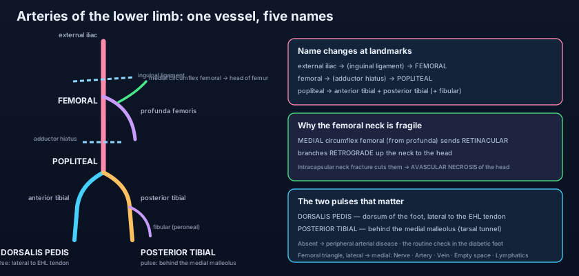
At the lower border of popliteus the single river finally forks, and each branch takes a different compartment. The popliteal artery ends by dividing into the anterior tibial artery and the tibioperoneal (tibiofibular) trunk. The anterior tibial artery does something no other artery in the limb does: it passes ABOVE the upper border of the interosseous membrane to reach the anterior compartment, then descends on the front of that membrane, between tibialis anterior and extensor hallucis longus, with the deep fibular nerve alongside it. At the ankle, midway between the two malleoli, it crosses under the extensor retinaculum and — name change five — becomes the dorsalis pedis artery. You feel it on the dorsum of the foot immediately LATERAL to the tendon of extensor hallucis longus; ask the patient to lift the big toe, the tendon stands up, and the pulse is beside it. The dorsalis pedis gives the arcuate artery to the toes, then dives between the first and second metatarsals as the deep plantar artery to join and complete the plantar arch on the sole. Meanwhile the tibioperoneal trunk divides into the fibular (peroneal) artery and the posterior tibial artery. The fibular artery runs down behind the fibula and is the vessel that supplies the lateral compartment — a compartment with no artery of its own, fed entirely by perforating branches reaching round the bone, a fact worth remembering when reading the muscles of the leg. The posterior tibial artery descends through the deep posterior compartment, and at the ankle passes behind the medial malleolus inside the tarsal tunnel, where it is palpable, before dividing into the medial and lateral plantar arteries. The lateral plantar artery is the larger, and it curves across the sole to form the deep plantar arch, which the deep plantar branch of the dorsalis pedis joins from above — so the two great arteries of the leg, one anterior and one posterior, shake hands under the foot.
The safety nets: cruciate and trochanteric anastomoses
The body does not trust a single pipe. Around the hip it builds two named ring-roads. The trochanteric anastomosis sits in the trochanteric fossa and is formed by the superior and inferior gluteal arteries meeting the two circumflex femoral arteries; it is the main source of the retinacular vessels to the femoral head. The cruciate anastomosis lies lower, at the level of the lesser trochanter, where four vessels meet in a rough cross: the inferior gluteal artery from above, the first perforating artery of the profunda from below, and the medial and lateral circumflex femoral arteries from either side. Together they mean that blood from the internal iliac system can reach the thigh even if the external iliac or femoral artery is blocked. The same principle repeats at the knee (the genicular anastomosis) and around the ankle and foot (the malleolar and plantar arches). It is the reason a patient can occlude the superficial femoral artery almost completely and still walk into clinic with a warm foot — the collaterals have had years to open. Compare this with the arteries of the upper limb, where the scapular anastomosis around the shoulder does exactly the same job for the arm: wherever a joint bends and a vessel could be pinched, evolution has built a bypass.
The four pulses of the lower limb are not an examination ritual — they are a map, and each one localises the block to a segment. Femoral (at the mid-inguinal point): present means the aortoiliac segment is open. Popliteal (deep in the fossa, knee slightly flexed, both thumbs in front and fingers pressing behind): present means the femoral and adductor canal segment is open. Posterior tibial (behind the medial malleolus): present means the posterior tibial artery has survived the calf. Dorsalis pedis (lateral to the extensor hallucis longus tendon on the dorsum): present means the anterior tibial has survived. Work down the limb and the first pulse that disappears tells you the level of disease. Remember too that the dorsalis pedis is congenitally absent or non-palpable in a small but real minority of perfectly healthy people — so an absent dorsalis pedis with a normal posterior tibial is a finding to interpret, not a diagnosis. The arteries' return journey — the deep veins that run with them and the superficial saphenous system — is traced in the veins and lymphatics of the lower limb.
The two-hundred-metre man: intermittent claudication. A narrowed superficial femoral artery delivers enough blood for a calf at rest but not for a calf that is working, so the muscle switches to anaerobic metabolism and cramps — and stops hurting the moment demand falls. The bedside number is the ankle–brachial pressure index (ABPI), the ankle systolic pressure divided by the arm's: normal is about 0.9 to 1.3, and below 0.9 means peripheral arterial disease. The cardiologist's doorway: because the femoral artery is large, superficial and lies over the head of the femur (a firm surface to compress against afterwards), it is the classic access route for cardiac catheterisation — a wire passed retrograde up the femoral, iliac and aorta to the heart. The bypassed leg: when the superficial femoral artery occludes in the adductor canal, a graft is sewn from the femoral artery above to the popliteal below — the femoral–popliteal bypass — literally rebuilding the missing stretch of river. The diabetic foot: diabetes damages the smallest vessels and the nerves together, so a patient can have palpable pulses and still develop a foot ulcer, because the microcirculation fails and neuropathy removes the pain that would have warned them. Never let a present pulse reassure you about a numb foot.
- Abdominal aorta → common iliac → external iliac (giving the inferior epigastric and deep circumflex iliac) → femoral, at the mid-inguinal point under the inguinal ligament.
- The internal iliac supplies the buttock and medial thigh via the superior gluteal, inferior gluteal and obturator arteries.
- In the femoral triangle, lateral → medial is NAVEL; the artery is LATERAL to the vein, and the femoral nerve lies OUTSIDE the femoral sheath.
- The profunda femoris is the main supply of the thigh: lateral and medial circumflex femoral + four perforating arteries.
- The medial circumflex femoral gives retinacular branches running RETROGRADE up the femoral neck — the sole supply of the adult femoral head.
- An INTRACAPSULAR neck of femur fracture tears them → avascular necrosis → replace the head; an intertrochanteric fracture spares them → fix it.
- Femoral → adductor (subsartorial/Hunter's) canal → adductor hiatus → popliteal artery.
- The popliteal artery is the DEEPEST structure in the popliteal fossa — hence a deep pulsatile aneurysm, and injury in knee dislocation or supracondylar femoral fracture.
- It gives the superior, middle and inferior genicular arteries forming the genicular anastomosis around the knee.
- At the lower border of popliteus it divides: anterior tibial (ABOVE the interosseous membrane → dorsalis pedis) and tibioperoneal trunk.
- The tibioperoneal trunk gives the fibular artery (the only supply of the lateral compartment) and the posterior tibial artery.
- Four pulses: femoral (mid-inguinal point), popliteal (deep in the fossa), posterior tibial (behind the medial malleolus), dorsalis pedis (lateral to the EHL tendon).
- Locating the femoral pulse at the mid-point of the inguinal ligament. It is at the MID-INGUINAL POINT — midway between the ASIS and the PUBIC SYMPHYSIS — which is not the same landmark as the midpoint of the ligament (ASIS to pubic tubercle), where the deep inguinal ring lies.
- Putting the femoral vein lateral to the artery. Lateral to medial the order is Nerve, Artery, Vein — the vein is MEDIAL to the pulse, which is exactly why a needle placed a finger's breadth medial to the pulse hits the vein.
- Assuming the fibular (peroneal) artery supplies the anterior compartment. It runs in the posterior compartment behind the fibula and feeds the LATERAL compartment by perforating branches; the anterior compartment belongs to the anterior tibial artery.
An 82-year-old woman sustains a displaced intracapsular fracture of the neck of the femur. Disruption of which vessel's branches best explains the high risk of avascular necrosis of the femoral head?
- One continuous vessel, renamed at each landmark: external iliac → femoral (under the inguinal ligament at the mid-inguinal point) → popliteal (through the adductor canal and adductor hiatus) → anterior tibial (→ dorsalis pedis) and the tibioperoneal trunk → fibular + posterior tibial (→ medial and lateral plantar arteries).
- The profunda femoris supplies the thigh (lateral and medial circumflex femoral + four perforators); the medial circumflex femoral sends retrograde retinacular branches up the femoral neck, so an intracapsular fracture causes avascular necrosis of the head.
- In the femoral triangle the order is NAVEL (artery LATERAL to vein, nerve OUTSIDE the sheath); the popliteal artery is the DEEPEST structure in the popliteal fossa and is at risk in knee dislocation.
- Four pulses map the limb — femoral, popliteal, posterior tibial (behind the medial malleolus), dorsalis pedis (lateral to the EHL tendon); ABPI below 0.9 means peripheral arterial disease, and rich anastomoses (cruciate, trochanteric, genicular) let a slow occlusion pass unnoticed while a sudden one gives the six Ps.
- Drake RL, Vogl AW, Mitchell AWM. Gray's Anatomy for Students — Lower limb: arteries of the thigh, leg and foot.
- Moore KL, Dalley AF, Agur AMR. Clinically Oriented Anatomy — Blood supply of the lower limb and the femoral triangle.
- Netter FH. Atlas of Human Anatomy — Arteries of the thigh, popliteal fossa, leg and sole.
- Last RJ. Last's Anatomy: Regional and Applied — The femoral artery, adductor canal and popliteal fossa.
- Snell RS. Clinical Anatomy by Regions — Peripheral arterial disease, femoral neck fracture and avascular necrosis.
- TeachMeAnatomy — Arterial Supply to the Lower Limb; The Femoral Triangle; The Popliteal Fossa.

